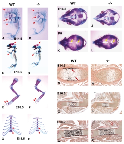
Fig. 2.
Bone formation is delayed in Gpr48-/- embryos. (A-H) Whole skeletal preparation of wild-type (A,C,E,G) and Gpr48-/- mutant (B,D,F,H) mice at E14.5 (A,B), E16.5 (C,D) and E18.5 (E-H). Arrows indicate the delayed Alizarin Red staining (of bone) in the skull, jaw, sternum, clavicle, phalange and limb in Gpr48-/- embryos. (I-L) Effects of Gpr48 on intramembranous bone formation and skull ossification. Top view of the mouse skull at E16.5 (I,J) and P0 (K,L) for wild-type (I,K) and Gpr48-/- (J,L) mice after Alcian Blue/Alizarin Red staining. Arrows indicate the widening of cranial sutures and the opened fontanelles in Gpr48-/- newborn mice. (M-R) von Kossa staining of femur of wild-type (M,O,Q) and Grp48-/- (N,P,R) embryos at E14.5 (M,N) (20×), E16.5 (O,P) (5×) and E18.5 (Q,R) (5×). Arrows indicate that wild-type mice already show von Kossa signal at E14.5, but this is absent from Gpr48-/- mice.

