ABSTRACT
Objective: To report a transoral freehand technique using continuous image guidance to biopsy a clival lesion. Methods: The patient was placed in a Mayfield head rest. Patient data were registered using preacquired magnetic resonance and computed tomography images, and then, under continuous image guidance, a bone biopsy instrument was advanced freehand through a transoral route into the clival lesion. Results: Bone biopsy instrumentation was successfully placed into the clival lesion. Biopsy forceps were placed through the working channel of the biopsy instrumentation and specimen was successfully retrieved from the clival mass. The patient tolerated the procedure well and the only complaint was dysphagia, which resolved in several days postoperatively. Conclusion: The transoral freehand technique with stereotactic navigation was found to be a simple, safe, fast, and effective way to biopsy a clival lesion.
Keywords: Stereotactic, transoral, clivus, image guidance
Lesions of the clivus are relatively rare. The most common destructive lesions to affect the clivus are chordomas and metastasis.1 Gaining surgical access to the clivus is technically challenging; however, transoral and transnasal approaches have been well documented in the literature.2,3,4,5,6,7,8 In the past, neurosurgeons have biopsied clival lesions using a transnasal or transoral open approach, with and without neuronavigation. Stereotactic biopsies have also been reported using a frame-based stereotactic technique.3 To the best of our knowledge, there is no report in the literature on freehand stereotactic biopsy using continuous image guidance. Here, the authors report a transoral freehand technique using continuous image guidance to biopsy a clival lesion in a 54-year-old woman with a history of breast cancer.
CASE REPORT
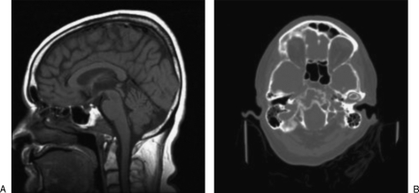
We examined a 54-year-old woman with a history of breast cancer who was referred by her oncologist after a lesion in the clivus was discovered by positron emission tomographic (PET) scan and confirmed by computed tomography (CT) and magnetic resonance imaging (MRI) during routine postsurgical follow-up (Fig. 1). She was neurologically intact and without complaint and underwent stereotactic biopsy for diagnosis.
Figure 1.
(A) Saggital T1 magnetic resonance imaging and (B) axial computed tomography scan demonstrating destructive clival lesion.
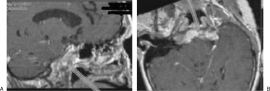
The patient was positioned supine on the operating room table with her head placed in a Mayfield fixation device in neutral position. Patient data were registered with the Stryker Navigation System II (Kalamazoo, MI) using previously acquired CT and MRI scans. The pharyngeal wall was disinfected with Betadine, and the patient received intravenous Ancef preoperatively. Two red rubber catheters were placed through the nose and out through the mouth for superior retraction of the soft palate. The Stryker bone biopsy instrumentation was registered with the Stryker image guidance system. The device, consisting of a sharp serrated tip with a hollow working channel, was advanced through the posterior pharyngeal wall and into the clivus under continuous image guidance. Once we were satisfied that the instrument had reached the center of the clival lesion (Fig. 2), small biopsy forceps were introduced through the working channel, and tissue from the lesion was obtained and sent to pathology. The patient tolerated the procedure well. Her only complaint was dysphagia, which resolved in ~6 days postoperatively. The pathology was consistent with metastatic breast carcinoma, and the patient underwent radiation therapy.
Figure 2.
Continuous image guidance using the Stryker navigation system. Biopsy instrumentation is shown entering the clival lesion in the (A) saggital and (B) axial planes.
DISCUSSION
Though there are many reports of transoral and transnasal stereotactic biopsy of clival lesions, to the best of our knowledge, this is the first report of the use of a freehand technique without a stereotactic frame. Patil and Chand3 reported four cases of transoral stereotactic biopsy of clival lesions in which they used a mouth gag and tongue depressor to keep the mouth open and the tongue out of the way. The procedure was performed on the CT table, and Ancef was given preoperatively. A stereotactic frame was used, with the probe holder adjusted on the arc and set to the appropriate trajectory (midline, through the soft palate and posterior pharyngeal wall). A sharp stylet was advanced through the stereotactic holder and into the target. This stereotactic approach was found to be safe and effective.
Vougioukas et al8 reported on the advantages of using neuronavigation to perform the open transoral approach. For the four patients in their study, they found that neuronavigation optimized preoperative planning and aided in performing safe wide resections of pathology located at the cranial base and craniocervical junction.
The transoral approach to the clivus, with or without neuronavigation, has been well reported and accepted by the neurosurgical community. Our novel freehand stereotactic technique with continuous image guidance was found to be safe, fast, and effective, with minimal patient discomfort, adequate sample obtained for pathology, and minimal operative time and setup. With the widespread availability of surgical image guidance systems, this technique is well suited for performing biopsies in patients with clival lesions.
REFERENCES
- Osborn A G. Diagnostic Neuroradiology. St. Louis: Mosby; 2008. p. 506.
- Menezes A H, VanGilder J C. Transoral-transpharyngeal approach to the anterior craniocervical junction. Ten-year experience with 72 patients. J Neurosurg. 1988;69:895–903. doi: 10.3171/jns.1988.69.6.0895. [DOI] [PubMed] [Google Scholar]
- Patil A A, Chand A. Modifications of transnasal and transoral stereotactic procedures–technical notes. Acta Neurochir (Wien) 1995;134:46–50. doi: 10.1007/BF01428501. [DOI] [PubMed] [Google Scholar]
- Pollack I F, Welch W, Jacobs G B, Janecka I P. Frameless stereotactic guidance. An intraoperative adjunct in the transoral approach for ventral cervicomedullary junction decompression. Spine. 1995;20:216–220. [PubMed] [Google Scholar]
- Seifert V, Laszig R. Transoral transpalatal removal of a giant premesencephalic clivus chordoma. Acta Neurochir (Wien) 1991;112:141–146. doi: 10.1007/BF01405143. [DOI] [PubMed] [Google Scholar]
- Spetzler R F, Hadley M N, Sonntag V K. The transoral approach to the anterior superior cervical spine. A review of 29 cases. Acta Neurochir Suppl (Wien) 1988;43:69–74. doi: 10.1007/978-3-7091-8978-8_16. [DOI] [PubMed] [Google Scholar]
- Veres R, Bago A, Fedorcsak I. Early experiences with image-guided transoral surgery for the pathologies of the upper cervical spine. Spine. 2001;26:1385–1388. doi: 10.1097/00007632-200106150-00024. [DOI] [PubMed] [Google Scholar]
- Vougioukas V I, Hubbe U, Schipper J, Spetzger U. Navigated transoral approach to the cranial base and the craniocervical junction: technical note. Neurosurgery. 2003;52:247–250. doi: 10.1097/00006123-200301000-00034. [DOI] [PubMed] [Google Scholar]


