Abstract
Wilson disease is an inherited disorder of human copper metabolism, characterized by gradual accumulation of copper in tissues, predominantly liver and brain. The gene defect lies in the Wilson disease protein ATP7B, a copper transporting ATPase highly active in hepatocytes. In the liver, ATP7B is essential for excretion of excess copper into the bile and for copper loading of ceruloplasmin in the Golgi apparatus. The extrahepatic role of ATP7B is not yet completely understood. We analysed the intestinal expression of ATP7B in mice using RT-PCR, Western blot and indirect immunofluorescence. We found abundant expression of ATP7B in stomach and small intestine, but not in colon. Using confocal microscopy we demonstrate a Golgi localization of ATP7B in enterocytes. In response to elevated copper, the Wilson disease protein shows an intracellular trafficking pattern in the intestinal polarized cell line CaCo-2, moving away from the Golgi apparatus to dispersed vesicles. This suggests a role for intestinal ATP7B in sequestration of copper in intracellular vesicles for maintenance of copper homeostasis in the enterocyte. In conclusion, the expression of ATP7B in the small intestine might represent an additional regulatory mechanism to fine-tune intestinal copper absorption.
Keywords: ATP7B, CaCo-2, gut, Wilson disease
Introduction
Wilson disease (WD) is a rare autosomal recessively inherited human disorder of copper metabolism (Scheinberg & Sternlieb, 1965; Gitlin, 2003; Ala et al. 2007) characterized by copper accumulation in various tissues, mainly in liver and brain, causing severe neurological defects and liver disease. WD is caused by mutations in the Wilson disease protein (WDP) gene encoding the transmembrane copper-transporting ATPase ATP7B (Bull et al. 1993; Petrukhin et al. 1993; Tanzi et al. 1993) highly expressed in the liver (Schaefer et al. 1999a,b). The WDP is critical for excretion of copper into the bile (Terada et al. 1999) but also supplies copper ions for the ferroxidase ceruloplasmin (Terada et al. 1998), which is the main copper-containing protein of serum (Hellman & Gitlin, 2002).
X-linked Menkes disease is another inherited copper transport defect in humans and is caused by mutations in the Menkes protein (MNK) ATP7A, a related intestinal P-type ATPase (Cox & Moore, 2002; Mercer & Llanos, 2003; Lutsenko et al. 2007). Mutations in MNK lead to a fatal condition of systemic copper deficiency due to impaired intestinal copper absorption (Danks, 1988; Kaler, 1998).
ATP7A and ATP7B belong to a family of cation-transporting P-type ATPases translocating ions across cell membranes by hydrolysis of ATP (Kuhlbrandt, 2004). ATP7B and ATP7A share a 57% sequence homology (Vulpe & Packman, 1995) and both change their intracellular location in response to alterations of the cellular copper level [as reviewed by Harris (2000) and Lutsenko et al. (2003)].
At low copper levels, ATP7B is localized to the late Golgi compartment (Roelofsen et al. 2000; Huster et al. 2003) where presumably copper loading of ceruloplasmin takes place. However, when copper is high, ATP7B shifts away from the Golgi apparatus to cytoplasmically dispersed vesicles which in polarized liver cells accumulate subapically (Hung et al. 1997; Schaefer et al. 1999a,b; Roelofsen et al. 2000; Guo et al. 2005; Cater et al. 2006). It is not clear how copper transported to these vesicles would finally reach the bile but it is generally assumed that the translocation of ATP7B is a necessary pre-condition for copper excretion (Forbes & Cox, 2000; Gitlin, 2003; Cater et al. 2007).
Like ATP7B, ATP7A resides in the trans-Golgi network (TGN) at low copper levels (Dierick et al. 1997). It has been postulated that ATP7A-laden vesicles are continually moving between the Golgi and plasma membrane (Camakaris et al. 1995; Petris et al. 1996; Petris & Mercer, 1999). Further studies suggested a translocation of ATP7A to the plasma membrane (PM) in response to elevated copper levels in non-polarized cell models (Petris et al. 1996; Petris et al. 2002; Cobbold et al. 2003; Pase et al. 2004) and to the basolateral membrane in polarized cell models (Greenough et al. 2004). However, reports concerning trafficking and especially the amount of ATP7A at the plasma membrane are ambiguous.
ATP7A immunoreactivity has been reported in the perinuclear region of mouse enterocytes when exposed to low copper and in vesicles located near the basolateral membrane of mice exposed to high Cu (Monty et al. 2005).
Using in vivo confocal colocalization studies, Nyase and coworkers (Nyasae et al. 2007) could not detect an overlap between ATP7A and basolateral markers in polarized CaCo-2 cells after copper loading. Only by using a biotinylation assay were 8–10% of ATP7A detectable at the basolateral membrane under these conditions. However, the reported data seem to be consistent with the model that ATP7A facilitates the translocation of copper ions across the basolateral membrane.
Based on the discrepancy in their trafficking pattern concerning the destination compartment (subapical vesicles for ATP7B vs. basolateral plasma membrane for ATP7A) it seemed likely that there are distinct functional roles for ATP7A and ATP7B. With this in mind, the tissue expression data for both ATPases provide important additional information.
In the murine embryo RNA in situ hybridization showed ATP7A expression in almost all tissues including the brain, heart, lung, liver, kidney and skin (Kuo et al. 1997). In adult liver, ATP7A expression could not be confirmed, but expression of MNKmRNA was reported in muscle, kidney, lung, brain; placenta and pancreas (Chelly et al. 1993; Mercer et al. 1993) as well as in heart, spleen and testis (Levinson et al. 1994; Mercer et al. 1994).
Recently, Ravia and coworkers reported ATP7A to be present on brush border membranes and basolateral membranes in rat duodenum (Ravia et al. 2005). This observation could only in part be confirmed by others, who did not observe a predominant apical PM localization of ATP7A in murine duodenum and upper jejunum (Nyasae et al. 2007) or in jejunum of ATP7A transgenic mice (Monty et al. 2005).
In contrast to ATP7A, the WDP gene is strongly expressed in the liver (Bull et al. 1993; Yamaguchi et al. 1993). Beyond the liver, ATP7B expression was reported for kidney (Moore & Cox, 2002; Linz et al. 2007) and the central nervous system (Kuo et al. 1997; Barnes et al. 2005). Expression of mRNA was demonstrated by in situ hybridization in heart, lung, respiratory epithelia and thymus, and was abundant in embryonic intestine (Kuo et al. 1997). ATP7B expression has also been described in sheep intestine (Lockhart et al. 2000).
Furthermore, ATP7B expression was described in mammary tissue (Michalczyk et al. 2000; Michalczyk et al. 2008). Recent studies revealed the presence of both APT7A and ATP7B in kidney (Linz et al. 2007) and in the syncytiotrophoblast of human placenta (Hardman et al. 2004). A coexpression of ATP7B and ATP7A has furthermore been reported for human placental Jeg-3 cells (Hardman et al. 2007a,b), OK cells and MDCK cells (Linz et al. 2007) and most recently for the polarized human mammary cell line PMC42-LA (Michalczyk et al. 2008).
Here we characterize ATP7B expression in murine intestine for the first time. Our results demonstrate significant expression of ATP7B in stomach and small intestine. Interestingly, the expression pattern of ATP7B partially overlapped that of ATP7A (Nyasae et al. 2007). We demonstrated the copper-dependent trafficking of ATP7B in non-polarized and polarized CaCo-2 cells, an established model for enterocytes of the small intestine. Our data imply a role for ATP7B in the regulation of enterocyte copper homeostasis, most likely by sequestration of excess copper in enterocytes or possibly facilitating apical excretion. As our data indicate a coexpression of ATP7B and ATP7A in the enterocyte, this could suggest that cross-talk between ATP7B and ATP7A might be of relevance for the regulation of intestinal copper absorption.
Materials and methods
Antibodies
The antibody against ATP7B was essentially prepared as previously described (Hung et al. 1997). Oligonucleotides were designed and utilized to amplify the region of the Wilson protein encoding amino acids 325–635. This region was amplified by PCR and subcloned in the pGEX-2T vector (Amersham Pharmacia Biotech). Escherichia coli BL21 cells harboring the expression plasmid were grown to an optical density of 1.5 at 600nm at 31°C and induced with isopropyl 1-thio-β-d-galactopyranoside. Cultures were harvested by centrifugation, resuspended in phosphate-buffered saline (PBS) containing 1% Triton X-100 and lysed using a high pressure cell crusher. The supernatant was incubated with glutathione-agarose beads. Bound glutathione S transferase (GST) fusion protein was thrombin cleaved to obtain the ATP7B-fragment. New Zealand White rabbits were immunized with 4×100mg of this recombinant protein (immunization was carried out according to standard procedures of EuroGentec, Belgium). Affinity purification of the antiserum was performed using AminoLink® Plus columns as recommended by the manufacturer (Pierce, Rockford, IL, USA). Recombinant protein was coupled to the columns using 0.1m sodium citrate, 0.05m sodium carbonate, pH 10. Remaining active sites were blocked by sodium cyanoborohydride solution (5m NaCNBH3 in 0.01m NaOH). An additional pre-elution step using elution buffer (0.2m glycine-HCl; pH 2.6) was performed to remove non-covalently bound recombinant protein. After washing of the column with PBS (pH 7.2), the antiserum was loaded to the column. After repeated washing of the column, the bound antibodies were eluted from the column using the elution buffer. Protein concentration of the eluate was determined using a mini Bradford assay. The eluted antibody solution was rebuffered in PBS 5% BSA (bovine serum albumin) using PD-10 columns. For long-term storage, 50% glycerol was added to the rebuffered antibody solution.
The monoclonal anti-NaK-ATPase-Ab (clone 9A-5) was obtained from Dianova (Hamburg, Germany). The monoclonal anti-human-placental alkaline phosphatase (PLAP)-Ab (clone 8B6) was obtained from DAKO (Glostrup, Denmark). The biotinylated Rer1 antibody was raised against the oligopeptide CTHGKRRYRGKEDAGKAFAS, affinity purified and biotinylated as described for gp27 (Fullekrug et al. 1999).
Cell culture and copper exposure
HepG2 (ATCC-No.: HB-8065) cells and CaCo-2 (ATCC-No.: HTB-37) cells were maintained under standard tissue culture conditions with the appropriate culture media. CaCo-2 cells were grown on coverslips till 80% confluent or on polyester filter inserts (Transwell Clear, 12-mm diameter, pore size 0.4µm; Corning, Bodenheim, Germany) for 4weeks and formation of a tight layer and polarization was monitored, respectively.
Before copper exposure, cells were washed twice in prewarmed PBS (pH 7.2) to remove culture medium. Cells were incubated for 2h either with prewarmed DMEM containing 100µm of the copper chelator bathocuproine disulfonic acid (BCS) or 100µm copper sulfate (CuSO4) alternatively. When filter-grown cells were used, solutions were applied to both the apical and basolateral chamber. After repeated washing in PBS, cells were fixed using paraformaldehyde (PFA) (4% PFA in PBS; pH 7.2; 10min).
Mice
The generation of the Atp7b−/– mice has been described previously (Buiakova et al. 1999). Mice were housed at the University of Heidelberg Animal Facility according to the national health guidelines on the use of laboratory and experimental animals. Food and water were provided ad libitum and no further treatment was performed. Mice were sacrificed at 4weeks of age. Pieces of mouse tissues of wild-type mice from a C57/Bl6 background and from ATP7B-knock-out mice were embedded in Tissue-Tek (Sakura Finetek, Tokyo, Japan) and flash frozen in liquid nitrogen. Cryosections were cut at 2–5µm, and then processed for immunofluorescence.
Immunofluorescence and immunohistochemistry
Immunofluorescence procedures (fixation with paraformaldehyde, permeabilization with 0.1% saponin), confocal image acquisition on a Leica TCS SP2 system and arrangement with Adobe Photoshop were essentially performed as described earlier (Fullekrug et al. 1999). Antibodies were incubated in PBS containing 0.1% saponin (Sigma), 0.5% gelatine (Teleostan gelatine, Sigma) and 5mgmL−1 BSA. Corresponding secondary antibodies (Dianova, Hamburg, Germany) were used conjugated to Cy3 or fluoroscein isothiocyanate (FITC). In some experiments phalloidin-TRITC (Jackson Immuno Research, Baltimore, MD, USA) was added in a 1:10000 dilution to the secondary antibody solution. Before mounting, nuclei were stained with Hoechst 33342 (Invitrogen). Coverslips were mounted in Prolong Gold™ antifade mounting medium (Molecular Probes, Leiden, the Netherlands).
For double-immunofluorescence of Rer1 (biotinylated rabbit anti-Rer1) and ATP7B (rabbit anti-ATP7B) incubations were performed sequentially: After initial ATP7B-immunofluorescence, active sites of the 2nd antibody were blocked using rabbit IgG (Dianova) and an additional fixation-step with paraformaldehyde (4% PFA in PBS; pH 7,2; 10min) was performed. Sections were then treated with the Avidin-Biotin-Kit (Vector Laboratories, Burlingame, CA, USA) according to the manufacturer's protocol to block endogenous biotin before incubation with biotinylated rabbit-anti-Rer1 antibody. The biotinylated Rer1-antibody was visualized using Cy2-conjugated streptavidin (Jackson ImmunoResearch).
ATP7B immunofluorescence in tissue sections was analysed with an Olympus IX50 fluorescence microscope equipped with a 60× oil immersion objective. Confocal images were taken on a TCS SP2 microscope (Leica Microsystems, Germany, objective: 63× magnification, oil immersion, NA 1.32) of sections or filters stained for double-immunofluorescence for colocalization studies. Double immunofluorescence images were taken sequentially, and parameters were adjusted so that all light intensities were in the recording range. The intensity of the laser beam and photo multiplier levels was adjusted for each slide and each fluorophore (Cy2, Cy3, FITC, TRITC); line averaging was set to 4 to optimize the signal-to-noise ratio. Images shown were derived from representative single confocal planes and arranged with Adobe Photoshop and Adobe Illustrator.
Immunoblotting
Equal amounts of protein (25µg) were separated by sodium dodecyl sulfate–polyacrylamide gel electrophoresis (SDS-PAGE) in 10% gels followed by electrophoretic transfer to nitrocellulose membranes, incubated with the affinity purified anti-ATP7B-antibody and visualized using enhanced chemiluminescence detection. To verify the specificity of the affinity purified anti-ATP7B-antibody, protein-inhibition experiments were performed with the ATP7B-fragment used to raise the antibody. No signal was detected after preincubation of the primary antibody solution with the ATP7B-fragment.
Quantification of ATP7B translocation
After copper exposure and ATP7B immunofluorescence, coverslips were analysed with an Olympus IX50 fluorescence microscope equipped with a 60× oil immersion objective. Counting was blinded, as the examiner did not know which treatment the cells had received. A cell was classified as ‘translocated’ when a diffuse vesicular staining was observed. Cells showing a tight perinuclear ATP7B-immunoreactivity without a vesicular pattern were classified as ‘not translocated’. Translocated cells were divided by the total number of cells (approximately 200 cells were counted for each coverslip; the translocation ratio was calculated per coverslip; two coverslips were counted per experiment). The standard deviation was calculated from two independent experiments.
Real-time PCR
Total RNA was isolated from tissues with the RNeasy Mini Kit including DNAse digestion. The Omniscript RT kit and random N6 primers were used for reverse transcription. All reagents and procedures for cDNA synthesis were from Qiagen (Hilden, Germany). Real-time PCR was performed using the LightCycler system (Roche, Mannheim, Germany). Transcripts were detected with SYBR Green I and normalized to β-actin as internal control. Light Cycler Relative Quantification Software Version 1.0 together with calibration plasmids pRRL-PGK-ATP7B (Merle et al. 2006), EST-m-atp7b (RZPD BQ768959 Berlin, Germany) was used for calculation of ATP7B/actin ratios. Primer sequences: mouse atp7b: ACA TGG TTG GGA TAC CTA TTG C, TTG AGC TGA AGA GAC GAG AGC; mouse β-actin: GAC GGC CAG GTC ATC ACT ATT G, CCA CAG GAT TCC ATA CCC AAG A; human ATP7B: CCA CAT GAA GCC CCT GAC, GTA CTG CTC CTC ATC CCT GC; human β-ACTIN: AGG ATG CAG AAG GAG ATC ACT G, GGG TGT AAC GCA ACT AAG TCA TAG.
Results
ATP7B is expressed in mouse upper intestine and CaCo-2 cells
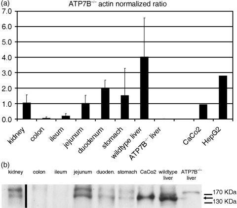
Extrahepatic mouse tissues were evaluated for their ATP7B expression by real-time PCR. As shown in Fig. 1a, analysis confirmed an abundant expression of ATP7B in liver and strong expression in kidney, stomach, duodenum and jejunum. Tissue samples derived from wild-type colon, wild-type ileum or ATP7B−/– liver did not show any signal above background for ATP7B. Caco-2 cells are an established model for small intestine and were evaluated for their mRNA content of hATP7B/WDP and showed a significant expression.
Fig. 1.
a) Real-time PCR quantification of ATP7B in murine tissues and human CaCo-2 cells. Mouse tissues were evaluated for their mRNA content of mATP7B. The results confirmed a strong expression of mATP7B in liver and moderate expression in kidney, stomach, duodenum and jejunum. Tissue samples derived from wild-type colon, wild-type ileum or liver of ATP7B−/– mice did not show a significant signal above background (*). Error bars indicate the standard deviation of three independent experiments (different mice). Caco-2 and HepG2 cells were evaluated for their mRNA content of hATP7B/ WDP and showed a significant expression (right columns). Arbitrary units were obtained by normalizing the signal to the internal control (values shown are 10−3 * signal ATP7B/signal actin). b) Immunoblot analysis of ATP7B in murine tissues and CaCo-2 cells. Tissues or cells were lysed, and 25µg of protein was separated by SDS-PAGE, transferred to nitrocellulose, incubated with the affinity-purified ATP7B antibody, and analysed by chemiluminescence. The lane with mouse kidney lysate is derived from a different blot.
Real-time PCR data were confirmed by Western blot analysis. As shown in Fig. 1b, a band at ∼160kDa was observed in wild-type mouse liver, kidney, stomach, duodenum, jejunum and CaCo-2 cell lysates analysed by immunoblotting with the affinity purified antibody specific to the human Wilson protein. No signal was detected in ileum and colon (Fig. 1b) or in esophagus, rectum and caecum (data not shown).
ATP7B is expressed in enterocytes in the upper intestine
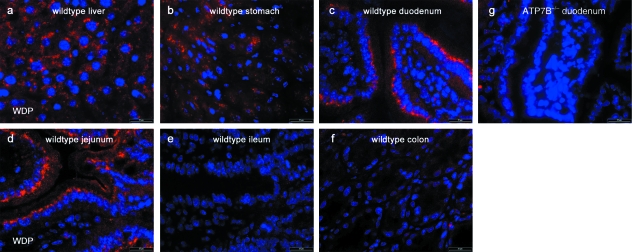
In mouse liver sections the staining pattern of the affinity purified polyclonal ATP7B antibody was in conformity with previously published results (Schaefer et al. 1999a,b), as abundant ATP7B immunoreactivity was found in the perinuclear region in hepatocytes (Fig. 2a). Surprisingly, strong ATP7B immunoreactivity was observed in the upper mouse intestine, where the ATP7B signal localized to a perinuclear, supranuclear region at the luminal side of the nucleus in enterocytes of duodenum (Fig. 2c) and jejunum (Fig. 2d). Surprisingly strong ATP7B immunoreactivity was also found in samples from mouse stomach (Fig. 2b).
Fig. 2.
Tissue localization of ATP7B in mouse intestine. a–d) ATP7B immunofluorescence in tissue derived from wild-type mice showed strong ATP7B immunoreactivity in liver, stomach, duodenum and jejunum. No signal above background was detected either in ileum (e) and colon (f) of wild-type mice or in duodenum derived from ATP7B−/– mice (g). Bars: 20µm.
No significant immunoreactivity was observed in other parts of the intestine. As shown in Fig. 2e–f, no ATP7B immunoreactivity could be visualized in distal ileum or colon. Esophageal samples and mouse tissues derived from caecum or rectum were ATP7B negative (data not shown).
Interestingly we observed strong ATP7B expression in areas of the intestine where Nyase and coworkers reported significant ATP7A expression recently (Nyasae et al. 2007). To exclude a significant cross-reactivity of the ATP7B-antibody with the Menkes protein, tissue sections derived from ATP7B−/– mice were stained. Here no signal beyond background was observed for the affinity purified anti-ATP7B antibody either in ATP7B−/– liver tissue or in tissue sections of small intestine from ATP7B−/– mice (ATP7B−/– duodenum; Fig. 2g).
ATP7B localizes to the Golgi apparatus of enterocytes
To analyse the subcellular localization of ATP7B, colocalization studies with apical and basolateral membrane markers and the Golgi-marker Rer1 were performed in sections from small intestine.
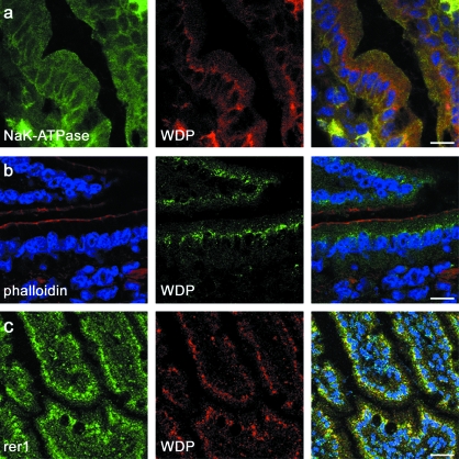
As actin is concentrated in the terminal web directly underneath the apical membrane we used phalloidin-TRITC, which binds to actin, to visualize the apical actin cortex. We could not detect an overlap of ATP7B immunoreactivity with the phalloidin staining of the apical actin cortex (Fig. 3b).
Fig. 3.
Localization of ATP7B in relation to marker proteins for the apical and basolateral plasma membrane and the Golgi apparatus. No overlap between ATP7B (mid lane) and the basolateral membrane marker NaK-ATPase (a) was observed. The apical actin cortex was visualized by phalloidin-TRITC (b, red) but showed no colocalization with ATP7B immunoreactivity. As is shown in (c), ATP7B colocalized with the Golgi marker Rer1. Cell nuclei were stained with Hoechst 33342 (blue). Bars: (a,b) 10µm, (c) 20µm.
No colocalization of ATP7B and the basolateral plasma membrane marker NaK-ATPase (Fig. 3a) was observed. But ATP7B immunoreactivity was found to overlap with the Golgi marker Rer1 (Fig. 3c).
Translocation of ATP7B is evident in intestinal CaCo-2 cells
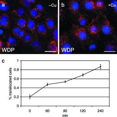
We used CaCo-2 cells, an established model of human small intestine to analyse the copper-dependent translocation of ATP7B. Under conditions of low copper, ATP7B localized to distinct perinuclear regions (Fig. 4a). Under elevated copper levels, a more diffuse small punctate staining pattern was observed (Fig. 4b).
Fig. 4.
a) Copper-dependent translocation of endogenous ATP7B in CaCo-2 cells. CaCo-2 cells were incubated for 2h either with the copper chelating agent BCS (A) or with 100µm CuSO4 (b). Cells were fixed and endogenous ATP7B stained by indirect immunofluorescence. Under conditions of low copper, ATP7B localized to distinct perinuclear regions (a). Under elevated copper levels, a more diffuse small puncta staining pattern was observed (b). Bars: 20µm. c) Time course of translocation of endogenous ATP7B in CaCo-2 cells. After an initial treatment with the copper chelator BCS for 1h, CaCo-2 cells were exposed to 100µm copper ions for variable amounts of time. Microscopy evaluation was done after indirect immunofluorescence staining of ATP7B, scoring Golgi pattern (not translocated) vs. dispersed staining (translocated). Data are expressed as mean±standard deviation.
The characteristic differences in the ATP7B distribution pattern allowed for quantification of translocated cells by scoring the cells as Golgi pattern (not translocated) vs. dispersed staining (translocated). With increasing copper exposure duration, more cells were found translocated as shown in the time course in Fig. 4c. Remarkably, after only 60min, up to 50% of the cells showed a diffuse vesicular distribution of ATP7B.
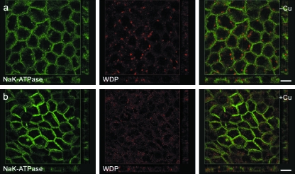
In some cells exposed to copper we noticed a slight enhancement of ATP7B immunoreactivity near the margins of the cells. To analyse whether this could be due to a plasma membrane localization of ATP7B we performed confocal colocalization studies in polarized CaCo-2 cells grown on filter inserts. In these polarized CaCo-2 cells the ATP7B signal was found predominantly in large puncta in the perinuclear region under conditions of copper depletion (Fig. 5a). As in non-polarized cells, the ATP7B distribution pattern changed after exposure to copper, resulting in small puncta dispersed throughout the cytoplasm (Fig. 5b). Under elevated copper conditions we did not find a significant accumulation of ATP7B immunoreactivity beneath either the apical or the basolateral membrane in CaCo-2 cells. Using confocal microscopy analysis we did not observe a colocalization of ATP7B with apical markers like PLAP (data not shown) or with basolateral markers as shown for NaK-ATPase in Fig. 5b.
Fig. 5.
Copper-dependent trafficking of ATP7B in polarized CaCo-2 cells. CaCo-2 cells were grown on filters till polarized and exposed for 2h to medium containing either 100µm CuSO4or 100µm BCS for 2h. Double immunofluorescence staining for ATP7B (red fluorescence) and NaK-ATPase (green fluorescence) was performed. Under conditions of low copper, ATP7B localized to distinct perinuclear regions, as shown in (a). In contrast, under elevated copper levels a more diffuse small puncta staining pattern was observed (b). Regardless of the copper level ATP7B and the basolateral marker NaK-ATPase were found segregated. Bars: 10µm.
Discussion
The clinical phenotype of ATP7B malfunction in Wilson disease causes symptoms mainly associated with liver and brain, although ATP7B expression is evident in many other tissues. Whereas in the liver, ATP7B is crucial for copper excretion in bile and loading of apo-ceruloplasmin, the function of extrahepatic ATP7B is not fully understood.
In this study we characterize the expression pattern of ATP7B in murine intestine and the intestinal cell line CaCo-2. Our data show a significant ATP7B expression in mouse small intestine, surprisingly in areas where abundant ATP7A expression has already been reported (Nyasae et al. 2007). To our knowledge there are no defined intestinal disease manifestations in either WD patients or animal models of the disease. Thus we found 41% of WD patients suffering from abdominal pain at initial presentation – a symptom rapidly diminishing under therapy (Stremmel et al. 1991). But it remains highly speculative to correlate these findings with WDP malfunction in intestine. Here, an approach to stain ATP7B in human material (e.g. biopsies at endoscopy) might be able to contribute to a further analysis. An examination of ATP7B localization in human duodenal biopsies might even be of some diagnostic value, e.g. ATP7B-mutations leading to an altered cellular localization could be identified that way.
Nevertheless, the strong expression of WDP in small intestine is remarkable and raises questions concerning the role of intestinal ATP7B. Intestinal copper uptake involves ctr1 and probably other transporters like DMT1 as apical transporters and ATP7A as the main basolateral copper transmembrane transporter (Lutsenko et al. 2007). A plausible role for ATP7B expression in small intestine could be fine tuning the regulation of dietary copper uptake as well as control of copper homeostasis of the enterocyte. This assumption would be in line with our finding of a predominant localization of ATP7B to the enterocytes which are facilitating copper absorption. In the polarized intestinal cell model CaCo-2 copper absorption has already been analysed using radioactive copper in a bi-chamber setting (Arredondo et al. 2000; Zerounian et al. 2003). The data from these studies support a model of an inverse relationship between copper availability and the capacity to absorb copper, consistent with homeostatic regulation (Zerounian et al. 2003).
In general, the translocation of ATP7B seems a necessary precondition for copper excretion. After copper exposure of non-polarized or polarized CaCo-2 cells we observed translocation of ATP7B to a diffuse vesicular compartment. In principle there are different plausible mechanisms for how ATP7B could facilitate copper export in the enterocyte. One is luminal excretion of excess copper. Most recently, Michalczyk and co-workers showed in 64Cu studies that ATP7B overexpression facilitates copper efflux from the apical surface in the polarized human mammary cell line PMC42-LA (Michalczyk et al. 2008). This observation hints at the conclusion that ATP7B facilitates apical copper efflux in different cells and that this conserved mechanism could find a correlate in the enterocyte as well. We could not detect accumulation of ATP7B-positive vesicles near or at the apical plasma membrane in enterocytes or CaCo-2 cells. But this does not rule out the possibility that a certain population of ATP7B-positive vesicles could be able to reach the apical membrane and facilitate copper excretion. But even if the main function of ATP7B-laden vesicles in the enterocyte is the sequestration of copper, total copper absorption could also be influenced. The life span of enterocytes is very limited and they are shed after a few days. In this way, copper stored in the shed enterocytes would be lost with the faeces. The mechanism of shedding of copper-laden enterocytes has been discussed to be of relevance in zinc therapy in WD as well. Zinc is an established oral treatment agent (Hoogenraad et al. 1979;Brewer et al. 1983), leading to decreased intestinal copper uptake, most likely by induction of metallothionein, a metal-binding protein, in the mucosal cells of the gut (Janssens et al. 1984;Yuzbasiyan-Gurkan et al. 1992;Reeves et al. 1996). Copper bound to zinc-induced metallothionein – or copper sequestered by ATP7B under physiological conditions – might then be lost by shedding.
But shedding would represent a mechanism unique to the enterocytes when compared to other extrahepatic tissues with significant coexpression of ATP7B and ATP7A. Recent studies have reported the coexpression of both proteins in retinal cells (Krajacic et al. 2006), placenta-derived cells (Hardman et al. 2007a,b), kidney and MDCK cells (Linz et al. 2007) and mammary tissues (Michalczyk et al. 2008). These data already support a new model that points to an involvement of both ATP7A and ATP7B in the control of cellular and systemic copper homeostasis with distinct functional roles. This recently postulated cross-talk (Linz et al. 2007; Lutsenko et al. 2007) between ATP7B and ATP7A might be of relevance for the regulation of intestinal copper absorption as well.
ATP7A and ATP7B seem to be able to substitute partially for each other, suggesting at least in part overlapping functions. For example, in ATP7B−/– mouse cerebellum the expression pattern of the ATP7B target gene ceruloplasmin shifts to cell types expressing ATP7A (Barnes et al. 2005). That observation suggests that ATP7A – at least in part – can restore copper delivery to ceruloplasmin. Further data suggest that ATP7A seems indeed capable of translocation of copper ions into the TGN (Hung et al. 1997; Payne & Gitlin, 1998). This would suggest that ATP7B might not to be mandatory for the delivery of copper ions to the TGN for apo-protein assembly in cells coexpressing ATP7A.
Conversely, expression of ATP7B partially restores the copper efflux in Menkes fibroblasts lacking ATP7A (La Fontaine et al. 1998), suggesting that ATP7A inactivity can in part be rescued by heterologous expression of ATP7B (La Fontaine et al. 1998; Lockhart et al. 2002). But as is evident from the Menkes disease phenotype, it seems likely that this copper transport capacity of ATP7B across the basolateral membrane in enterocytes is very limited – if ATP7B is involved in basolateral copper transport at all.
As both ATPases facilitate cellular copper efflux, their distinct functional roles seem to be defined by, on the one hand, basolateral-directed transport via ATP7A and, on the other, apical (or canalicular) transport by ATP7B. The role of ATP7B in maintaining body copper homeostasis might thus be a dual one, involving canalicular biliary excretion and fine-tuning of intestinal copper absorption. Furthermore, on the cellular level in the enterocyte, ATP7B might be involved in avoidance of copper overload due to excess copper. A plausible mechanism could be the sequestration of copper in intracellular vesicles for luminal excretion or storage in the enterocyte that is lost by shedding. In conclusion, the presence of ATP7B in enterocytes in small intestine is likely to contribute to maintenance of cellular copper homeostasis and thus modulate intestinal copper uptake.
Acknowledgments
We thank Sabine Tuma, Simone Staffer and Karin Bents for excellent technical assistance and Mark Schaefer for helpful discussions on this manuscript. We thank Günther Giese at the microscopy facility of the MPI for Medical Research, Heidelberg. This work was supported by a grant from the Dietmar Hopp Foundation (to W.S.) and a Young Investigator Grant of the Medical Faculty of the University of Heidelberg (to K.H.W.).
References
- Ala A, Walker AP, Ashkan K, Dooley JS, Schilsky ML. Wilson's disease. Lancet. 2007;369:397–408. doi: 10.1016/S0140-6736(07)60196-2. [DOI] [PubMed] [Google Scholar]
- Arredondo M, Uauy R, Gonzalez M. Regulation of copper uptake and transport in intestinal cell monolayers by acute and chronic copper exposure. Biochim Biophys Acta. 2000;1474:169–176. doi: 10.1016/s0304-4165(00)00015-5. [DOI] [PubMed] [Google Scholar]
- Barnes N, Tsivkovskii R, Tsivkovskaia N, Lutsenko S. The copper-transporting ATPases, menkes and wilson disease proteins, have distinct roles in adult and developing cerebellum. J Biol Chem. 2005;280:9640–9645. doi: 10.1074/jbc.M413840200. [DOI] [PubMed] [Google Scholar]
- Brewer GJ, Hill GM, Prasad AS, Cossack ZT, Rabbani P. Oral zinc therapy for Wilson's disease. Ann Intern Med. 1983;99:314–319. doi: 10.7326/0003-4819-99-3-314. [DOI] [PubMed] [Google Scholar]
- Buiakova OI, Xu J, Lutsenko S, et al. Null mutation of the murine ATP7B (Wilson disease) gene results in intracellular copper accumulation and late-onset hepatic nodular transformation. Hum Mol Genet. 1999;8:1665–1671. doi: 10.1093/hmg/8.9.1665. [DOI] [PubMed] [Google Scholar]
- Bull PC, Thomas GR, Rommens JM, Forbes JR, Cox DW. The Wilson disease gene is a putative copper transporting P-type ATPase similar to the Menkes gene. Nat Genet. 1993;5:327–337. doi: 10.1038/ng1293-327. [DOI] [PubMed] [Google Scholar]
- Camakaris J, Petris MJ, Bailey L, et al. Gene amplification of the Menkes (MNK; ATP7A) P-type ATPase gene of CHO cells is associated with copper resistance and enhanced copper efflux. Hum Mol Genet. 1995;4:2117–2123. doi: 10.1093/hmg/4.11.2117. [DOI] [PubMed] [Google Scholar]
- Cater MA, La Fontaine S, Shield K, Deal Y, Mercer JF. ATP7B mediates vesicular sequestration of copper: insight into biliary copper excretion. Gastroenterology. 2006;130:493–506. doi: 10.1053/j.gastro.2005.10.054. [DOI] [PubMed] [Google Scholar]
- Cater MA, La Fontaine S, Mercer JF. Copper binding to the N-terminal metal-binding sites or the CPC motif is not essential for copper-induced trafficking of the human Wilson protein (ATP7B) Biochem J. 2007;401:143–153. doi: 10.1042/BJ20061055. [DOI] [PMC free article] [PubMed] [Google Scholar]
- Chelly J, Tumer Z, Tonnesen T, et al. Isolation of a candidate gene for Menkes disease that encodes a potential heavy metal binding protein. Nat Genet. 1993;3:14–19. doi: 10.1038/ng0193-14. [DOI] [PubMed] [Google Scholar]
- Cobbold C, Coventry J, Ponnambalam S, Monaco AP. The Menkes disease ATPase (ATP7A) is internalized via a Rac1-regulated, clathrin- and caveolae-independent pathway. Hum Mol Genet. 2003;12:1523–1533. doi: 10.1093/hmg/ddg166. [DOI] [PubMed] [Google Scholar]
- Cox DW, Moore SD. Copper transporting P-type ATPases and human disease. J Bioenerg Biomembr. 2002;34:333–338. doi: 10.1023/a:1021293818125. [DOI] [PubMed] [Google Scholar]
- Danks DM. Copper deficiency in humans. Annu Rev Nutr. 1988;8:235–257. doi: 10.1146/annurev.nu.08.070188.001315. [DOI] [PubMed] [Google Scholar]
- Dierick HA, Adam AN, Escara-Wilke JF, Glover TW. Immunocytochemical localization of the Menkes copper transport protein (ATP7A) to the trans-Golgi network. Hum Mol Genet. 1997;6:409–416. doi: 10.1093/hmg/6.3.409. [DOI] [PMC free article] [PubMed] [Google Scholar]
- Forbes JR, Cox DW. Copper-dependent trafficking of Wilson disease mutant ATP7B proteins. Hum Mol Genet. 2000;9:1927–1935. doi: 10.1093/hmg/9.13.1927. [DOI] [PubMed] [Google Scholar]
- Fullekrug J, Suganuma T, Tang BL, et al. Localization and recycling of gp27 (hp24gamma3): complex formation with other p24 family members. Mol Biol Cell. 1999;10:1939–1955. doi: 10.1091/mbc.10.6.1939. [DOI] [PMC free article] [PubMed] [Google Scholar]
- Gitlin JD. Wilson disease. Gastroenterology. 2003;125:1868–1877. doi: 10.1053/j.gastro.2003.05.010. [DOI] [PubMed] [Google Scholar]
- Greenough M, Pase L, Voskoboinik I, et al. Signals regulating trafficking of Menkes (MNK; ATP7A) copper-translocating P-type ATPase in polarized MDCK cells. Am J Physiol Cell Physiol. 2004;287:C1463–C1471. doi: 10.1152/ajpcell.00179.2004. [DOI] [PubMed] [Google Scholar]
- Guo Y, Nyasae L, Braiterman LT, Hubbard AL. NH2-terminal signals in ATP7B Cu-ATPase mediate its Cu-dependent anterograde traffic in polarized hepatic cells. Am J Physiol Gastrointest Liver Physiol. 2005;289:G904–G916. doi: 10.1152/ajpgi.00262.2005. [DOI] [PubMed] [Google Scholar]
- Hardman B, Manuelpillai U, Wallace EM, et al. Expression and localization of Menkes and Wilson copper transporting ATPases in human placenta. Placenta. 2004;25:512–517. doi: 10.1016/j.placenta.2003.11.013. [DOI] [PubMed] [Google Scholar]
- Hardman B, Michalczyk A, Greenough M, et al. Distinct functional roles for the Menkes and Wilson copper translocating P-type ATPases in human placental cells. Cell Physiol Biochem. 2007a;20:1073–1084. doi: 10.1159/000110718. [DOI] [PubMed] [Google Scholar]
- Hardman B, Michalczyk A, Greenough M, et al. Hormonal regulation of the Menkes and Wilson copper-transporting ATPases in human placental Jeg-3 cells. Biochem J. 2007b;402:241–250. doi: 10.1042/BJ20061099. [DOI] [PMC free article] [PubMed] [Google Scholar]
- Harris ED. Cellular copper transport and metabolism. Annu Rev Nutr. 2000;20:291–310. doi: 10.1146/annurev.nutr.20.1.291. [DOI] [PubMed] [Google Scholar]
- Hellman NE, Gitlin JD. Ceruloplasmin metabolism and function. Annu Rev Nutr. 2002;22:439–458. doi: 10.1146/annurev.nutr.22.012502.114457. [DOI] [PubMed] [Google Scholar]
- Hoogenraad TU, Koevoet R, de Ruyter Korver EG. Oral zinc sulphate as long-term treatment in Wilson's disease (hepatolenticular degeneration) Eur Neurol. 1979;18:205–211. doi: 10.1159/000115077. [DOI] [PubMed] [Google Scholar]
- Hung IH, Suzuki M, Yamaguchi Y, et al. Biochemical characterization of the Wilson disease protein and functional expression in the yeast Saccharomyces cerevisiae. J Biol Chem. 1997;272:21461–21466. doi: 10.1074/jbc.272.34.21461. [DOI] [PubMed] [Google Scholar]
- Huster D, Hoppert M, Lutsenko S, et al. Defective cellular localization of mutant ATP7B in Wilson's disease patients and hepatoma cell lines. Gastroenterology. 2003;124:335–345. doi: 10.1053/gast.2003.50066. [DOI] [PubMed] [Google Scholar]
- Janssens AR, Bosman FT, Ruiter DJ, Van den Hamer CJ. Immunohistochemical demonstration of the cytoplasmic copper-associated protein in the liver in primary biliary cirrhosis: its identification as metallothionein. Liver. 1984;4:139–147. doi: 10.1111/j.1600-0676.1984.tb00919.x. [DOI] [PubMed] [Google Scholar]
- Kaler SG. Metabolic and molecular bases of Menkes disease and occipital horn syndrome. Pediatr Dev Pathol. 1998;1:85–98. doi: 10.1007/s100249900011. [DOI] [PubMed] [Google Scholar]
- Krajacic P, Qian Y, Hahn P, et al. Retinal localization and copper-dependent relocalization of the Wilson and Menkes disease proteins. Invest Ophthalmol Vis Sci. 2006;47:3129–3134. doi: 10.1167/iovs.05-1601. [DOI] [PubMed] [Google Scholar]
- Kuhlbrandt W. Biology, structure and mechanism of P-type ATPases. Nat Rev Mol Cell Biol. 2004;5:282–295. doi: 10.1038/nrm1354. [DOI] [PubMed] [Google Scholar]
- Kuo YM, Gitschier J, Packman S. Developmental expression of the mouse mottled and toxic milk genes suggests distinct functions for the Menkes and Wilson disease copper transporters. Hum Mol Genet. 1997;6:1043–1049. doi: 10.1093/hmg/6.7.1043. [DOI] [PubMed] [Google Scholar]
- La Fontaine SL, Firth SD, Camakaris J, et al. Correction of the copper transport defect of Menkes patient fibroblasts by expression of the Menkes and Wilson ATPases. J Biol Chem. 1998;273:31375–31380. doi: 10.1074/jbc.273.47.31375. [DOI] [PubMed] [Google Scholar]
- Levinson B, Vulpe C, Elder B, et al. The mottled gene is the mouse homologue of the Menkes disease gene. Nat Genet. 1994;6:369–373. doi: 10.1038/ng0494-369. [DOI] [PubMed] [Google Scholar]
- Linz R, Barnes NL, Zimnicka AM, et al. The intracellular targeting of copper-transporting ATPase ATP7A in a normal and Atp7b−/– kidney. Am J Physiol Renal Physiol. 2007;294:F53–61. doi: 10.1152/ajprenal.00314.2007. [DOI] [PubMed] [Google Scholar]
- Lockhart PJ, La Fontaine S, Firth SD, et al. Correction of the copper transport defect of Menkes patient fibroblasts by expression of two forms of the sheep Wilson ATPase. Biochim Biophys Acta. 2002;1588:189–194. doi: 10.1016/s0925-4439(02)00164-3. [DOI] [PubMed] [Google Scholar]
- Lockhart PJ, Wilcox SA, Dahl HM, Mercer JF. Cloning, mapping and expression analysis of the sheep Wilson disease gene homologue. Biochim Biophys Acta. 2000;1491:229–239. doi: 10.1016/s0167-4781(00)00054-3. 1–3. [DOI] [PubMed] [Google Scholar]
- Lutsenko S, Barnes NL, Bartee MY, Dmitriev OY. Function and regulation of human copper-transporting ATPases. Physiol Rev. 2007;87:1011–1046. doi: 10.1152/physrev.00004.2006. [DOI] [PubMed] [Google Scholar]
- Lutsenko S, Tsivkovskii R, Walker JM. Functional properties of the human copper-transporting ATPase ATP7B (the Wilson's disease protein) and regulation by metallochaperone Atox1. Ann N Y Acad Sci. 2003;986:204–211. doi: 10.1111/j.1749-6632.2003.tb07161.x. [DOI] [PubMed] [Google Scholar]
- Mercer JF, Llanos RM. Molecular and cellular aspects of copper transport in developing mammals. J Nutr. 2003;133(5) Suppl. 1:1481S–1484S. doi: 10.1093/jn/133.5.1481S. [DOI] [PubMed] [Google Scholar]
- Mercer JF, Livingston J, Hall B, et al. Isolation of a partial candidate gene for Menkes disease by positional cloning. Nat Genet. 1993;3:20–5. doi: 10.1038/ng0193-20. [DOI] [PubMed] [Google Scholar]
- Mercer JF, Grimes A, Ambrosini L, et al. Mutations in the murine homologue of the Menkes gene in dappled and blotchy mice. Nat Genet. 1994;6:374–378. doi: 10.1038/ng0494-374. [DOI] [PubMed] [Google Scholar]
- Merle U, Encke J, Tuma S, et al. Lentiviral gene transfer ameliorates disease progression in Long-Evans cinnamon rats: An animal model for Wilson disease. Scand J Gastroenterol. 2006;41:974–982. doi: 10.1080/00365520600554790. [DOI] [PubMed] [Google Scholar]
- Michalczyk AA, Rieger J, Allen KJ, et al. Defective localization of the Wilson disease protein (ATP7B) in the mammary gland of the toxic milk mouse and the effects of copper supplementation. Biochem J. 2000;352(2):565–571. [PMC free article] [PubMed] [Google Scholar]
- Michalczyk A, Bastow E, Greenough M, et al. ATP7B expression in human breast epithelial cells is mediated by lactational hormones. J Histochem Cytochem. 2008;56:389–399. doi: 10.1369/jhc.7A7300.2008. [DOI] [PMC free article] [PubMed] [Google Scholar]
- Monty JF, Llanos RM, Mercer JF, Kramer DR. Copper exposure induces trafficking of the Menkes protein in intestinal epithelium of ATP7A transgenic mice. J Nutr. 2005;135:2762–2766. doi: 10.1093/jn/135.12.2762. [DOI] [PubMed] [Google Scholar]
- Moore SD, Cox DW. Expression in mouse kidney of membrane copper transporters Atp7a and Atp7b. Nephron. 2002;92:629–634. doi: 10.1159/000064075. [DOI] [PubMed] [Google Scholar]
- Nyasae L, Bustos R, Braiterman L, Eipper B, Hubbard A. Dynamics of endogenous ATP7A (Menkes protein) in intestinal epithelial cells: copper-dependent redistribution between two intracellular sites. Am J Physiol Gastrointest Liver Physiol. 2007;292:G1181–G1194. doi: 10.1152/ajpgi.00472.2006. [DOI] [PubMed] [Google Scholar]
- Pase L, Voskoboinik I, Greenough M, Camakaris J. Copper stimulates trafficking of a distinct pool of the Menkes copper ATPase (ATP7A) to the plasma membrane and diverts it into a rapid recycling pool. Biochem J. 2004;378(3):1031–1037. doi: 10.1042/BJ20031181. [DOI] [PMC free article] [PubMed] [Google Scholar]
- Payne AS, Gitlin JD. Functional expression of the Menkes disease protein reveals common biochemical mechanisms among the copper-transporting P-type ATPases. J Biol Chem. 1998;273:3765–3770. doi: 10.1074/jbc.273.6.3765. [DOI] [PubMed] [Google Scholar]
- Petris MJ, Mercer JF. The Menkes protein (ATP7A; MNK) cycles via the plasma membrane both in basal and elevated extracellular copper using a C-terminal di-leucine endocytic signal. Hum Mol Genet. 1999;8:2107–2115. doi: 10.1093/hmg/8.11.2107. [DOI] [PubMed] [Google Scholar]
- Petris MJ, Mercer JF, Culvenor JG, et al. Ligand-regulated transport of the Menkes copper P-type ATPase efflux pump from the Golgi apparatus to the plasma membrane: a novel mechanism of regulated trafficking. EMBO J. 1996;15:6084–6095. [PMC free article] [PubMed] [Google Scholar]
- Petris MJ, Voskoboinik I, Cater M, et al. Copper-regulated trafficking of the Menkes disease copper ATPase is associated with formation of a phosphorylated catalytic intermediate. J Biol Chem. 2002;277:46736–46742. doi: 10.1074/jbc.M208864200. [DOI] [PubMed] [Google Scholar]
- Petrukhin K, Fischer SG, Pirastu M, et al. Mapping, cloning and genetic characterization of the region containing the Wilson disease gene. Nat Genet. 1993;5:338–343. doi: 10.1038/ng1293-338. [DOI] [PubMed] [Google Scholar]
- Ravia JJ, Stephen RM, Ghishan FK, Collins JF. Menkes copper ATPase (Atp7a) is a novel metal-responsive gene in rat duodenum, and immunoreactive protein is present on brush-border and basolateral membrane domains. J Biol Chem. 2005;280:36221–36227. doi: 10.1074/jbc.M506727200. [DOI] [PMC free article] [PubMed] [Google Scholar]
- Reeves PG, Briske-Anderson M, Newman SM, Jr. High zinc concentrations in culture media affect copper uptake and transport in differentiated human colon adenocarcinoma cells. J Nutr. 1996;126:1701–1712. doi: 10.1093/jn/126.6.1701. [DOI] [PubMed] [Google Scholar]
- Roelofsen H, Wolters H, Van Luyn MJ, et al. Copper-induced apical trafficking of ATP7B in polarized hepatoma cells provides a mechanism for biliary copper excretion. Gastroenterology. 2000;119:782–793. doi: 10.1053/gast.2000.17834. [DOI] [PubMed] [Google Scholar]
- Schaefer M, Hopkins RG, Failla ML, Gitlin JD. Hepatocyte-specific localization and copper-dependent trafficking of the Wilson's disease protein in the liver. Am J Physiol. 1999a;276(3):G639–G646. doi: 10.1152/ajpgi.1999.276.3.G639. 1. [DOI] [PubMed] [Google Scholar]
- Schaefer M, Roelofsen H, Wolters H, et al. Localization of the Wilson's disease protein in human liver. Gastroenterology. 1999b;117:1380–1385. doi: 10.1016/s0016-5085(99)70288-x. [DOI] [PubMed] [Google Scholar]
- Scheinberg IH, Sternlieb I. Wilson's disease. Annu Rev Med. 1965;16:119–134. doi: 10.1146/annurev.me.16.020165.001003. [DOI] [PubMed] [Google Scholar]
- Stremmel W, Meyerrose KW, Niederau C, et al. Wilson disease: clinical presentation, treatment, and survival. Ann Intern Med. 1991;115:720–726. doi: 10.7326/0003-4819-115-9-720. [DOI] [PubMed] [Google Scholar]
- Tanzi RE, Petrukhin K, Chernov I, et al. The Wilson disease gene is a copper transporting ATPase with homology to the Menkes disease gene. Nat Genet. 1993;5:344–350. doi: 10.1038/ng1293-344. [DOI] [PubMed] [Google Scholar]
- Terada K, Aiba N, Yang XL, et al. Biliary excretion of copper in LEC rat after introduction of copper transporting P-type ATPase, ATP7B. FEBS Lett. 1999;448:53–56. doi: 10.1016/s0014-5793(99)00319-1. [DOI] [PubMed] [Google Scholar]
- Terada K, Nakako T, Yang XL, et al. Restoration of holoceruloplasmin synthesis in LEC rat after infusion of recombinant adenovirus bearing WND cDNA. J Biol Chem. 1998;273:1815–1820. doi: 10.1074/jbc.273.3.1815. [DOI] [PubMed] [Google Scholar]
- Vulpe CD, Packman S. Cellular copper transport. Annu Rev Nutr. 1995;15:293–322. doi: 10.1146/annurev.nu.15.070195.001453. [DOI] [PubMed] [Google Scholar]
- Yamaguchi Y, Heiny ME, Gitlin JD. Isolation and characterization of a human liver cDNA as a candidate gene for Wilson disease. Biochem Biophys Res Commun. 1993;197:271–277. doi: 10.1006/bbrc.1993.2471. [DOI] [PubMed] [Google Scholar]
- Yuzbasiyan-Gurkan V, Grider A, Nostrant T, Cousins RJ, Brewer GJ. Treatment of Wilson's disease with zinc: X. Intestinal metallothionein induction. J Lab Clin Med. 1992;120:380–386. [PubMed] [Google Scholar]
- Zerounian NR, Redekosky C, Malpe R, Linder MC. Regulation of copper absorption by copper availability in the Caco-2 cell intestinal model. Am J Physiol Gastrointest Liver Physiol. 2003;284:G739–G747. doi: 10.1152/ajpgi.00415.2002. [DOI] [PubMed] [Google Scholar]


