Abstract
Two nucleoside inhibitors of DNA methylation, azacitidine and decitabine, are now standard of care for the treatment of the myelodysplastic syndrome, a deadly form of leukemia. These old drugs, developed as cytotoxic agents and nearly abandoned decades ago were resurrected by the renewed interest in DNA methylation. They have now provided proof of principle for epigenetic therapy, the final chapter in the long saga to provide legitimacy to the field of epigenetics in cancer. But challenges remain; we don’t understand precisely how or why the drugs work or stop working after an initial response. Extending these promising findings to solid tumors face substantial hurdles from drug uptake to clinical trial design. We do not know yet how to select patients for this therapy and how to move it from life extension to cure. The epigenetic potential of DNA methylation inhibitors may be limited by other epigenetic mechanisms that are also worth exploring as therapeutic targets. But the idea of stably changing gene expression in-vivo has transformative potential in cancer therapy and beyond.
Introduction
Multicellular life relies on epigenetic processes to specialize the function of groups of cells for optimal physiology. Be it for development, differentiation, stemness or sex chromosome dosage compensation, stable, cell specific regulation of gene expression is essential for normal function[1]. Multiple mechanisms have evolved to perform these essential functions – DNA methylation, a bewildering array of histone modifications, RNA-based regulation etc. It took a while to recognize that these same processes are intimately involved in the pathophysiology of disease states, but research into epigenetic deregulation is now as common place as research into genetic etiology in various medical conditions.
It is easy to understand how cancer can be an epigenetic disease. Cells have built in machinery to turn off and on gene expression permanently. Usurping these processes to deregulate functional pathways is likely simpler than generating mutations or chromosomal aberrations (unless DNA repair is deficient and genetic instability is present). Indeed, early cancer research focused on the fact that neoplastic cells have aberrant gene expression and differentiation states, pointing to shared mechanisms between normal development and cancer[2]. In retrospect, the fact that cancer was viewed as a genetic disease for so long was likely an artifact of focus and technology: Chromosomal changes began to be detectable before we understood epigenetics at a molecular level, and the early focus on familial cancer and rapidly induced viral tumors favored the discovery of mutational carcinogenesis, which has largely colored our views over the past three decades[3, 4]. But it became clear in the past decade that genetic changes cannot fully explain cancer and, indeed, whole genome analyses are revealing surprisingly few shared mutational events in cases that lack genetic instability[5, 6]. In parallel, deciphering the mediators of cellular epigenetic inheritance has led to an explosion of information on epigenetic alterations in cancer, and it is now accepted that these play an important part in defining the transformed phenotype[2, 7].
In mammals, as in most (but not all) complex organisms, DNA methylation is an essential component of the epigenetic machinery of the cells. By stably regulating gene expression and providing a mechanism for sustaining these expression patterns through mitosis, DNA methylation fulfills the basic requirements of an epigenetic code[8]. Thus, DNA methylation is necessary and sufficient to explain some of the peculiar patterns of gene expression observed in mature organisms, from imprinting to X-inactivation to germ cell restricted gene expression. Not surprisingly then, altered DNA methylation is also a central component of the molecular nature of neoplasia, and there is ample evidence documenting the central role this process plays in deregulating key pathways important to the transformed phenotype. Much remains to be learned about the causes of DNA methylation abnormalities in cancer; for the most part, methylation appears to be gene specific. In some cases, a rare methylation event appears in cancer due to selection[2], while in others methylation anomalies are downstream of an oncogenic event[9]. This is discussed in more detail in this issue[10]. Current knowledge indicates that all malignancies have a mixture of genetic and epigenetic defects, and no purely genetic or epigenetic neoplasms have been identified. Most remarkably, interfering with maintenance of DNA methylation reactivates silenced gene expression and reverses much of this pathway deregulation, leading to therapeutically desirable effects such as differentiation, apoptosis, enhanced recognition by the immune system etc[11, 12]. DNA methylation inhibitors have made it to the clinic, and are now part of the standard of care in certain forms of leukemias.
Inhibiting DNA Methylation
We all start life thanks to inhibition of DNA methylation. As soon as embryogenesis begins, a massive decrease in DNA methylation reprograms the epigenome and creates a nearly blank slate on which development and differentiation can be written[13]. Thus, a decrease in DNA methylation is compatible with life, at least in embryogenesis. Nuclear transplantation-induced reprogramming can also erase (if incompletely) DNA methylation in adult cells[14] and, when applied to cancer, appears to reverse the malignant phenotype, even in the face of genetic alterations[15]. Outside of epigenetic reprogramming, inhibition of DNA methylation can only be achieved by genetic or pharmacologic targeting of DNA methyltransferase enzymes. Given that DNA methylation is a post-DNA synthesis event that needs to be sustained by the presence of methylating enzymes, cellular replication in the face of reduced levels of these enzymes results in significant demethylation in daughter cells, accompanied by gene reactivation[12]. When applied to cancer cells, this approach does have a therapeutic ratio: Normal cells tend to survive hypomethylation while cancer cells tend to be killed (or at least stop proliferating) when this happens, perhaps because cancer cells are dependent on critical gene silencing for survival (while normal cells do not).
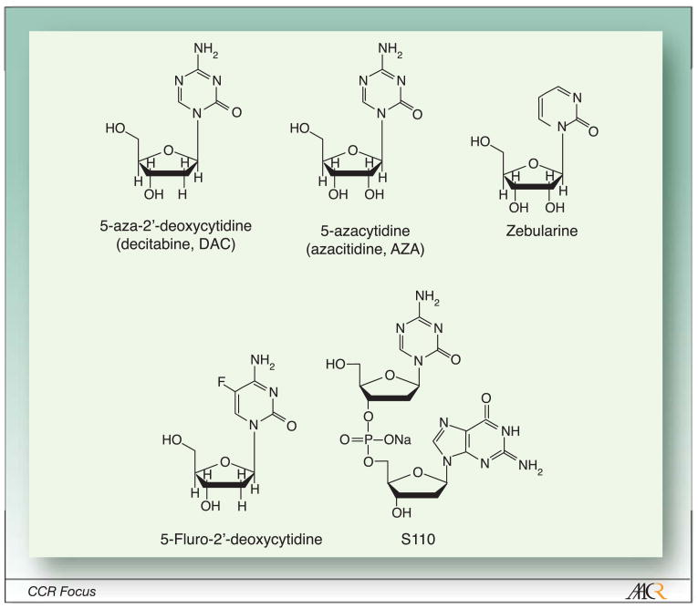
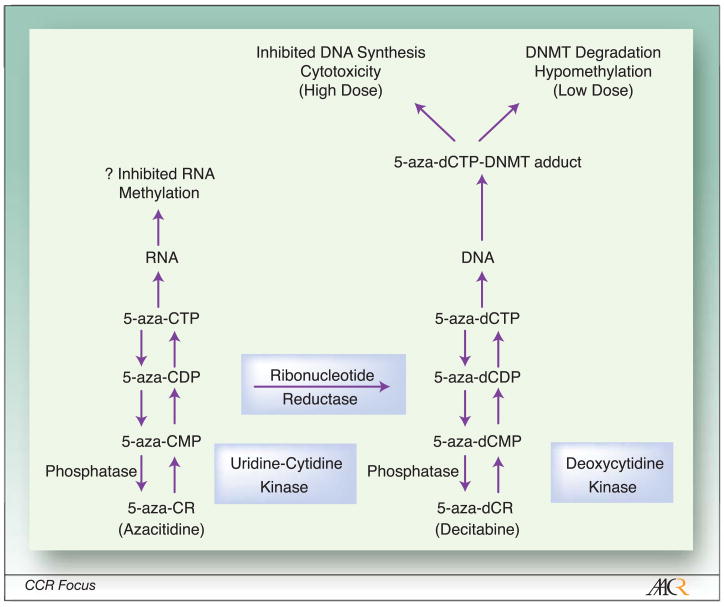
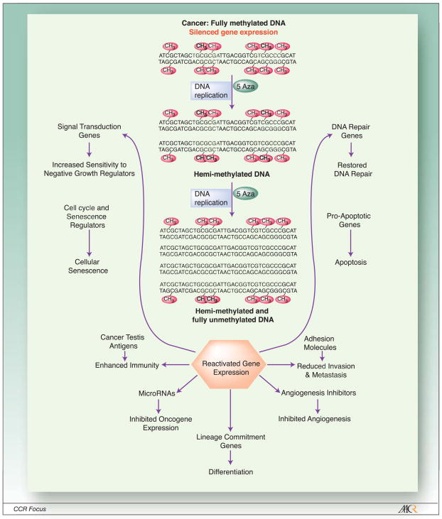
Drugs that inhibit DNA methylation were discovered by pure serendipity[12]. Cytosine analogs developed as cytotoxic anti-cancer agents in the 1960’s and tested in the clinic in the 1970’s were found to induce peculiar differentiation phenotypes in-vitro[16]. This DNA hypomethylating property is limited to cytosine analogs with 5′ modifications of the ring (Fig. 1). Other cytosine or nucleoside analogs do not affect DNA methylation directly. Eventually, this property of the two main analogs, 5-azacytidine (AZA) and 5-aza-2′-deoxycytidine (DAC) was traced to their ability to incorporate into DNA, trap DNA methyltransferases and target these enzymes for degradation[11, 17] (Fig. 2). DNA synthesis in the absence of these enzymes then results in hypomethylation in the daughter cells and eventually to reactivation of silenced gene expression (Fig. 3). Several other 5′ modified nucleoside analogues have been described (Fig. 1) and either in pre-clinical studies or in early stage clinical trials[18].
Figure 1.
Chemical structure of nucleoside DNA Methylation Inhibitors. AZA and DAC are FDA approved for the treatment of MDS. Zebularine has not been pursued clinically because of toxicity in pre-clinical models. 5-FC is in early stage clinical trials, while S110 is still undergoing pre-clinical testing.
Figure 2.
Mechanisms of action of hypomethylating nucleoside analogues. Azacitidine and decitabine are efficiently incorporated into cells by specialized transporters, following which their metabolism diverge. They are phosphorylated by different enzymes, eventually to 5-aza-CTP which incorporates into RNA and has poorly defined effects there and 5-aza-dCTP which incorporates into DNA. A fraction of 5-aza-CDP is also converted to 5-aza-dCDP. Once incorporated into DNA, 5-aza-dCTP forms irreversible covalent bonds with DNMTs, which result in bulky DNA-protein adducts and inhibition of DNA synthesis. At high doses, this results in cell death (and is therefore a cytotoxic intervention). At lower doses, the complexes are excised and degraded by the proteosome. DNA is repaired, following which DNA synthesis resumes in the absence of DNMTs, resulting in hypomethylation of newly synthesized DNA.
Figure 3.
Pleiotropic Therapeutic Effects of DNA Methylation Inhibition and Gene Reactivation in Cancer. DNA methylation is maintained post replication by the action of DNA methyltransferases. DAC and AZA lead to degradation of the main DNA methyltransferases, and continued replication results in passive demethylation that eventually results in reactivated gene expression. Activated gene expression, in turn, has effects on multiple different pathways, each of which could contribute to a clinical response.
Various non-nucleoside analogue drugs were also reported to inhibit DNA methylation, including Procainamide, Hydralazine, EGCG and others. However, their ability to induce DNA hypomethylation in-vitro has been questioned[19, 20], and they are “weak” hypomethylators at best. Some confusion may have arisen through equating inhibition of DNA methyltransferase activity and actual hypomethylation induction. DNA methyltransferase activity is proliferation dependent and cell cycle regulated[21, 22]. Thus, any insult that leads to proliferation arrest also leads to measurable decreases in DNA methyltransferase activity, but in this setting inhibited activity has not been conclusively shown to affect DNA methylation per-se. Any clinical effect of these weak “demethylating” drugs may require prolonged exposure to high doses of the compounds and the uncertainties over their potency have limited enthusiasm for clinical trials in neoplasia.
Other than nucleoside analogs and “weak” hypomethylators, there has been interest in the development of small molecules targeting DNA methyltransferases[20]. Those have theoretical advantages in that the DNA incorporation requirement of nucleoside analogs makes these drugs exquisitely S-phase specific, a considerable barrier in some malignancies (see below). However, the three known DNA methyltransferases have redundant functions to a certain extent, and it is clear that inhibiting more than one is required to optimally activate tumor-suppressor genes[23]. This may explain why, despite considerable effort, no small molecule inhibitor of DNA methyltransferases has made it to the clinic yet. MG98, an antisense compound directed at DNMT1 is effective in-vitro, but human clinical trials have shown disappointing results so far[24].
It is important to recognize that while inhibiting DNA methylation is a molecularly precise targeted therapy approach, the downstream effects on neoplastic behavior are quite non-specific (Figs. 2 and 3). The trapping of DNMTs onto DNA creates bulky adducts that can inhibit DNA synthesis and eventually result in cell death by cytotoxicity[25]. In fact, in-vitro experiments showed that at high doses of the compounds, inhibition of DNA synthesis masks effects on DNA methylation, resulting in a U shaped dose-response for hypomethylation induction[26]. Even at optimal (low) doses for hypomethylation induction, if one considers reactivated genes, a case could be made (and data be found) for effects of the inhibitors on multiple pathways[2, 7] including senescence (via P16 activation, for example), apoptosis (via activation of pro-apoptotic genes), differentiation (e.g. responsiveness to retinoic acid), stem cell renewal (by abrogating self-renewal signals), invasion (by upregulating inhibitors of motility), angiogenesis (through angiogenesis inhibitors such as THBS1), immune recognition (by activation of cancer testis antigens) etc. With the recent discovery of many microRNAs (miRNA) silenced by DNA methylation in cancer[27], one needs to add to the list effects of the drugs on down-regulation of oncogenes such as BCL6, CDK6 and other growth promoters. Finally, given the promiscuity of hypermethylation in cancer, one also needs to keep in mind the possibility that hypomethylation can activate those rare oncogenes known to be silenced in cancer[2] (e.g. COX2, EGFR etc.). This non-specificity makes predictions of clinical outcomes in-vivo quite difficult, and indeed likely tumor and patient specific. In other words, in some patients responses may be predominantly apoptosis mediated and rapid, while in others a differentiation effect is seen. However, it is likely that a mixture of effects reflecting the sum total of the pathways activated is in fact the reality in most cases.
Clinical Results
AZA and DAC have had two lives as anti-neoplastic agents. Early clinical trials in the 1970’s and 1980’s were based on the presumed cytotoxic effects of the drugs. Tested mostly at very high doses for relatively short exposure times, the drugs were found to be sporadically effective, more so in hematologic malignancies than in solid tumors, but also quite toxic[28]. The ratio of efficacy to toxicity was deemed unfavorable, and their usage was largely abandoned after the US FDA rejected an application for registration of AZA. This was not particularly surprising in hindsight. The drugs only work as epigenetic modifiers when given at low doses, given that high doses will inhibit DNA synthesis which will short-circuit their DNA hypomethylating effect. In-vitro, differentiation after AZA is only observed at low doses[17], and hypomethylation after DAC (in-vitro) is U shaped, with no effects on DNA methylation at all at high doses[29] (which are achievable in-vivo). Thus, the traditional phase I/II sequence of drug testing failed in this case because of the unusual dose-response properties of the drugs. Sadly, this failure may not be limited to this class of agents, a sobering fact considering that even today, most drugs including targeted agents still follow the classical phase I/II paradigm of clinical testing at maximally tolerated dose.
The hypomethylating nucleoside analogs came back to life over the past decade through the persistence of a few investigators[30, 31] and the renewed interest in DNA methylation as a therapeutic target brought about by basic investigations[7]. Through a convergence of serendipity (the desire to treat older patients who cannot tolerate high doses) and mechanism of action based dose finding trials[32], both AZA and DAC were tested in relatively large studies at low to moderate doses and over multiple cycles of administration, thus optimizing their epigenetic modulation potential. Following promising phase II studies, AZA was tested in two separate phase III studies[30, 33] in the myelodysplastic syndrome (MDS). Response rates ranging from 30 to 60% were observed, with documented improved survival compared to either supportive care or cytotoxic chemotherapy. Those trials used an open-ended treatment approach where patients were encouraged to continue receiving the drug until progression or death. DAC also had promising early studies in MDS, and phase III studies confirmed responses and modulation of disease (for example delay in AML progression) but failed to show substantial effects on survival[34, 35]. As opposed to the AZA studies, the DAC phase III studies limited the number of cycles patients received. When using an open ended approach similar to that of AZA trials[36, 37], DAC resulted in a very high response rate (40% complete response, over 70% total response), survival comparable to that seen in the AZA trials and superior to that observed with cytotoxic chemotherapy. It remains to be seen whether AZA (which incorporates into both RNA and DNA) is truly clinically different from DAC (which incorporates only in DNA) as the two drugs have not been directly compared. The results of recent clinical trials in leukemia and solid tumors are summarized in Table 1.
Table 1.
Selected recent clinical results with DNA methylation inhibitors.
| Drug | Dosage | Indication | Results | Comments | Reference |
|---|---|---|---|---|---|
| Azacitidine | 75 mg/m2 SQ, daily x7, q4 weeks | MDS (symptomatic) | CR 7%, PR 16%, HI 37% | Improved progression-free survival compared to supportive care | [30] |
| Azacitidine | 75 mg/m2 SQ, daily x7, q4 weeks | MDS (advanced) | CR 17%, PR 12%, HI 20% | Improved survival compared to conventional therapy | [33] |
| Azacitidine + Phenylbutyrate |
Aza 25–75 mg/m2 SQ, daily for 5–14 days, q4 weeks + Phenylbutyrate 375 mg/kg/d IV, CI x7 days beginning on the final day of aza | MDS and AML | CR 14%, PR 3%, HI 21% | Tumor suppressor gene hypomethylation more pronounced in responders | [44] |
| Azacitidine + Valproic acid + ATRA |
Aza 75 mg/m2 SQ, daily x7, q4 weeks + Valproic acid 50–75 mg/kg PO days 1–7 + ATRA 45 mg/m2 PO days 3–7 | Poor prognosis or relapsed or refractory leukemias | CR 22%, CRi 5%, Other responses 13% | Higher response rate in previously untreated patients. Correlation between Valproic acid levels and response | [41] |
| Azacitidine + VPA |
Aza 20–95 mg/m2 SQ, daily x10, q4 weeks + VPA titrated to plasma levels of 75 to 100 μg/mL |
Relapsed refractory solid tumors | SD 25% | [55] | |
| Decitabine | 15 mg/m2 IV over 3 hours q8h for 9 doses, q6 weeks | MDS (advanced) | CR 24%, PR 10%, HI 14% | [56] | |
| Decitabine | 15 mg/m2 IV over 3 hours q8h for 9 doses, q6 weeks | MDS (advanced) | CR 9%, PR 8%, HI 13% | Improved progression-free survival compared to supportive care | [57] |
| Decitabine | 5–20 mg/m2 IV over 1 hour daily x10 | Poor prognosis or relapsed or refractory leukemias | CR 18%, PR 6%, HI 8% | Greater response rate at lower doses. Flat hypomethylation dose-response past 150 mg/m2 total dose | [32, 42] |
| Decitabine | 3 dose schedules; most patients treated at 20 mg/m2 IV over 1 hour daily x5, q4 weeks | MDS (advanced) | CR 34%, PR 1%, CRi+HI 37% | Improved survival compared to chemotherapy in a case-control study. Association between P15 reactivation and response | [36, 37] |
| Decitabine | 15–20 mg/m2 IV over 1 hour daily x10 (some patients also received Valproic acid) | AML | CR 16%, CRi 16%, PR 12% | Correlation between ER reactivation and response. Addition of valproic acid did not impact responses. | [45] |
| Decitabine | 2.5–20 mg/m2 IV over 1 hour daily x5–10 days | Relapsed or refractory solid tumors | PR 4%, Minor response 11%, SD 46% (at 4 weeks) | No correlation between tumor and peripheral blood mononuclear cell hypomethylation after therapy | (Stewart et. al., CCR, in press) |
| Decitabine + Valproic acid |
DAC 15 mg/m2 IV over 1 hour daily x10 + Valproic acid 25–50 mg/kg PO days 1–10 | Poor prognosis or relapsed or refractory leukemias | CR 19%, CRi 4% | Higher response rate in previously untreated patients. Correlation between Valproic acid levels and response. | [58] |
| Decitabine + Interleukin 2 |
DAC 0.1–0.3 mg/kg SQ d days/week for 2 weeks + IL2 600,000 IU/kg IV q8h x 14 doses on weeks 3 and 6 | Metastatic melanoma and renal tumors | CR 6%, PR 12%, Minor response 12%, SD 18% | [59] | |
| Decitabine + Carboplatin |
DAC 45–135 mg/m2 IVx1, q4 weeks + Carbo AUC 5–6 IV d8 |
Relapsed or refractory solid tumors | PR 3%, SD 9% | Hypomethylation of MAGE1A documented in tumors | [60] |
While the drugs result in measurable improvements in disease burden, quality of life, survival etc., the patterns of responses are quite different from what traditional cytotoxic therapies achieve in MDS. For example, chemotherapy induces rapid responses in MDS (most after one cycle, infrequently after two cycles, and non-responders after two cycles rarely if ever respond to subsequent cytotoxics). AZA and DAC are very different. Responses are rare after one cycle and improve over time[30, 33]. It is not unusual to see patients with no evidence of therapeutic efficacy for 3 months achieve complete remission with continued therapy[38]. The side-effect profile is also different. Usual cytotoxic side-effects (mucositis, hair loss, diarrhea, renal failure etc.) are rare with the hypomethylating agents, the toxicity of which is mainly on bone marrow function (neutropenia is dose-limiting). Finally, studies suggest that the quality of responses may be different with hypomethylators – survival in remission, for example, appears to be better with AZA and DAC compared to traditional chemotherapy[33, 36]. All these have convinced many investigators that the mechanism of action of the drugs is indeed different from what one would see with chemotherapy, even low-dose chemotherapy.
It is instructive to reflect on why DNA methylation inhibitors, drugs that should work across a spectrum of malignancies, were tested and FDA-approved in a relatively rare disease, MDS. There is a priori no special reason why MDS should be an epigenetically responsive disease. Data so far suggest no more or less DNA methylation changes in MDS compared to other cancers, and many patients have chromosomal abnormalities as well[39]. An empiric explanation lies in recent observations. Responses to these drugs are most apparent (i) in patients who are not previously treated with drugs and thus have not had a chance to develop drug resistance and (ii) after several cycles of therapy. Thus, the most efficient testing would have been in a slowly evolving disease where no standard of care exists, a situation unique to MDS at the time the drugs were being reintroduced. With that realization, investigators are taking a second look at these drugs in other cancers. Early data suggests substantial activity in acute myeloid leukemia (AML) for example, if testing is done in previously untreated patients (Table 1).
Translational Results
Much effort has been devoted to molecular evaluation, prediction and understanding of clinical responses to hypomethylating agents. In various leukemias where this therapy has been tested (MDS, AML, chronic myelogenous leukemia), pre-treatment molecular characteristics (clinical factors, chromosomal changes, mutations, gene methylation) have not been able to accurately predict the chances of response[40]. This could be due to the limited set of markers/genes examined so far. It is also possible that critical factors to response do not lie in the intrinsic molecular nature of the neoplasm but in the patient and tumor-specific pharmacologic disposition of the drugs. In any case, this remains an open area of investigation.
An interesting and complex issue is whether drug-induced epigenetic modulation of the neoplastic cells in-vivo happens, and whether it is important to clinical responses. The complexity lies in the dynamic nature of the changes, in the clonal shifts that occur while on therapy, and in the variability of current technology to measure changes in gene methylation and expression. An ideal study would sample sorted/purified neoplastic cells everyday (or nearly so) throughout a whole treatment course to capture the entire ranges of observed effects. For the most part, this is neither practical nor really feasible as clonal shifts within a neoplasm can be impossible to detect. Indeed, if therapy is really effective acutely, the most hypomethylated cells may have died and would therefore not be available for sampling and analysis. With these acknowledged limitations, what can be gleaned from various studies is that (i) global DNA methylation measured directly (5-methyl-Cytosine content) or indirectly (repetitive element methylation) decreases shortly after treatment is started, nadirs around 10 days and recovers back to baseline 28–35 days after treatment start[38, 40, 41]; (ii) tumor-suppressor gene methylation decreases in some patients but not all after therapy[42–44]; recovery to baseline is also variable; and (iii) gene expression of silenced genes (e.g. P15, ER etc.) is induced shortly after treatment in some patients[37, 45] with, again, variable drift back to baseline.
In terms of correlations between methylation/epigenetic modulation and response, several patterns emerge: (i) Global methylation changes are not consistently associated with responses[40]. In CML, there is even an inverse relation[46], which may be related to the clonal shifts described earlier. (ii) Tumor-suppressor gene methylation change at early time points was associated with response in some studies but not all[40, 44, 45], likely because of the complexity of this analysis described earlier. (iii) Gene expression induction generally correlates with response better than gene demethylation[37, 45]. (iv) Sustained changes in methylation/gene expression do correlate with response[43], but they are confounded by clonal elimination. Of interest, simultaneous tracking of DNA methylation and gene mutation confirmed that epigenetic modulation precedes clonal elimination in some cases[38, 43].
The lack of consistent association between epigenetic patterns at baseline, DNA hypomethylation induction and response to therapy has raised the issue of the precise in-vivo mechanism of action of the drugs. As mentioned earlier, AZA and DAC do induce dose-dependent cytotoxicity and it is possible that some (or all) of the responses seen are related to this effect. In our opinion, this is unlikely because observed clinical responses are not consistent with a cytotoxic effect in that (i) increasing doses of the active agents has not resulted in increased responses as one would expect with a pure cytotoxic drug[32], (ii) the kinetics of responses (early hypomethylation, delayed clonal elimination) are also inconsistent with direct cytotoxicity[38], (iii) responses have clearly been seen in patients highly resistant to the classical cytotoxic drug cytarabine[32] which shares pharmacologic disposition with DAC and (iv) survival with DAC or AZA is clearly superior to survival achieved with combination cytotoxic therapy[33, 36]. Of course, it is likely that cytotoxicity plays a role in the responses seen in some patients, but the uniqueness of the clinical outcomes after DAC or AZA strongly suggest a contribution by epigenetic effects of the drugs.
Barriers to Effective Epigenetic Therapy
With molecular and clinical proof of principle at hand, it is clear that hypomethylation therapy is here to stay. In order for it to achieve the broad impact that the extent of DNA methylation abnormalities in cancer suggest, several barriers need to be overcome: Drug delivery, appropriate clinical testing, understanding resistance, and understanding sensitivity.
Drug delivery is perhaps the single most daunting barrier to effective translation of the leukemia findings to solid tumors. There is no question (from mouse models to anecdotal responses in clinical trials) that hypomethylating drugs can induce responses in patients with solid tumors (Table 1). However, an important issue is delivering sustained levels of drug and overcoming the S-phase dependency of the therapy. Thus, in tumors with a low replicating pool of cells, drug incorporation will likely be less than that in normal hematopoietic cells, exposing patients to toxicity with little chances of a response. Indeed, in a phase I study, we observed little correlation between hypomethylation induction in blood and in (solid) tumor biopsies (Stewart D., submitted). Current efforts to improve on this situation include alternate delivery methods for the nucleoside analogues, oral preparations[47], a DAC-containing dinucleotide with improved stability[48] and, potentially, small molecules that can inhibit DNA methylases without requiring DNA incorporation[20].
Even if drugs were identified with optimal penetration into solid tumors (or perhaps cancers identified with a high enough S phase for current drugs to work), the issue of optimal clinical testing needs careful thought. Based on the MDS experience, patience and front-line testing are required to optimize the chances of observing clinical efficacy. Unfortunately, many solid tumor phase I/II study designs rely on heavily pre-treated patients and single course of administration for evaluation. This strategy has failed before for hypomethylating agents and may fail again if the same designs are utilized. There is no reason why multi-drug resistant cancers would not also be resistant to hypomethylating drugs. It is therefore important to identify populations of patients where such therapy can be tested relatively early, and over at least 2–4 courses. Possibilities include an adjuvant setting, or testing this approach prior to standard therapy in incurable cancers where conventional therapy offers relatively little prolongation of life.
An issue that has not yet been given enough attention is the mechanism of primary and secondary resistance to existing DNA methylation inhibitors. It is clear that not all patients respond equally to AZA or DAC. We do not understand why some patients are resistant to these drugs. In-vitro, resistance appears to be primarily pharmacologic[49], and this needs to be tested in-vivo as well. Interestingly, the correlation between gene expression induction and response also identifies another potential source of resistance[40]. It is at least theoretically possible that hypomethylation does not lead to gene reactivation because of some other down-stream defect intrinsic to the cancers. A detailed understanding of in-vitro and in-vivo links between hypomethylation and gene reactivation would help in this regard. Even more mysterious is the mechanism of secondary failure to these drugs. Clinically, there is no evidence yet that AZA or DAC are curative, and relapses are the norm, even in the face of continued drug exposure[33]. Preliminary data suggests that relapsed MDS has less methylation that prior to therapy initiation, thus potentially pointing to methylation independent clonal evolution. This issue will be important to understand as we move towards attempts to cure cancer using epigenetic therapy.
Understanding sensitivity is perhaps even more pressing than understanding resistance. Of course, cancer researchers remain puzzled as to why many therapies work when they do work (why are testicular cancer and pediatric leukemia so curable?), but this is even more pressing for targeted therapies where at least hypotheses can be formulated and tested. Fig. 3 outlines a number of potential mechanisms by which epigenetic therapy can lead to clonal elimination of neoplasms. It remains to be seen whether these possibilities can be deciphered and distinguished in-vivo, but if they can, the information will help guide the next generation of clinical trials. For example, if immune modulation is important, then combinations of epigenetic therapy with immunotherapy may be indicated. If a major effect on stem cell renewal is observed, then perhaps epigenetic therapy may have the greatest impact after removing the cancer bulk.
The Next Questions
Beyond the issues raised by the early clinical trials and the application of this knowledge to other malignancies, the question of epigenetic therapy will undoubtedly gain traction in the next few years. It behooves us to be sure that DNA methylation inhibition works, at least in large part, by actually inhibiting DNA methylation in cancers (rather than, say, cytotoxicity, or affecting normal stem cells etc.). It is essential to determine its safety in the long run as we move it to healthier populations of patients (in the adjuvant setting rather than the metastatic setting, for example). And it is important to put it in the context of all epigenetic processes. Much has been made, for example, of the in-vitro synergy between DNA methylation inhibitors and histone deacetylase inhibitors[50] and this is discussed elsewhere in this issue[51]. But this synergy is partly dependent on sensitivity to methylation inhibitors. Thus, it may not be the case that it overcomes clinical resistance to AZA or DAC, which will be required for the combinations to be successful in the clinic. Randomized studies are ongoing to address the issue. Much research has also been invested in deciphering other components of the epigenetic code. Histone modifications by methylases and demethylases are key for some epigenetic processes[52]. Polycomb group proteins which affect gene expression via a key modification of histone H3 (lysine 27 trimethylation) can powerfully regulate gene expression independently of DNA methylation, and early data suggest that this pathway is deregulated in cancer and a promising target for therapeutic intervention[53]. Finally, excitement over epigenetic reprogramming is growing as a tool of developing stem cells for regenerative medicine. DNA methylation inhibitors can facilitate this process in-vitro[54], and it is tantalizing to think that this form of therapy could have applications well beyond cancer in the future.
Acknowledgments
Work in the author’s laboratory is supported by National Institutes of Health grants CA100632, CA108631, CA098006 and CA105346. JPI is an American Cancer Society Clinical Research professor supported by a generous gift from the F. M. Kirby Foundation.
Reference List
- 1.Reik W. Stability and flexibility of epigenetic gene regulation in mammalian development. Nature. 2007;447:425–32. doi: 10.1038/nature05918. [DOI] [PubMed] [Google Scholar]
- 2.Toyota M, Issa JP. Epigenetic changes in solid and hematopoietic tumors. Semin Oncol. 2005;32:521–30. doi: 10.1053/j.seminoncol.2005.07.003. [DOI] [PubMed] [Google Scholar]
- 3.Vogelstein B, Kinzler KW. Cancer genes and the pathways they control. Nat Med. 2004;10:789–99. doi: 10.1038/nm1087. [DOI] [PubMed] [Google Scholar]
- 4.Kinzler KW, Vogelstein B. Lessons form hereditary colorectal cancer. Cell. 1996;87:159–70. doi: 10.1016/s0092-8674(00)81333-1. [DOI] [PubMed] [Google Scholar]
- 5.Wood LD, Parsons DW, Jones S, et al. The genomic landscapes of human breast and colorectal cancers. Science. 2007;318:1108–13. doi: 10.1126/science.1145720. [DOI] [PubMed] [Google Scholar]
- 6.Ley TJ, Mardis ER, Ding L, et al. DNA sequencing of a cytogenetically normal acute myeloid leukaemia genome. Nature. 2008;456:66–72. doi: 10.1038/nature07485. [DOI] [PMC free article] [PubMed] [Google Scholar]
- 7.Jones PA, Baylin SB. The epigenomics of cancer. Cell. 2007;128:683–92. doi: 10.1016/j.cell.2007.01.029. [DOI] [PMC free article] [PubMed] [Google Scholar]
- 8.Bird A. DNA methylation patterns and epigenetic memory. Genes Dev. 2002;16:6–21. doi: 10.1101/gad.947102. [DOI] [PubMed] [Google Scholar]
- 9.Gazin C, Wajapeyee N, Gobeil S, Virbasius CM, Green MR. An elaborate pathway required for Ras-mediated epigenetic silencing. Nature. 2007;449:1073–7. doi: 10.1038/nature06251. [DOI] [PMC free article] [PubMed] [Google Scholar]
- 10.McCabe MT, Brandes JC, Vertino PM. Cancer DNA methylation: molecular mechanisms and clinical implications. Clin Cancer Res. 2009 doi: 10.1158/1078-0432.CCR-08-2784. In press. [DOI] [PMC free article] [PubMed] [Google Scholar]
- 11.Yoo CB, Jones PA. Epigenetic therapy of cancer: past, present and future. Nat Rev Drug Discov. 2006;5:37–50. doi: 10.1038/nrd1930. [DOI] [PubMed] [Google Scholar]
- 12.Issa JP. DNA methylation as a therapeutic target in cancer. Clin Cancer Res. 2007;13:1634–7. doi: 10.1158/1078-0432.CCR-06-2076. [DOI] [PubMed] [Google Scholar]
- 13.Reik W, Dean W, Walter J. Epigenetic reprogramming in mammalian development. Science. 2001;293:1089–93. doi: 10.1126/science.1063443. [DOI] [PubMed] [Google Scholar]
- 14.Rideout WM, III, Eggan K, Jaenisch R. Nuclear cloning and epigenetic reprogramming of the genome. Science. 2001;293:1093–8. doi: 10.1126/science.1063206. [DOI] [PubMed] [Google Scholar]
- 15.Hochedlinger K, Blelloch R, Brennan C, et al. Reprogramming of a melanoma genome by nuclear transplantation. Genes Dev. 2004;18:1875–85. doi: 10.1101/gad.1213504. [DOI] [PMC free article] [PubMed] [Google Scholar]
- 16.Taylor SM, Jones PA. Multiple new phenotypes induced in 10T1/2 and 3T3 cells treated with 5-azacytidine. Cell. 1979;17:771–9. doi: 10.1016/0092-8674(79)90317-9. [DOI] [PubMed] [Google Scholar]
- 17.Jones PA, Taylor SM. Cellular differentiation, cytidine analogs and DNA methylation. Cell. 1980;20:85–93. doi: 10.1016/0092-8674(80)90237-8. [DOI] [PubMed] [Google Scholar]
- 18.Beumer JH, Parise RA, Newman EM, et al. Concentrations of the DNA methyltransferase inhibitor 5-fluoro-2′-deoxycytidine (FdCyd) and its cytotoxic metabolites in plasma of patients treated with FdCyd and tetrahydrouridine (THU) Cancer Chemother Pharmacol. 2008;62:363–8. doi: 10.1007/s00280-007-0603-8. [DOI] [PubMed] [Google Scholar]
- 19.Chuang JC, Yoo CB, Kwan JM, et al. Comparison of biological effects of non-nucleoside DNA methylation inhibitors versus 5-aza-2′-deoxycytidine. Mol Cancer Ther. 2005;4:1515–20. doi: 10.1158/1535-7163.MCT-05-0172. [DOI] [PubMed] [Google Scholar]
- 20.Stresemann C, Brueckner B, Musch T, Stopper H, Lyko F. Functional diversity of DNA methyltransferase inhibitors in human cancer cell lines. Cancer Res. 2006;66:2794–800. doi: 10.1158/0008-5472.CAN-05-2821. [DOI] [PubMed] [Google Scholar]
- 21.Issa JP, Vertino PM, Wu J, et al. Increased cytosine DNA-methyltransferase activity during colon cancer progression. J Natl Cancer Inst. 1993;85:1235–40. doi: 10.1093/jnci/85.15.1235. [DOI] [PubMed] [Google Scholar]
- 22.Robertson KD, Keyomarsi K, Gonzales FA, Velicescu M, Jones PA. Differential mRNA expression of the human DNA methyltransferases (DNMTs) 1, 3a and 3b during the G(0)/G(1) to S phase transition in normal and tumor cells. Nucleic Acids Res. 2000;28:2108–13. doi: 10.1093/nar/28.10.2108. [DOI] [PMC free article] [PubMed] [Google Scholar]
- 23.Rhee I, Bachman KE, Park BH, et al. DNMT1 and DNMT3b cooperate to silence genes in human cancer cells. Nature. 2002;416:552–6. doi: 10.1038/416552a. [DOI] [PubMed] [Google Scholar]
- 24.Klisovic RB, Stock W, Cataland S, et al. A phase I biological study of MG98, an oligodeoxynucleotide antisense to DNA methyltransferase 1, in patients with high-risk myelodysplasia and acute myeloid leukemia. Clin Cancer Res. 2008;14:2444–9. doi: 10.1158/1078-0432.CCR-07-1320. [DOI] [PubMed] [Google Scholar]
- 25.Juttermann R, Li E, Jaenisch R. Toxicity of 5-aza-2′-deoxycytidine to mammalian cells is mediated primarily by covalent trapping of DNA methyltransferase rather than DNA demethylation. Proc Natl Acad Sci U S A. 1994;91:11797–801. doi: 10.1073/pnas.91.25.11797. [DOI] [PMC free article] [PubMed] [Google Scholar]
- 26.Qin T, Jelinek J, Si J, Shu J, Issa JP. Mechanisms of resistance to 5-aza-2′-deoxycytidine in human cancer cell lines. Blood. 2008 doi: 10.1182/blood-2008-02-140038. [DOI] [PMC free article] [PubMed] [Google Scholar]
- 27.Saito Y, Liang G, Egger G, et al. Specific activation of microRNA-127 with downregulation of the proto-oncogene BCL6 by chromatin-modifying drugs in human cancer cells. Cancer Cell. 2006;9:435–43. doi: 10.1016/j.ccr.2006.04.020. [DOI] [PubMed] [Google Scholar]
- 28.Santini V, Kantarjian HM, Issa JP. Changes in DNA methylation in neoplasia: pathophysiology and therapeutic implications. Ann Intern Med. 2001;134:573–86. doi: 10.7326/0003-4819-134-7-200104030-00011. [DOI] [PubMed] [Google Scholar]
- 29.Qin T, Youssef EM, Jelinek J, Yang AS, Garcia-Manero G, Issa JP. Combination effects of Cytarabine and Decitabine in human leukemic cell lines. 2007 doi: 10.1158/1078-0432.CCR-06-2762. In press. [DOI] [PubMed] [Google Scholar]
- 30.Silverman LR, Demakos EP, Peterson BL, et al. Randomized controlled trial of azacitidine in patients with the myelodysplastic syndrome: a study of the cancer and leukemia group B. J Clin Oncol. 2002;20:2429–40. doi: 10.1200/JCO.2002.04.117. [DOI] [PubMed] [Google Scholar]
- 31.Wijermans P, Lubbert M, Verhoef G, et al. Low-dose 5-aza-2′-deoxycytidine, a DNA hypomethylating agent, for the treatment of high-risk myelodysplastic syndrome: a multicenter phase II study in elderly patients. J Clin Oncol. 2000;18:956–62. doi: 10.1200/JCO.2000.18.5.956. [DOI] [PubMed] [Google Scholar]
- 32.Issa JP, Garcia-Manero G, Giles FJ, et al. Phase I study of low-dose prolonged exposure schedules of the hypomethylating agent 5-aza-2′-deoxycytidine (Decitabine) in hematopoietic malignancies. Blood. 2004;103:1635–40. doi: 10.1182/blood-2003-03-0687. [DOI] [PubMed] [Google Scholar]
- 33.Fenaux P, Mufti GJ, Hellstrom-Lindberg E, et al. Efficacy of azacitidine compared with that of conventional care regimens in the treatment of higher-risk myelodysplastic syndromes: a randomised, open-label, phase III study. Lancet Oncol. 2009 doi: 10.1016/S1470-2045(09)70003-8. [DOI] [PMC free article] [PubMed] [Google Scholar]
- 34.Kantarjian H, Issa JP, Rosenfeld CS, et al. Decitabine improves patient outcomes in myelodysplastic syndromes: results of a phase III randomized study. Cancer. 2006 doi: 10.1002/cncr.21792. [DOI] [PubMed] [Google Scholar]
- 35.Wijermans P, Suciu S, Baila L, et al. Low Dose Decitabine Versus Best Supportive Care in Elderly Patients with Intermediate or High Risk MDS Not Eligible for Intensive Chemotherapy: Final Results of the Randomized Phase III Study (06011) of the EORTC Leukemia and German MDS Study Groups. Blood. 2008;112:90. [Google Scholar]
- 36.Kantarjian HM, O’Brien S, Huang X, et al. Survival advantage with decitabine versus intensive chemotherapy in patients with higher risk myelodysplastic syndrome: comparison with historical experience. Cancer. 2007 doi: 10.1002/cncr.22508. [DOI] [PMC free article] [PubMed] [Google Scholar]
- 37.Kantarjian H, Oki Y, Garcia-Manero G, et al. Results of a randomized study of 3 schedules of low-dose decitabine in higher-risk myelodysplastic syndrome and chronic myelomonocytic leukemia. Blood. 2007;109:52–7. doi: 10.1182/blood-2006-05-021162. [DOI] [PubMed] [Google Scholar]
- 38.Oki Y, Jelinek J, Shen L, Kantarjian HM, Issa JP. Induction of hypomethylation and molecular response after decitabine therapy in patients with chronic myelomonocytic leukemia. Blood. 2008;111:2382–4. doi: 10.1182/blood-2007-07-103960. [DOI] [PMC free article] [PubMed] [Google Scholar]
- 39.List AF, Vardiman J, Issa JP, Dewitte TM. Myelodysplastic syndromes. Hematology (Am Soc Hematol Educ Program) 2004:297–317. doi: 10.1182/asheducation-2004.1.297. [DOI] [PubMed] [Google Scholar]
- 40.Oki Y, Aoki E, Issa JP. Decitabine--bedside to bench. Crit Rev Oncol Hematol. 2007;61:140–52. doi: 10.1016/j.critrevonc.2006.07.010. [DOI] [PubMed] [Google Scholar]
- 41.Soriano AO, Yang H, Faderl S, et al. Safety and clinical activity of the combination of 5-azacytidine, valproic acid and all-trans retinoic acid in acute myeloid leukemia and myelodysplastic syndrome. Blood. 2007 doi: 10.1182/blood-2007-03-078576. [DOI] [PubMed] [Google Scholar]
- 42.Yang AS, Doshi KD, Choi SW, et al. DNA methylation changes after 5-aza-2′-deoxycytidine therapy in patients with leukemia. Cancer Res. 2006;66:5495–503. doi: 10.1158/0008-5472.CAN-05-2385. [DOI] [PubMed] [Google Scholar]
- 43.Daskalakis M, Nguyen TT, Nguyen C, et al. Demethylation of a hypermethylated P15/INK4B gene in patients with myelodysplastic syndrome by 5-Aza-2′-deoxycytidine (decitabine) treatment. Blood. 2002;100:2957–64. doi: 10.1182/blood.V100.8.2957. [DOI] [PubMed] [Google Scholar]
- 44.Gore SD, Baylin S, Sugar E, et al. Combined DNA methyltransferase and histone deacetylase inhibition in the treatment of myeloid neoplasms. Cancer Res. 2006;66:6361–9. doi: 10.1158/0008-5472.CAN-06-0080. [DOI] [PubMed] [Google Scholar]
- 45.Blum W, Klisovic RB, Hackanson B, et al. Phase I study of decitabine alone or in combination with valproic acid in acute myeloid leukemia. J Clin Oncol. 2007;25:3884–91. doi: 10.1200/JCO.2006.09.4169. [DOI] [PubMed] [Google Scholar]
- 46.Issa JP, Gharibyan V, Cortes J, et al. Phase II study of low-dose decitabine in patients with chronic myelogenous leukemia resistant to imatinib mesylate. J Clin Oncol. 2005;23:3948–56. doi: 10.1200/JCO.2005.11.981. [DOI] [PubMed] [Google Scholar]
- 47.Garcia-Manero G, Stoltz ML, Ward MR, Kantarjian H, Sharma S. A pilot pharmacokinetic study of oral azacitidine. Leukemia. 2008;22:1680–4. doi: 10.1038/leu.2008.145. [DOI] [PubMed] [Google Scholar]
- 48.Yoo CB, Jeong S, Egger G, et al. Delivery of 5-aza-2′-deoxycytidine to cells using oligodeoxynucleotides. Cancer Res. 2007;67:6400–8. doi: 10.1158/0008-5472.CAN-07-0251. [DOI] [PubMed] [Google Scholar]
- 49.Qin T, Jelinek J, Si J, Shu J, Issa JP. Mechanisms of resistance to 5-aza-2′-deoxycytidine in human cancer cell lines. Blood. 2008 doi: 10.1182/blood-2008-02-140038. [DOI] [PMC free article] [PubMed] [Google Scholar]
- 50.Cameron EE, Bachman KE, Myohanen S, Herman JG, Baylin SB. Synergy of demethylation and histone deacetylase inhibition in the re- expression of genes silenced in cancer. Nat Genet. 1999;21:103–7. doi: 10.1038/5047. [DOI] [PubMed] [Google Scholar]
- 51.Bots M, Johnstone RW. Rational combinations using HDAC inhibitors. Clin Cancer Res. 2009 doi: 10.1158/1078-0432.CCR-08-2786. In press. [DOI] [PubMed] [Google Scholar]
- 52.Kouzarides T. Chromatin modifications and their function. Cell. 2007;128:693–705. doi: 10.1016/j.cell.2007.02.005. [DOI] [PubMed] [Google Scholar]
- 53.Kondo Y, Shen L, Cheng AS, et al. Gene silencing in cancer by histone H3 lysine 27 trimethylation independent of promoter DNA methylation. Nat Genet. 2008;40:741–50. doi: 10.1038/ng.159. [DOI] [PubMed] [Google Scholar]
- 54.Mikkelsen TS, Hanna J, Zhang X, et al. Dissecting direct reprogramming through integrative genomic analysis. Nature. 2008;454:49–55. doi: 10.1038/nature07056. [DOI] [PMC free article] [PubMed] [Google Scholar]
- 55.Braiteh F, Soriano AO, Garcia-Manero G, et al. Phase I study of epigenetic modulation with 5-azacytidine and valproic acid in patients with advanced cancers. Clin Cancer Res. 2008;14:6296–301. doi: 10.1158/1078-0432.CCR-08-1247. [DOI] [PMC free article] [PubMed] [Google Scholar]
- 56.Wijermans PW, Lubbert M, Verhoef G, Klimek V, Bosly A. An epigenetic approach to the treatment of advanced MDS; the experience with the DNA demethylating agent 5-aza-2′-deoxycytidine (decitabine) in 177 patients. Ann Hematol. 2005;84 Suppl 1:9–17. doi: 10.1007/s00277-005-0012-1. [DOI] [PubMed] [Google Scholar]
- 57.Kantarjian H, Issa JP, Rosenfeld CS, et al. Decitabine improves patient outcomes in myelodysplastic syndromes: results of a phase III randomized study. Cancer. 2006;106:1794–803. doi: 10.1002/cncr.21792. [DOI] [PubMed] [Google Scholar]
- 58.Garcia-Manero G, Kantarjian HM, Sanchez-Gonzalez B, et al. Phase 1/2 study of the combination of 5-aza-2′-deoxycytidine with valproic acid in patients with leukemia. Blood. 2006;108:3271–9. doi: 10.1182/blood-2006-03-009142. [DOI] [PMC free article] [PubMed] [Google Scholar]
- 59.Gollob JA, Sciambi CJ, Peterson BL, et al. Phase I trial of sequential low-dose 5-aza-2′-deoxycytidine plus high-dose intravenous bolus interleukin-2 in patients with melanoma or renal cell carcinoma. Clin Cancer Res. 2006;12:4619–27. doi: 10.1158/1078-0432.CCR-06-0883. [DOI] [PubMed] [Google Scholar]
- 60.Appleton K, Mackay HJ, Judson I, et al. Phase I and pharmacodynamic trial of the DNA methyltransferase inhibitor decitabine and carboplatin in solid tumors. J Clin Oncol. 2007;25:4603–9. doi: 10.1200/JCO.2007.10.8688. [DOI] [PubMed] [Google Scholar]


