Abstract
Human natural killer (NK) cells are central in immune defense against tumor and virally infected cells. Ziram is used as an accelerating agent in latex production and as an agricultural fungicide. Previous studies showed that continuous exposure to ziram inhibits NK lytic function. Additionally, they showed that a brief (1h) exposure to ziram caused persistent loss of lytic function. This study examined whether decreases in lytic function were accompanied by decreases in the target-binding function of NK cells and found that some, but not all, exposures to ziram caused significant decreases in binding function. Ziram exposures that caused a loss of binding function were examined for effects on expression of key NK cell surface proteins needed for binding to targets . Exposure to 2 μM ziram followed by 24 or 48 h in ziram- free media decreased CD16 expression but no other exposures caused decreases in cell surface proteins. As decreases in ATP could be in part responsible for loss of lytic function, the effect of ziram exposures on ATP levels of NK cells were examined. Certain ziram exposures decreased ATP levels in NK cells, but a decrease in ATP was not necessarily associated with a decrease in lytic function. However, the results indicate that ziram –induced losses of lytic function cannot be fully explained by alteration in binding, cell surface protein expression, or ATP levels
Keywords: NK cells, Binding function, CD16 expression, ATP levels
INTRODUCTION
Dithiocarbamate fungicides are used in agriculture for protection of crops and seeds (Franekic et al., 1994). Ziram is a dithiocarbamate used to treat a variety of fungal diseases in crops around the world such as potatoes, nuts, some fruits, and grain. In industry ziram is used as an accelerating agent in the production of latex rubber (IRAC, 1991). Human exposure may occur by coming into contact with latex rubber, ingesting treated crops, or via inhalation (Caldas et al., 2001; Nettis et al., 2002). There are no published studies measuring blood levels of Ziram in humans. While it appears that it may be metabolized by hepatic enzymes, its metabolism and excretion are not clearly defined. Rats fed 30 mg/kg of ziram for a two year period showed some accumulation in their livers (0.03 mg) (Hazardous Substance Databank, 1993). Ziram has been shown to cause a positive response in patch tests, which are used to determine potential allergic reactions, to a mixture of latex rubber vulcanizing chemicals including ziram (De Jong et al., 2002). Although ziram has tested negative for mutagenic activity in human lymphocyte cultures (Zensen et al., 2001), chromosomal changes have been seen in workers exposed to ziram for 3−5 years, indicating some risk of mutations (Edwards, et al., 1991). Ziram increases the concanavalin A stimulated production of interferon gamma (INF γ) and Interleukin 4 (IL4) in murine a vascular lymph node cells (De Jong et al., 2002).
Human Natural Killer (NK) cells are lymphocytes that are capable of killing tumor cells, virally infected cells, and antibody-coated cells. NK cells play a central role in immune defense against viral infection and formation of primary tumors (Lotzova, 1993; Vivier et al., 2004). NK cells are responsible for limiting the spread of blood-borne metastases as well as limiting the development of primary tumors (Lotzova, 1993). NK cells are defined by the absence of the T cell receptor/CD3 complex and by the presence of the CD56 and /or CD16 on the cell surface (Lotzova, 1993). These cells are the front line of immune response against tumor and virally infected cells due to their ability to lyse appropriate target cells with out prior sensitization. Interference with NK-cell function by any compound could increase the risk of viral infection and tumor formation.
Our previous studies have shown that purified NK cells treated with certain concentrations of ziram are less efficient at killing tumor cells (K562). Ziram is effective in blocking the cytotoxic function of highly purified NK cells at concentrations as low as 125 nM and these effects increase with time (Wilson et al., 2004; Whalen et al., 2003). In addition there is a persistent loss of lytic function, after a 1 h exposure to 2.5 μM ziram followed by 24 h, 48 h, or 6 days in ziram-free media (Taylor et al., 2005). It is now important to address the mechanism by which ziram is producing the loss of lytic function in NK cells.
The current study examined whether ziram interferes with the ability of NK cells to bind to target cells, since binding is a necessary first step in the lytic process. Additionally, it investigated whether any decreases in binding function were accompanied by decreases in expression of certain NK cell surface proteins. Finally, the effects of ziram on ATP levels in NK cells were studied, as decreases in ATP could account for a loss of lytic function. Effects of both chronic and acute exposure were examined as their mechanisms may differ and, as mentioned above, we have seen lasting effects of an acute ( 1h ) exposure to ziram on NK lytic function. This study will further elucidate the effects of exposure to this fungicide on human immune function.
MATERIALS AND METHODS
Isolation of NK cells
Peripheral blood from healthy adult volunteer donors was used for this study. Blood samples (buffy coats) were obtained from a Red Cross donation facility (American Red Cross, Portland, OR). Donors gave informed consent to the Red Cross and our laboratory was approved for receipt of blood products by the Institutional Review Board of the American Red Cross. Highly purified NK cells were obtained using a rosetting procedure. Buffy coats were mixed with 0.8 mL of RossetteSep™ human NK cell enrichment antibody cocktail (StemCell Technologies, Vancouver, British Colombia, Canada) per 45 mL of buffy coat. The mixture was incubated for 25 min at room temperature with periodic mixing. Following the incubation, 4 mL of the mixture was layered onto 4 mL Ficoll-Hypaque (1.077 g/mL; Sigma, St. Louis, MO, USA) and centrifuged at 1200 g for 20 min. The cell layer was collected and washed twice with PBS and stored in complete media (RPMI-1640 supplemented with 10% heat-activated BCS, 2 mM L-glutamine and 50 U penicillin G with 50 μg streptomycin/ml) at 1×10⁶ cells/mL. The resulting cell preparation was > 95% CD16+ and 0 % CD3+ according to fluorescence microscopy (Whalen and Loganathan, 2000).
Chemical Preparation
Ziram was purchased from Wako Pure Chemicals Industries Limited (Osaka, Japan). Dimethlysulfoxide (DMSO) was obtained from Sigma-Aldrich (St. Louis, MO, USA). Stock solutions of the compounds were made in DMSO. The compounds were diluted in gelatin media (0.5% gelatin replaced the calf serum in complete medium) to achieve the desired concentrations so that the final concentrations of DMSO did not exceed 0.01%.
Cell Treatment and Cell Viability
NK cells were separated by centrifugation from complete medium (defined above) and transferred to complete medium containing 0.5% gelatin in place of the 10% bovine serum. This was done in order to avoid binding of the hydrophobic compound to the serum albumin, which could interfere with its delivery to the cells. NK cells were then exposed to ziram continuously for 24 h, 48 h, or 6 days. Additionally, NK cells were exposed to ziram for 1 h followed by 24 h or 48 h in ziram-free media. Ziram concentrations ranged from 2 to 0.5 μM.
Cell viability was determined by trypan blue exclusion. Cell number and viability, assessed at the beginning and end of each exposure period, did not vary significantly among experimental conditions for any of the concentrations.
Conjugation Assay
The percentage of target cells with bound NK cells was determined at two effector to target ratios 12:1 and 6:1. The NK cells were treated as described above for the cytotoxicity assay. Tumor cells were then incubated with an appropriate number of control or Ziram exposed lymphocytes in round-bottomed microtitre plates (20,000 tumor cells/well). Each condition was tested in triplicate. The cells were incubated at 37° C, air/CO2, 19:1, for 10 min and then placed on ice. Following the incubation period, the cells were gently resuspended with a micropipette, placed in a hemocytometer and viewed under a light microscope. The number of target cells with two or more lymphocytes bound to their surface was counted as well as the total number of targets to determine the percentage of tumor cells with lymphocytes bound. The minimum number of targets counted per determination was 50−100 (Whalen et al., 1992).
Flow Cytometry
NK cells were exposed to the appropriate concentrations of ziram for the appropriate length of time. Following exposure to ziram, the cells were washed and prepared for analysis on a FACScan (Becton Dickinson, San Jose, CA). The cells were washed twice with ice-cold PBS and 100 μl of cell suspension (250,000−500,000 cells) was labeled with 20 μl of one of the following antibodies: anti- CD2, CD11a, CD11b, CD11c, CD16 , CD18, CD56 (Pharmingen, San Diego, CA). Anti- CD2, CD11a, and CD16 were FITC-conjugated antibodies. Anti- CD11b, CD11c, CD18, and CD56 were phycoerythrin (PE)-conjugated. Appropriate FITC- and PE-conjugated isotype control antibodies were used. The antibody-containing cell suspension was incubated for a minimum of 30 minutes on ice, in the dark. Following the incubation period the cells were washed twice with ice-cold PBS (0.5−1 mL) and suspended in 250 μl of ice-cold 1% paraformaldehyde in PBS. Samples were analyzed using the FACScan flow cytometer from Becton Dickinson Immunocytometry Systems, Inc (BDIS). Instrument performance was standardized daily using Calibrite beads (BDIS) and the same instrument settings were used for all acquisitions of stained cells. The assays were sufficiently uniform to use the same FSC, SSC, and FL settings. All cell surface marker analyses were gated on the lymphocyte population. The acquisition and analysis software for flow cytometry data was CELLQuest v 3.3 from BDIS running on a G4 Apple computer (Whalen et al., 2001).
ATP Assay
Following exposures of cells to the various concentrations of compounds; 250,000 NK cells were added to 300 μL of somatic cell ATP releasing reagent (Sigma-Aldrich). To measure ATP, 100 μL of the lysed cell suspension was added to 100 μL of a Luciferin/Luciferase mixture (Sigma-Aldrich). The light emission was measured using the Kodak Imaging system (Kodak, Rochester NY). ATP levels were determined from a standard curve (Dudimah et al., 2007). Statistical analysis of the data was carried out utilizing ANOVA followed by pair wise comparisons of control versus exposed data at using a t-test. Additionally, Type I statistical error was also accounted for by using a sequential Bonferroni tests (Holm, 1979).
RESULTS
Effects of continuous exposure to ziram on NK cell tumor binding function
A 24 h exposure to 0.5 μM ziram caused no significant decrease in tumor binding function (p> 0.1) while exposure to 1 μM ziram caused an approximately 35% decrease in tumor binding function of NK cells (Figure 1A, p<0.01). The effects of 48 h exposures to 1 μM or 0.5 μM ziram on NK cells tumor binding function are shown in figure 1B. A 48 h exposure to 0.5 μM ziram caused no significant decrease in tumor binding function (p> 0.1) and 48h exposure to 1 μM ziram caused a roughly 40% decrease in the binding function of NK cells (Figure 1B, p< 0.01).
Figure 1.
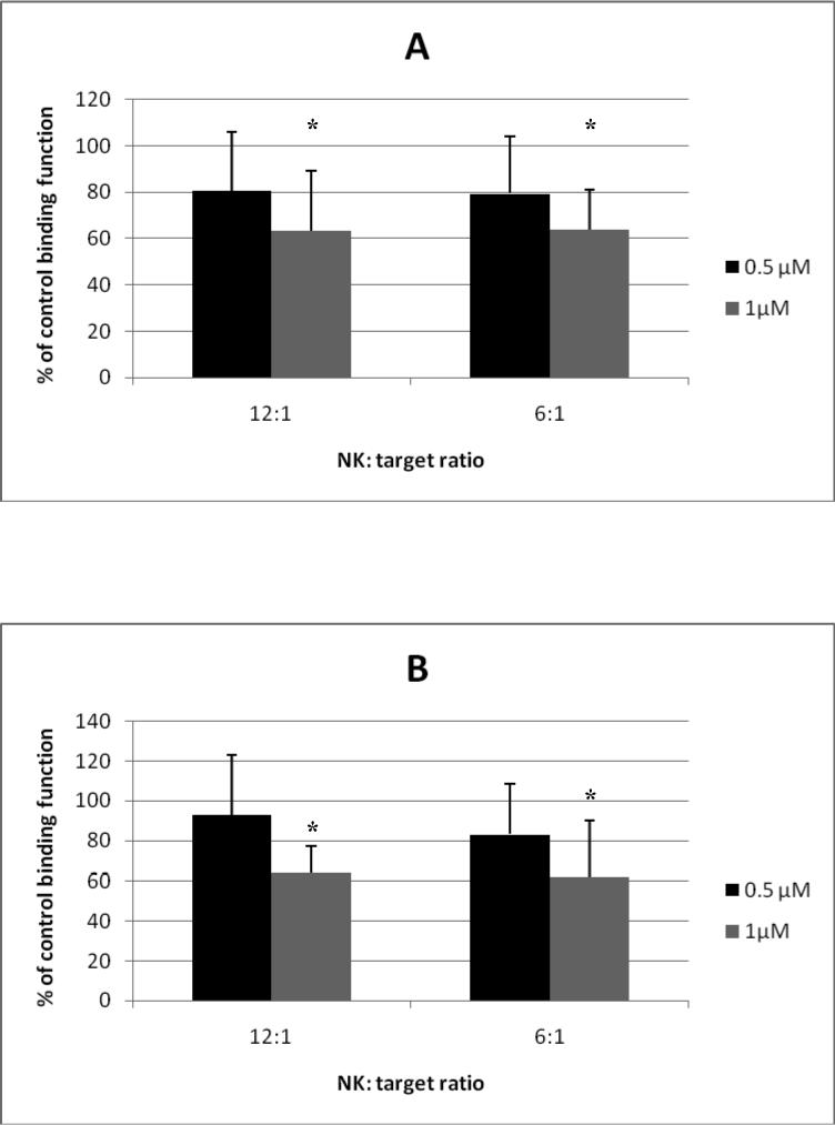
Effects of 24 h and 48 h continuous exposures to ziram on the ability of NK cells to bind K562 targets. A.) 24 h exposure of NK cells to 0.5 μM or 1 μM ziram . Results are mean±S.D. (n=6 for 0.5 μM; n=24 for 1 μM ). B.) 48 h exposure of NK cells to 0.5 μM or 1 μM ziram . Results are mean±S.D. (n=6 for 0.5 μM; n=18 for 1 μM). * indicates that the decrease in binding was statistically significant (p<0.01).
Effects of continuous exposures to ziram on cell-surface protein expression
24 h and 48 h exposures to 1 μM ziram caused no significant decrease in the expression of CD2, CD11a, CD11b, CD11c, CD16, CD18, or CD56 on NK cells.
Effects of 1h exposures to ziram followed by 24h or 48h in ziram-free media on NK tumor binding function
Effects of 1 h exposures to 1 μM or 2 μM ziram followed by 24 h in ziram-free media on NK tumor binding function are shown in figure 2A. A 1 h exposure to 1 μM ziram followed by 24 h in ziram-free media caused no significant decrease in tumor binding function (p> 0.5), while a 1 h exposure to 2 μM ziram followed by 24 h in ziram free-media caused a 70% decrease in tumor binding function (Figure 2A, p<0.001). Effects of 1 h exposures to 1 μM or 2 μM ziram followed by 48 h in ziram-free media on NK tumor binding function are shown in figure 2B. A1 h exposure to 1 μM ziram followed by 48 h in ziram-free media caused an approximately 30% decrease in tumor binding function (Figure 2B, p< 0.05) and 1 h exposure to 2 μM followed by 48 h in ziram-free media caused about a 70% decrease in tumor binding function of NK cells (Figure 2B, p < 0.001). The decrease in binding function was greater when NK cells were continuously exposed to 1 μM ziram for 24 and 48 h (35% and 40%, respectively, Figure 1) than when the exposure was for 1h followed by 24 or 48 h in ziram-free media (0% and 30%, respectively). The 30% loss of binding function seen 48 h after a 1 h exposure to ziram indicated that events occurring within the first 60 minutes of exposure may trigger processed that lead to a loss of binding function over time.
Figure 2.
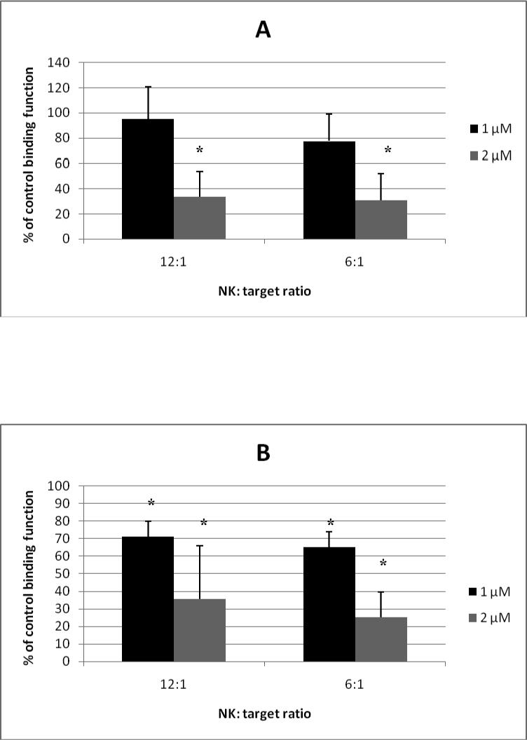
Effects of a 1h exposure to ziram followed by 24 h or 48 h in ziram-free media on the ability of NK cells to bind to K562 targets. A.) 1 h exposure to 1 μM or 2 μM ziram followed by 24 h in ziram-free media. Results are mean±S.D. (n=3 for 1 μM; n=18 for 2 μM ). B.) 1 h exposure to 1 μM or 2 μM ziram followed by 48 h in ziram-free media. Results are mean±S.D. (n=3 for 1 μM; n=15 for 2 μM). * indicates that the decrease in binding was statistically significant (p<0.01).
Effects of 1 h exposures followed by 24 h or 48 h in ziram-free media cell- surface protein expression
Of the cell surface proteins examined, only CD16 expression was decreased by exposure of NK cells to ziram (Figure 3). A 1 h exposure to 1 μM ziram followed by 24 h in ziram-free media caused no significant decrease in CD16 expression, while a 1 h exposure to 2 μM ziram followed by 24 h in ziram-free media caused a 70% decrease in CD16 expression (Figure 3A, p< 0.001, Figure 3C is a representative experiment). Effects of 1 h exposures to 2 μM or 1 μM ziram followed by 48 h in ziram-free media on CD16 expression are shown in Figure 3B. 1 h exposure to 1 μM ziram followed by 48 h in ziram-free media caused no significant decrease in CD16 expression (p>0.5), however, a 1h exposure to 2 μM ziram followed by 48 h in ziram-free media cause a 60% decrease in NK CD16 expression (Figure 3B, p<0.001, Figure 3D is a representative experiment).
Figure 3.
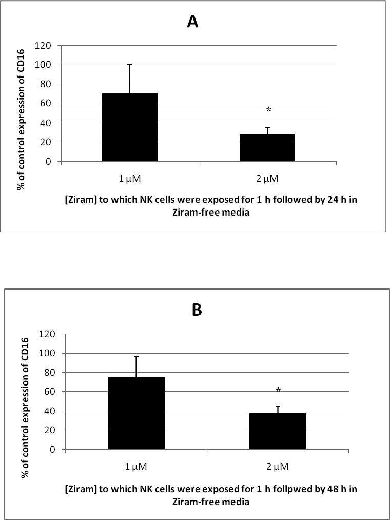
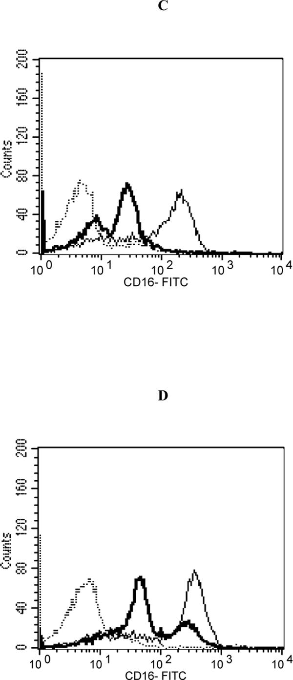
Effects of 1h exposure to ziram followed by 24 h or 48 h in ziram-free media on CD16 expression in NK cells. A.) 1 h exposure to 1 μM or 2 μM ziram followed by 24 h in ziram-free media. Results are mean±S.D. (n=4 for 1 μM; n=5 for 2 μM ). B.) 1 h exposure to 1 μM or 2 μM ziram followed by 48 h in ziram-free media. Results are mean±S.D. (n=4 for 1 μM; n=4 for 2 μM). * indicates that the decrease in CD16 expression was statistically significant (p<0.01). A.) Histogram from a representative experiment of a 1 h exposure to 2 μM ziram followed by 24 h in ziram-free media: Dashed line = IgG (isotype) control; thin solid line = control NK cells stained with anti-CD16 antibody; bold line = ziram-exposed cells stained with anti-CD16 antibody; y axis = cell number; x axis = fluorescence intensity . B.) Histogram from a representative experiment of a 1 h exposure to 2 μM ziram followed by 48 h in ziram-free media: Dashed line = IgG control; thin solid line = control NK cells stained with anti-CD16 antibody; bold line = ziram-exposed cells stained with anti-CD16 antibody; y axis = cell number; x axis = fluorescence intensity .
Effects of 24 h, 48 h, and 6 day exposure to ziram on ATP levels of NK cells
Effects of 24 h exposures to 0.25 μM, 0.5 μM, or 1 μM ziram on ATP levels in NK cells are shown in figure 4A . A 24 h exposure to 1 μM caused an approximately 50% decrease in ATP levels in NK cells (Figure 4A, p<0.00001). Decreases of about 18% were seen with 24 h exposures to 0.25 μM and 0.5 μM ziram (p<0.05). Effects of 48 h exposures to 0.25 μM, 0.5 μM, or 1 μM ziram on NK ATP levels are shown in figure 4B. A 48 h exposure to 1 μM caused a 48% decrease in ATP levels and 0.5 μM caused a 29% decrease ( Figure 4B, p<0.001). However, exposure to 0.25 μM ziram caused no decrease in ATP levels. Effects of a 6 day exposure to 0.25 μM, 0.5 μM, or 1 μM ziram on ATP levels of NK cells are shown in figure 4C. 6 day exposures to caused no significant decrease in ATP levels of NK cells (p>0.05).
Figure 4.
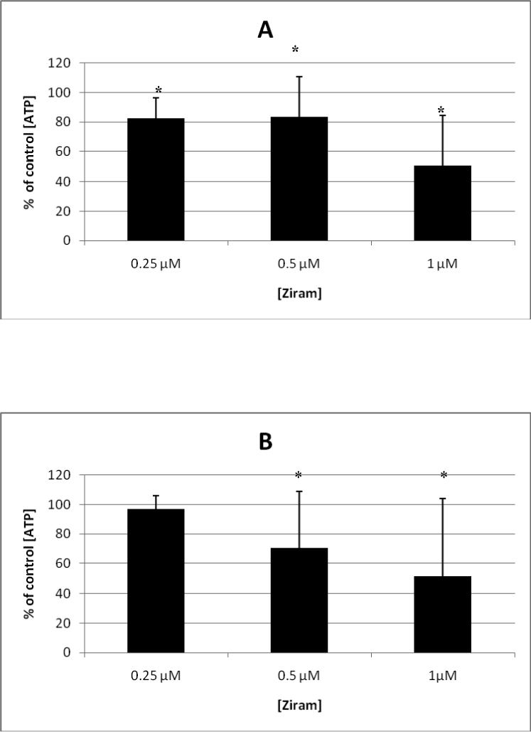
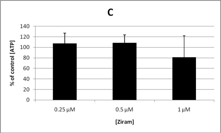
Effects of 24 h, 48 h, and 6 day continuous exposures ziram on the ATP levels in NK cells. A.) 24 h exposure of NK cells to 0.25 μM, 0.5 μM or 1 μM ziram . Results are mean±S.D. (n= 15 for 0.25 μM, 0.5 μM, and 1 μM). B.) 48 h exposure of NK cells to 0.25 μM, 0.5 μM or 1 μM ziram. Results are mean±S.D. (n=9 for 0.25 μM; n=32 for 0.5 μM and 1 μM). C.) 6 day exposure of NK cells to 0.25 μM, 0.5 μM or 1 μM ziram. Results are mean±S.D. (n=18 for 0.25 μM, 0.5 μM and 1 μM). * indicates that the decrease in ATP was statistically significant (p<0.01).
Effect of Glucose addition on ziram-induced decreases in ATP levels of NK cells
While ziram might interfere with mitochondrial ATP production, ATP production via the glycolytic pathway might sustain ATP levels until glucose supplies began to decline. To examine this possibility, we tested whether glucose supplementation of the culture medium affected the ziram-induced decreases in ATP levels seen at 48 h. Glucose was added at a final concentration of 2 g/L to control and ziram-exposed NK cells after 24 h to cells receiving a 48 h exposure to ziram. Cells from the same donor were exposed to ziram without glucose supplementation. The data from these experiments showed that Ziram was able to decrease ATP levels even when glucose was added to the cell culture (data not shown).
DISCUSSION
The purpose of the current study was to examine the effects of ziram on the ability of NK cells to bind to their targets and to express cell surface markers that are known to be involved in NK binding to targets as possible causes of the ziram-induced decreases in lytic function. Additionally, the effect of ziram exposures on ATP levels of NK cells were studied in order to determine if decreases in ATP might in part explain losses of lytic function and/or binding function. Since NK cell binding to their targets is primary in achieving target lysis, it was important to examine if NK cell binding function was diminished by exposure to ziram. NK cells use specific cell surface markers to recognize and bind to their targets so if a decrease in binding was observed it was imperative to examine whether this was accompanied by decreases in cell surface marker expression. Decreases in the intracellular concentration ATP in NK cells as a consequence of ziram exposures have the potential to underly any changes in binding and/or cell surface marker expression as well as the already established decrease in lytic function, and thus were examined.
It has been shown that a continuous exposures of NK cells to 1 μM ziram for 24 h or 48 h causes about a 40% decrease in lytic function after 24 h and a nearly 80% decrease after 48 h (Wilson et al., 2004). The current study showed that there was a decrease in binding function after these same exposures of about 35 % after 24 h and about 40% after 48 h. However, the previous study also showed that there were decreases in lytic function after both 24 h and 48 h exposures to 0.5 μM ziram and after a 48 h exposure to 0.25 μM ziram (Wilson et al., 2004), and these exposures had no effect on the ability of the NK cell to bind to targets. Thus, it appears that lack of ability of NK cells to bind to their target is not the reason for the loss of lytic function that has been seen with continuous exposures to concentrations of ziram below 1 μM. Certain cell surface markers are known to be involved in NK binding/lytic function. CD11a, CD18, CD16, and CD56 have all been shown to be important in binding to target cells (Lotzova, 1993). Studies using flow cytometry to monitor the effects of butyltin (BT) exposures on NK cell surface markers showed decreases in CD16 and CD56 following exposures to BTs and these decreases corresponded to decreases in the ability of NK cells to bind to target cells (Odman-Ghazi et al., 2003; Whalen et al., 2001). However, when expression of cell surface proteins important to target binding were examined in NK cells exposed to 1 μM ziram for either 24 or 48 h there was no decreased expression of any of the seven proteins studied.
Previous studies have also shown that the lytic function of NK cells was decreased by greater than 90% when NK cells were exposed to 2.5 μM ziram followed by 24 h or 48 h in ziram- free media (Taylor et al., 2005). The current results showed that a 1 h exposures to 2 μM ziram followed by 24 h or 48 h in ziram-free media caused a significant decrease in NK cell binding function with an accompanying decrease in the expression of CD16. So, it appears the loss of binding function seen with these particular ziram exposures was associated with a decrease in CD16 expression.
It is essential to examine NK cell ATP levels after exposure to ziram to see if there is a link between loss of NK lytic function and ATP levels in NK cells. As ATP is essential to cellular function , ziram-induced decreases in ATP might account, at least in part, for loss of NK cell function. Ziram, is a carbamate fungicide and this class of compound inhibits the enzyme aconitase , which converts citrate to isocitrate in the Krebs cycle (Owens, 1963) , which would result in decreased ATP. The current results showed that ATP levels in NK cells were decreased by about 18% with 24 h exposures to 0.25 μM and 0.5 μM ziram and by about 50% with 1 μM. Previous studies have shown that a 24 h exposure to 1 μM ziram decreased the lytic function of NK cells by about 40% (Wilson et al., 2004). Thus, there appears to be an association of decreased ATP levels with decreased lytic function when NK cells were exposed to 1 μM ziram for 24 h. In a past study, using rotenone and oligomycin to decrease ATP levels in NK cells, we found that a decrease in ATP of about 20% for 24 h or 48 h caused an approximately 50% decrease in lytic function (Dudimah et al., 2007) The current results also showed a 48 h exposure to 1μM ziram caused a decrease in ATP levels of about 48% and 0.5 μM decreased ATP levels by about 29%. Previously, each of these concentrations has been shown to decrease lytic function by 76% and 64%, respectively (Wilson et al., 2004). However, 0.25 μM ziram decreased lytic function by 48% after 48 h, but causes no decrease in ATP levels. Additionally, it is known that concentrations as low as 0.125 μM ziram were able to decrease lytic function by about 65% after a 6 day exposure (Wilson et al., 2004). However, there were no significant decreases in ATP levels at any of the ziram concentrations tested after 6 days. Thus, it appears that decreases levels of ATP in NK cells cannot fully account for the losses of lytic function seen with ziram exposures. Further, it appears that the negative effect of ziram on ATP levels is lost over the course of a 6 day period.
Since we saw a decrease in ATP levels, we examined whether the addition of glucose to NK cells during the exposure to ziram would replenish NK ATP levels (via the glycolytic pathway). The results showed that glucose supplementation had no impact on the ziram-induced decreases in ATP level. This suggests that ATP generation by the glycolytic pathway is not sufficient to overcome the negative effects of ziram on NK cell ATP levels.
In summary these results indicate that: (1) ziram exposure can decrease the target-binding function of NK cells; (2) decreased binding function does not necessarily accompany the loss of lytic function seen with ziram exposures; (3) decreased expression of the cell surface proteins examined cannot account for the ziram-induced loss of binding function; (4) certain ziram exposures decrease the levels of ATP in NK cells; (5) decreases in ATP do not necessarily accompany loss of lytic function; (6) glucose supplementation cannot overcome ziram-induced decreases in ATP levels.
Table 1.
Effect of Ziram on the expression of CD16 on purified NK cells
| Treatment Intensity | % positive Cells | Mean Flourescence |
|---|---|---|
| CD16 | CD16 | |
| Control | 77±8 | 282±111 |
| 2 μM Ziram | ||
| 1h | 60±9 | 78±31 |
| followed by 24h | ||
| in Ziram-free media | ||
| Control | 80±7 | 255±124 |
| 2 μM Ziram | ||
| 1h | 74±8 | 93±34 |
| followed by 48h | ||
| in Ziram-free me |
Results are the mean +/−S.D. of at least three separate experiments using different donors.
ACKNOWLEDGMENTS
This research was supported by Grant S06GM008092-32 from the National Institutes of Health.
REFERENCES
- Caldas ED, Conceicao MH, Miranda MCC, deSouza LCKR, Lima JF. Determination of Dithiocarbamate Fungicide residues in Food by a Spectrophotometric Method Using a Vertical Disulfide Reaction System. J. Agricultural and Food Chemistry. 2001;49:4521–4525. doi: 10.1021/jf010124a. [DOI] [PubMed] [Google Scholar]
- De Jong WH, Tentji M, Spiekstra SW, Vandebriel RJ, Van Loveren H. Determination of the sensitizing activity of the rubber contact sensitisers TMTD, ZDMC, MBT, and DEA in a modified local lymph node assay and the effect of sodium dodecyl sulfate pretreatment on local lymph node responses. Toxicology. 2002;176:123–134. doi: 10.1016/s0300-483x(02)00131-2. [DOI] [PubMed] [Google Scholar]
- Dudimah FD, Odman-Ghazi SO, Hatcher F, Whalen MM. Effect of tributyltin (TBT) on ATP levels in human natural killer (NK) cells: relationship to TBT-induced decreases in NK function. J. Appl. Toxicol. 2007;27:86–94. doi: 10.1002/jat.1202. [DOI] [PubMed] [Google Scholar]
- Edwards IR, Ferry DG, Temple WA. Fungicides and related compounds. In: Hayes WJ, Laws ET, editors. Handbook of pesticide toxicology. Vol. 3. Academic Press; New York: 1991. Classes of pesticides. [Google Scholar]
- Franekic J, Bratulic N, Pavlica M, Papes D. Genotoxicity of dithiocarbamates and their metabolites. Mutation Research. 1994;325:65–74. doi: 10.1016/0165-7992(94)90003-5. [DOI] [PubMed] [Google Scholar]
- Hazardous Substance Databank, TOXNET . National Library of Medicine. Medlars, Management Section; Bethesda, MD: 1993. [Google Scholar]
- Holm S. A simple sequentially rejective multiple test procedure. Scand. J. Statistics. 1979;6:65–70. [Google Scholar]
- IRAC Monographs on the Evalution of Carcinogenic Risks to Humans, Occupational Exposures in Insecticides Application, and some Pesticides. Vol. 53, Lyon. 1991:423–438. [PMC free article] [PubMed] [Google Scholar]
- Lotzova E. Definition and function of natural killer cells. Nature Immunology. 1993;12:177–193. [PubMed] [Google Scholar]
- Nettis E, Assennato G, Ferrannini A, Tursi A. Type I allergy to natural rubber latex and type IV allergy to rubber chemicals in health care workers with glove-related skin symptoms. Clinical and Experimental Allergy. 2002;32:441–447. doi: 10.1046/j.1365-2222.2002.01308.x. [DOI] [PubMed] [Google Scholar]
- Odman-Ghazi SO, Hatcher F, Whalen MM. Expression of Functionally Relevant Cell Surface Markers in Dibutyltin-exposed Human Natural Killer Cells. Chemico-Biological Interactions. 2003;146:1–18. doi: 10.1016/s0009-2797(03)00069-3. [DOI] [PubMed] [Google Scholar]
- Owens R. Chemistry and physiology of fungicidal action. Annual Review Phytopatholology. 1963;1:77–100. [Google Scholar]
- Whalen MM, Loganthan BG. Toxicol. Appl. Pharmacol. 2000;171:141–148. doi: 10.1006/taap.2000.9121. [DOI] [PubMed] [Google Scholar]
- Taylor TR, Tucker T, Whalen MM. Persistent inhibition of human natural killer cell function by ziram and pentachlorophenol. Environmental Toxicology. 2005;20:418–424. doi: 10.1002/tox.20127. [DOI] [PubMed] [Google Scholar]
- Vivier E, Nunès JA, Vely F. Natural Killer Cell Signaling Pathways. Science. 2004;306:1517–1519. doi: 10.1126/science.1103478. [DOI] [PubMed] [Google Scholar]
- Whalen MM, Doshi RN, Brankhurst AD. Effetcs of pertussis toxin treatment on human natural killer cell function. Immunology. 1992;76:402–407. [PMC free article] [PubMed] [Google Scholar]
- Whalen MM, Ghazi S, Loganathan BG, Hatcher F. Expression of CD16, CD18, and CD56 in tributlytin-exposed human natural killer cells. Chemico-Biological Interactions. 2001;139:159–176. doi: 10.1016/s0009-2797(01)00297-6. [DOI] [PubMed] [Google Scholar]
- Whalen MM, Loganthan BG, Yamashita N, Saito T. Immunomodulation of human natural killer cell cytotoxic function by triazine and carbamate pesticides. Chemico- Biological Interactions. 2003;145:311–319. doi: 10.1016/s0009-2797(03)00027-9. [DOI] [PubMed] [Google Scholar]
- Wilson S, Dzon L, Reed A, Puritt M, Whalen MM. Effects invitro exposure to low levels of organotin and carbamate pesticides on human natural killer cell cytotoxic function. Environmental Toxicology. 2004;19:554–563. doi: 10.1002/tox.20061. [DOI] [PubMed] [Google Scholar]
- Zenzen V, Fauth E, Zankl H, Janzowski C, Eisenbrand G. Mutagenic and cytotoxicity effectiveness of zinc dimethyl and zinc diisonolydithiocarbamate in human lymphocyte cultures. Mutation Research. 2001;497:89–99. doi: 10.1016/s1383-5718(01)00238-8. [DOI] [PubMed] [Google Scholar]


