Abstract
The prevalence of internal carotid artery (ICA) morphological variations (MV), their characteristics, and their possible association with carotid stenosis, vascular risk factors, and previous transient ischemic attack or ischemic stroke was investigated in a consecutive series of patients.
Within a seven-month period, 1217 patients (557 men and 660 women; mean age [± SD] 62.7±18.1 years) consecutively referred to the Laboratory of Neurosonology, University of L’Aquila, Italy, underwent a neck vessel examination using a high-resolution B-mode ultrasound device with a 7.5 MHz linear phased array probe.
ICA MV were present in 319 (26.2%) patients; they were unilateral in 201 patients (63.0%) and bilateral in 118 patients (37.0%). Patients with ICA MV were older than those without ICA MV (66.3±19.9 years versus 61.4±18.0 years, P<0.0001) and were mostly women (62.4%, P=0.0008). Tortuosity was present in 195 (44.6%) arteries, kinking in 236 arteries (54.0%) and coiling in six arteries (1.4%). Carotid stenosis was found in 270 patients (22.2%). Among patients with ICA stenosis, MV were found in 134 patients (49.6%). Mean neck length was similar in patients with and without ICA MV (12.1±2.4 cm versus 12.6±2.9 cm, P=0.4). In the multivariate logistic regression analysis, the presence of ICA MV was associated with female sex and older age.
Tortuosity and kinking were frequently encountered during neurovascular examination. Their presence was usually related to aging and female sex, and did not imply any additive risk for stroke, although further studies are needed to clarify this point.
Keywords: Coiling, Duplex sonography, Internal carotid artery, Kinking, Stroke, Tortuosity
The internal carotid artery (ICA) usually runs straight in the neck. Morphological variations (MV) of its course are often found, and are classified as tortuosity, kinking or coiling (1,2). Despite their common occurrence, conclusive evidence about their prevalence, characteristics and relationship with the presence of cerebrovascular disease is still lacking.
The prevalence of ICA MV, their characteristics and their possible association with carotid stenosis, vascular risk factors, and previous transient ischemic attack (TIA) or ischemic stroke was investigated in a consecutive series of patients.
METHODS
Within a seven-month period, 1217 patients consecutively referred to the Laboratory of Neurosonology, University of L’Aquila, Italy, underwent a neck vessel examination using a high-resolution B-mode ultrasound device (AU5 Epi, Esaote Biomedica, Italy) with a 7.5 MHz linear phased array probe. Patients were referred for previous TIA or ischemic stroke in 155 cases (12.7%), dizziness in 381 cases (31.3%), syncope in 81 cases (6.7%), atherothrombotic risk factors – such as arterial hypertension or diabetes mellitus – in 301 cases (24.7%), follow-up of already diagnosed ICA stenosis in 231 cases (19.0%), and unspecified or nonvascular causes in the remaining 68 cases (5.6%).
Anterior, lateral, posterolateral sagittal, and transverse views were used to evaluate the presence of ICA MV and the degree of carotid stenosis. MV were classified as tortuosity, kinking or coiling (1,2). Tortuosity was defined as any elongation, or S- or C-shaped curve of the vessel. Kinking was defined as an acute angulation of one or more segments of the vessel, usually associated with functional or organic narrowing. Coiling was defined as a redundant elongation of the vessel, creating an exaggerated S-shaped curve or circular configuration (1,2). Kinking was graded as type I in the presence of an angle ranging from 60° to 90°, type II when the angle was between 30° and 60°, and type III (sharp) when the angle was less than 30° (3). According to their distance from the carotid bifurcation, ICA MV were classified as proximal when they were located up to 4 cm from the bifurcation and distal when located more than 4 cm from the bifurcation (1,2). Presence and degree of any ICA stenosis were assessed according to the measured maximum flow velocity, and to the arterial narrowing as a percentage diameter reduction at the maximum of the stenosis. This was calculated according to the formula (1−A/B)×100%, where A represented the tightest diameter of the stenosis, and B represented the suspected initial vessel diameter. Neck length was measured as the distance between the mandibular angle and the superior margin of the clavicle, with the patient supine and the neck in maximal extension. The side on which the measurement was taken was randomly selected.
Multivariate logistic regression analyses were used to compare characteristics of patients with and without ICA MV. Two-sided values of P<0.05 were considered to be statistically significant. The SPSS statistical software program (version 10.1, SPSS Inc, USA) was used for all analyses.
RESULTS
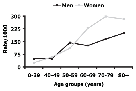
Of 1217 patients (557 men and 660 women, mean age [± SD] 62.7±18.1 years), ICA MV were present in 319 patients (26.2%); they were unilateral in 201 patients (63.0%) and bilateral in 118 patients (37.0%). Patients with ICA MV were older than those without ICA MV (66.3±19.9 years versus 61.4±18.0 years, P<0.0001) and were mostly women (62.4%, P=0.0008). As shown in Figure 1, the prevalence of ICA MV increased similarly in both sexes in all age groups up to 50 to 59 years. In the age groups 60 to 69 years and 70 to 79 years, ICA MV were more common in women than in men (95% CI 186.1 to 269.8, versus 89.9 to 163.3 for women and men, respectively, aged 60 to 69 years, 95% CI 253.4 to 340.0, versus 127.1 to 202.7 for women and men, respectively, aged 70 to 79 years).
Figure 1.
Prevalence of internal carotid artery morphological variations according to age and sex
As shown in Table 1, tortuosity was present in 195 arteries (44.6%), kinking in 236 arteries (54.0%) and coiling in six arteries (1.4%). Kinking was classified as type I in 183 arteries, type II in 36 arteries and type III in 17 arteries. Carotid stenosis was found in 270 patients (22.2%); in 183 patients the degree of stenosis was lower than 50%, and in 87 patients it was 50% or greater. Among patients with ICA stenosis, MV were found in 134 patients (49.6%). Two hundred eighty-three MV (64.8%) were proximal to the carotid bifurcation, and 154 (35.2%) were distal (P=0.0001). Mean neck length was 12.3±2.7 cm, and was similar in patients with and without ICA MV (12.1±2.4 cm versus 12.6±2.9 cm, P=0.4).
TABLE 1.
Distribution of internal carotid artery morphological variations, distance from carotid bifurcation and association with internal carotid artery stenosis
| Internal carotid artery morphological variations
|
||||
|---|---|---|---|---|
| Tortuosity, n (%) | Kinking, n (%) | Coiling, n (%) | Total, n | |
| Location relative to carotid bifurcation* | ||||
| Proximal | 139 (71.3) | 138 (58.5) | 6 (100) | 283 |
| Distal | 56 (28.7) | 98 (41.5) | – | 154 |
| Total | 195 (44.6) | 236 (54.0) | 6 (1.4) | 437 |
| Internal carotid artery stenosis | ||||
| Absent | 152 (77.9) | 185 (78.4) | 4 (66.7) | 341 |
| <50% | 35 (17.9) | 44 (18.6) | – | 79 |
| ≥50% | 8 (4.2) | 7 (3.0) | 2 (33.3) | 17 |
| Total | 195 (100) | 236 (100) | 6 (100) | 437 |
Internal carotid artery morphological variations were classified as proximal when they were located up to 4 cm from the carotid bifurcation, and distal when located more than 4 cm from the bifurcation
Tortuosity and kinking were more frequently found on the left than on the right side (61.5% on the left side versus 38.5% on the right for tortuosity; 57.2% on the left side versus 42.8% on the right for kinking, P<0.0001). The most common associations were represented by bilateral kinking (n=42), tortuosity and kinking (total n=39), and by bilateral tortuosity (n=35) (Table 2).
TABLE 2.
Distribution by side of internal carotid artery (ICA) morphological variations
| Right ICA
|
|||||
|---|---|---|---|---|---|
| Left ICA | Tortuosity | Kinking | Coiling | Straight | Total |
| Tortuosity | 35 | 18 | 1 | 66 | 120 |
| Kinking | 21 | 42 | – | 72 | 135 |
| Coiling | – | – | 1 | 1 | 2 |
| Straight | 19 | 41 | 2 | 898 | 960 |
| Total | 75 | 101 | 4 | 1037 | 1217 |
In the multivariate logistic regression analysis (Table 3), the presence of ICA MV was associated with female sex and older age.
TABLE 3.
Risk factors associated with internal carotid artery (ICA) morphological variations
| Multivariate analysis
|
|||
|---|---|---|---|
| Risk factor | OR | 95% CI | P |
| Age | 1.04 | 1.02–1.06 | <0.0001 |
| Female sex | 1.78 | 1.21–2.64 | 0.004 |
| <50% ICA stenosis | 1.43 | 0.89–2.31 | 0.1 |
| ≥50% ICA stenosis | 0.57 | 0.29–1.12 | 0.1 |
| Arterial hypertension | 1.20 | 0.81–1.78 | 0.4 |
| Diabetes mellitus | 0.85 | 0.49–1.47 | 0.6 |
| Previous ischemic stroke or TIA | 0.96 | 0.61–1.52 | 0.9 |
| Dizziness | 0.89 | 0.43–1.83 | 0.7 |
| Neck length | 0.97 | 0.90–1.04 | 0.4 |
TIA Transient ischemic attack
DISCUSSION
Previous studies (3–7) reported a 4% to 66% prevalence of MV; other studies using ultrasound duplex scanning reported a 32% to 58% prevalence of MV (5,6,8). Using a high-resolution B-mode ultrasound device, we found a 26.2% prevalence of ICA MV. Disagreement in prevalence rates may be explained by different diagnostic techniques, selection criteria of the study population and classification of MV.
In the present series, tortuosity and kinking were commonly reported ICA MV (6,8); kinking was the most common of the MV (54.0%), mostly represented by type I, closely followed by tortuosity (44.6%), while coiling was the most rare. Bilateral kinking represented the most common association, followed by tortuosity and type I kinking, and by bilateral type I kinking. MV were mostly proximal to the carotid bifurcation (1,2).
ICA MV distribution according to side was reported differently among studies. In the present series, kinking and tortuosity were more frequently found on the left side; the present findings may depend on the different anatomical origin of the right and left common carotid arteries, favouring MV in the arteries originating directly from the aortic arch.
As suggested by previous studies (5,6,8,9), we also found an overall higher prevalence of MV in women than in men. However, up to the age of 60 years, we did not find any difference between sexes, while from the age of 60 years onward, prevalence of MV was higher in women than in men. As a possible explanation for the relationship between age and sex, not excluding a selection bias related to competitive comorbid conditions in men, we hypothesized a more severe alteration in vessel wall elasticity in older women, depending on hormonal processes.
The etiology of ICA MV is still controversial, and different hypotheses are worthy of consideration. In a few cases, it was postulated that fibromuscular dysplasia, which is rare compared with the frequency of MV (10), has a possible etiological role. It was also thought that MV may be related to shorter neck lengths, even if we did not find any correlation with this feature (5). MV were also found in infants, and was thought to be congenital; coiling according to this hypothesis was frequently ascribed to embryological causes (4,7,11). An increased prevalence of kinking in patients with arterial hypertension was also reported (8,9,12); in those cases, raised endoluminal pressure and increased parietal tension caused by arterial hypertension were believed to favour the development of MV.
Because a few cases of MV were detected in patients less than 40 years of age, we cannot exclude that, in the present series, some MV were congenital. However, the increased prevalence of MV in the oldest age groups provided more support for the hypothesis that MV mostly develop later in life, probably because of reduced elasticity and degeneration of the arterial vessel wall (8,13). If this is the case, we should hypothesize a possible association of MV with aneurysms; however, this association has never been reported. MV were also reported in other arterial districts and in athletes, specifically in the iliac arteries (12). This occurrence suggests that MV may be promoted by repeated muscle flexion, extension, rotation and stretching that acts on the arterial wall – a hypothesis that needs to be studied.
Despite studies suggesting the low importance of ICA MV as a contributing factor in carotid plaque formation and a cause of major neurological symptoms (8,9), MV were also regarded as a possible cause of cerebral infarction, even in the absence of atherosclerosis. In some instances, patients with such variations were considered candidates for surgery (11,12,14–16). In the present series, the multivariate analysis did not show any relationship among ICA MV with vascular risk factors, ICA stenosis, or history of previous stroke or TIA, emphasizing the role of older age and female sex. We are aware that population-based studies that include patients from young to old age, with extensive follow-up, are needed to elucidate the causes and the possible consequences of ICA MV.
CONCLUSION
As suggested by our data, tortuosity and kinking are frequently encountered during neurovascular examination. Their presence is usually related to aging and female sex, and does not imply any additive risk for stroke, although further studies are needed to clarify this point.
REFERENCES
- 1.Weibel J, Fields WS. Tortuosity, coiling and kinking of the internal carotid artery. I. Etiology and radiographic anatomy. Neurology. 1965;15:7–18. doi: 10.1212/wnl.15.1.7. [DOI] [PubMed] [Google Scholar]
- 2.Weibel J, Fields WS. Tortuosity, coiling and kinking of the internal carotid artery. II. Relationship of morphological variation to cerebrovascular insufficiency. Neurology. 1965;15:462–8. doi: 10.1212/wnl.15.5.462. [DOI] [PubMed] [Google Scholar]
- 3.Metz H, Murray-Leslie RM, Bannister RG, Bull JW, Marshall J. Kinking of the internal carotid artery. Lancet. 1961;1:424–6. doi: 10.1016/s0140-6736(61)90004-6. [DOI] [PubMed] [Google Scholar]
- 4.Leipzig TJ, Dohrmann GJ. The tortuous or kinked carotid artery: Pathogenesis and clinical considerations. A historical review. Surg Neurol. 1986;25:478–86. doi: 10.1016/0090-3019(86)90087-x. [DOI] [PubMed] [Google Scholar]
- 5.Macchi C, Gulisano M, Giannelli F, Catini C, Pratesi C, Pacini P. Kinking of the human internal carotid artery: A statistical study in 100 healthy subjects by echocolor Doppler. J Cardiovasc Surg (Torino) 1997;38:629–37. [PubMed] [Google Scholar]
- 6.Pancera P, Ribul M, De Marchi S, Arosio E, Lechi A. Prevalence of morphological alterations in cervical vessels: A colour duplex ultrasonographic study in a series of 3300 subjects. Int Angiol. 1998;17:22–7. [PubMed] [Google Scholar]
- 7.Paulsen F, Tillmann B, Christofides C, Richter W, Koebke J. Curving and looping of the internal carotid artery in relation to the pharynx: Frequency, embryology and clinical implications. J Anat. 2000;197:373–81. doi: 10.1046/j.1469-7580.2000.19730373.x. [DOI] [PMC free article] [PubMed] [Google Scholar]
- 8.Del Corso L, Moruzzo D, Conte B, et al. Tortuosity, kinking, and coiling of the carotid artery: Expression of atherosclerosis or aging? Angiology. 1998;49:361–71. doi: 10.1177/000331979804900505. [DOI] [PubMed] [Google Scholar]
- 9.Pancera P, Ribul M, Presciuttini B, Lechi A. Prevalence of carotid artery kinking in 590 consecutive subjects evaluated by Echocolordoppler. Is there a correlation with arterial hypertension? J Intern Med. 2000;248:7–12. doi: 10.1046/j.1365-2796.2000.00611.x. [DOI] [PubMed] [Google Scholar]
- 10.Begelman SM, Olin JW. Fibromuscular dysplasia. Curr Opin Rheumatol. 2000;12:41–7. doi: 10.1097/00002281-200001000-00007. [DOI] [PubMed] [Google Scholar]
- 11.Desai B, Toole JF. Kinks, coils, and carotids: A review. Stroke. 1975;6:649–53. doi: 10.1161/01.str.6.6.649. [DOI] [PubMed] [Google Scholar]
- 12.Schep G, Bender MH, van de Tempel G, Wijn PF, de Vries WR, Eikelboom BC. Detection and treatment of claudication due to functional iliac obstruction in top endurance athletes: A prospective study. Lancet. 2002;359:466–73. doi: 10.1016/s0140-6736(02)07675-4. [DOI] [PubMed] [Google Scholar]
- 13.Vannix RS, Joergenson EJ, Carter R. Kinking of the internal carotid artery. Clinical significance and surgical management. Am J Surg. 1977;134:82–9. doi: 10.1016/0002-9610(77)90288-4. [DOI] [PubMed] [Google Scholar]
- 14.Illuminati G, Calió FG, Papaspyropoulos V, Montesano G, D’Urso A. Revascularization of the internal carotid artery for isolated, stenotic, and symptomatic kinking. Arch Surg. 2003;138:192–7. doi: 10.1001/archsurg.138.2.192. [DOI] [PubMed] [Google Scholar]
- 15.Koskas F, Bahnini A, Walden R, Kieffer E. Stenotic coiling and kinking of the internal carotid artery. Ann Vasc Surg. 1993;7:530–40. doi: 10.1007/BF02000147. [DOI] [PubMed] [Google Scholar]
- 16.Oliviero U, Scherillo G, Casaburi C, et al. Prospective evaluation of hypertensive patients with carotid kinking and coiling: An ultrasonographic 7-year study. Angiology. 2003;54:169–75. doi: 10.1177/000331970305400205. [DOI] [PubMed] [Google Scholar]


