Abstract
The results of surgical bypass and endarterectomy in Takayasu's arteritis (TA) were reported to be poor compared to usual atherosclerosis patients. However, if ischemic symptoms due to occlusive disease were severe, surgical procedures were inevitable. We report surgical experience of 5 patients with TA. Five women (ranged from 26 to 58 yr) were operated between June 1998 and May 2004. Three patients showed occlusion of main branches of aortic arch and had symptoms of cerebral ischemia. One patient showed near total occlusion in the midabdominal aorta and had symptoms of orthopnea and uncontrolled hypertension. One patient showed total occlusion of abdominal aorta at the level of aortic bifurcation and had a symptom of severe claudication on both legs. Bypasses from the ascending aorta to the carotid artery were performed in 3 cases. Bypass from the thoracic aorta to the left common iliac artery was performed in one case and endarterectomy of abdominal aorta in one case. The ischemic symptoms related with arterial occlusion were resolved after surgery. And the symptoms of cardiac failure disappeared. The symptomatic TA frequently required arterial reconstruction. The symptomatic improvement and excellent mid-term patency could be expected after arterial reconstruction and endarterectomy.
Keywords: Vascular Diseases; Takayasu's Arteritis; Bypass; Endarterectomy, Carotid
INTRODUCTION
Takayasu's arteritis (TA) is a nonspecific granulomatous inflammatory arteriopathy of unknown cause that results in occlusion or less common aneurysmal degeneration of large and medium-sized elastic arteries. The disease was first described in 1908 by Takayasu (1), a Japanese ophthalmologist, in a young female patient with retinal neovascularization and absent radial pulse. Subsequent descriptions of the disease have emphasized the 'pulseless' syndrome, with involvement of the brachiocephalic arteries. Its incidence is low, being reported in 1/3,000 autopsies in Japan and as low as 2.6 cases per one million in the United States (2, 3). The female-to-male ratio was reported as 8-10:1 (4).
The clinical feature was dependent on the organ involved. The patients presented with claudication if the lower abdominal aorta was involved, hypertension if the renal arteries were involved, stroke if the cerebral vascular vessels were involved or angina and myocardial infarction symptoms if the coronary arteries were involved.
Arteriography was the best modality for the diagnosis of TA with magnetic resonance angiography and CT scans (5-7). The disease did not just affect the arterial bifurcations but usually involved the entire length of arteries. The disease had a very peculiar distribution with the subclavian, axillary, carotids, renals and infra-abdominal aortas.
The clinical presentations of TA was not the same as atherosclerosis. The patients with atherosclerosis were usually elderly while patients with TA were young, but despite their youth, they still had severe cardiac, renal and pulmonary problems. Therefore these patients should undergo extensive medical evaluation prior to surgery. It was preferable to avoid surgery during the acute phase of the illness. The surgical procedure always consisted of a bypass operation to vessels normal on angiography proximal and distal to the occlusive or stenotic lesion. The aim of this report was to evaluate the effectiveness and safety of the operation performed for symptomatic TA.
MATERIALS AND METHODS
Patients
From June 1998 to May 2004, we operated on 5 patients with TA. The patients' age ranged 28 to 58 yr, all patients were female. For evaluation of diseased arterial system, conventional angiography or CT angiography was performed in 4 patients, MR angiography in 1 patient. The mid-abdominal aorta was involved in one patient. The aortic bifurcation and both common iliac artery was involved in one patients. In one patient all branches of the aortic arch were involved. Both common carotid artery and left vertebral artery were involved in one patient, both common carotid artery, right vertebral artery, and left subclavian artery were involved in one patient. The indications for revascularization or endarterectomy were disabling cerebral ischemic symptoms (such as cerebral infarction, repeated syncopal attacks, dizziness, visual disturbance), severe claudications of lower extremities and cardiac failure (Table 1).
Table 1.
Patient profiles
L/E, lower extremity; RVH, renovascular hypertension; SMA, superior mesenteric artery; ICA, internal carotid artery; F/U, follow-up.
Ascendo-bicarotid bypass
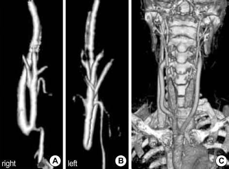
Operation was performed for the patients who had totally occluded bilateral common carotid arteries with 12×6 mm Y-shaped polytetrafluoroethylene (PTFE) graft (Fig. 1). Two patients who underwent ascendo-bicarotid bypass had no ischemic symptom of both upper extremities. Firstly, an oblique skin incision along the anterior border of the sternocleidomastoid muscle was made bilaterally. Rubber bands were passed around the bilateral common, internal, and external carotid arteries. A median sternotomy was then made, the ascending aorta was exposed and the body of Y-shaped PTFE graft was sutured with the partial ascending aorta clamped condition. The limbs of the PTFE graft were positioned along the course of the original carotid artery. After arteriotomy was made in the patent portion of the both internal carotid artery (ICA), bilateral limbs of the PTFE graft were anastomosed to the both ICA (Fig. 1).
Fig. 1.
Preoperative angiography shows patent right (A) and left (B) internal carotid arteries respectively. (C) Postoperative CT-angiography shows ascendo-bicarotid bypass.
Ascendo-right ICA bypass
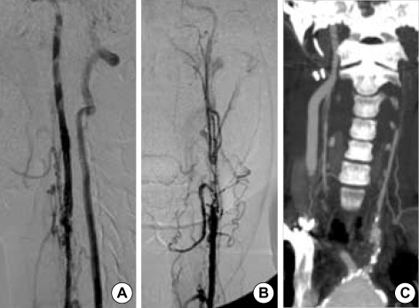
Operation was performed for the patient who had totally occluded both common carotid arteries. Also extracranial portion of the left internal carotid artery in this patient was so narrow that we were not able to anastomose it. This patient had no ischemic symptom of both upper extremities. An oblique skin incision along the anterior border of the right sternocleidomastoid muscle and a median sternotomy were then made. We performed ascendo-right ICA bypass with a 7 mm external supported PTFE graft.
Thoraco-left iliac bypass
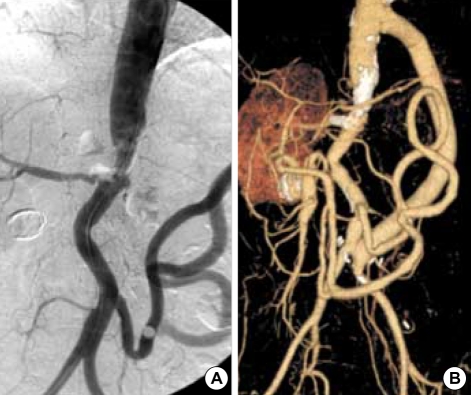
This patient suffered from orthopnea and uncontrolled hypertension because of renovascular origin. Preoperative vascular imaging study revealed nearly total occlusion in the midabdominal aorta. Thoracoabdominal incision through the 7th intercostals space for exposure of lower thoracic aorta and left common iliac artery was made. The retroperitoneal plane was entered by stripping the peritoneum away from the abdominal wall using blunt finger dissection. To enhance exposure, the peritoneum was dissected from the abdominal wall as far superiorly and inferiorly as possible. Division of left diaphragm was made. The lower thoracic aorta was exposed and 12 mm Dacron graft was sutured with the partial thoracic aorta clamped condition. The distal end of Dacron graft was sutured to the left common iliac artery (Fig. 3). For the renal arterial stenosis, we had performed percutaneous renal arterial stenting before operation.
Fig. 3.
(A) Preoperative angiography shows no visualization of superior mesenteric artery and left renal artery, a stenotic right renal artery, and a enlarged inferior mesenteric artery. (B) Postoperative CT angiography shows patent thoraco-left iliac bypass graft and stent in the right renal artery.
Aortoiliac endarterectomy
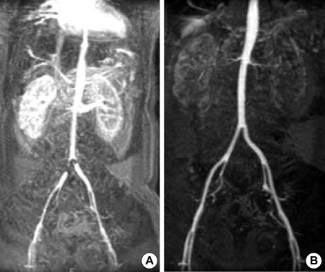
This operation was performed for the patient who was admitted for severe ischemic pain of both lower extremities. The midline incision of the abdomen was made. After exposing the lower abdominal aorta and both common iliac arteries, arteriotomy from the lower abdominal aorta to right common iliac artery was made. Endarterectomy was done easily using the dissector. Thereafter, arteriotomy was closed with 4-0 prolene (Fig. 4).
Fig. 4.
MR angiography demonstrates stenosis of the aortic bifurcation and both common iliac arteries (A), patent aortoiliac system postoperatively (B).
RESULTS
Cerebral ischemic symptoms and visual disturbance disappeared in all 3 patients after the ascendo-carotid bypass. One out of these 3 patients complained of migraine-like headache for 3 days after the operation. This symptom was relieved with no specific medication. There was no other postoperative complication. The symptoms of cardiac failure disappeared after thoraco-left iliac bypass. All patients were followed up at an interval of every 6 to 12 months with CT angiography. The patients of aortic endarterectomy showed no restenosis and all grafts were patent during the follow up periods (range; 4-75 months).
DISCUSSION
TA is an idiopathic, non-specific inflammatory disease involving the aorta and its main branches; histologically it is characterized by fibrosis in the intima and the adventitia, and degeneration and disintegration in the media.
Microscopic finding may reveal a heavy cellular infiltration of the media consisting mainly of lymphocytes and a variable mixture of histiocytes and plasma cells. Giant cells of western body type may be present and are occasionally numerous (8, 9). We could obtain the tissue only in patient who was performed aortoiliac endarterectomy. We could not obtain the tissue in other 4 patients because we performed bypass surgery. The pathologic finding of our patient showed that a heavy cellular infiltration of the media consisted mainly of lymphocyte and intimal thickening with myxoid degeneration. Three types of this entity were classified histologically: granulomatous inflammatory, diffuse productive inflammatory, and fibrosing (2).
The cause of TA remains obscure, but autoimmune and allergic responses to previous infections have been considered. Franklin and Morrison demonstrated a reaction between lymphocytes and bacterial protein antigen and considered that the inflammatory cells that were produced may destroy the surrounding fragile elastic fibers via their enzymes (11). Several studies demonstrated various human leucocyte antigen associations with TA, suggesting a genetic predisposition for the disease (11-14). The strong predilection for women, geographical incidence, and occasional familial occurrence may also indicate a role for genetic factors (15).
A large series of patients with this inflammatory arteriopathy have now been reported from Japan and Mexico, and the natural history, pathology, and arterial involvement have been better defined (16, 17). The clinical course of TA includes a prodromal "prepulseless phase," which is manifested by a variety of constitutional symptoms including pyrexia, weight loss, anorexia, night sweats, malaise, arthralgias, and myalgias. Since these symptoms are nonspecific and protean, they are rarely discernible as a discrete prodromal phase (16, 17). A "pulseless phase" follows months to years later and is attended by peripheral pulse deficits and symptomatic occlusive or aneurysmal arterial disease. The arterial involvement may be confined to the aortic arch as in Takayasu's original patient, or involve multiple aortic segments, or the entire aorta, its major branches, and the pulmonary artery as well (18). Arterial pathology varies from a fulminant acute inflammatory process to extensive post-inflammatory mural fibrosis (19).
Corticosteroids have been the primary therapy for TA, since dramatic symptomatic improvement and restoration of absent pulses were reported to follow its use (20). However, the efficacy of corticosteroids in TA has not been universally acknowledged, since most patients with symptomatic arterial insufficiency do not manifest dramatic clinical improvement. We used corticosteroid in all patients when they were in the acute phase of disease. Certainly, as in temporal arteritis, a reversible phase of this disease exquisitely sensitive to corticosteroid therapy may exist.
Vascular reconstruction in patient in patients with TA was approached with considerable trepidation in the past, particularly during the acute phase because of fear of occlusion of the involved vessel or graft, or anastomotic disruption (21). However, Ahn et al. reported the bypass surgery to the 18 patients with TA, and there was no motality (22). Mwipatayi et al. reported 115 surgical procedure with overall operative mortality in 4% (23). They confirmed that surgery was both safe and effective. There are distinct differences between the lesions of TA and those of atherosclerosis. Atherosclerotic type endarterectomy has not been feasible in this group of patients with TA because of involvement of the long segment of artery in the patients with TA. The choice of operative procedure depends on the number of arteries involved, the extent of arterial occlusive lesions and the clinical manifestations. But endarterectomy is usually indicated in patients with TA involving short segment, high flow artery (i.e., aortoiliac occlusive disease). We had reported 2 cases of abdominal aortic endarterectomy in TA and Leriche's syndrome (24).
In conclusion, surgical treatment of symptomatic TA was highly effective and safe. Symptomatic improvement and excellent long-term graft patency could be expected after arterial reconstruction.
Fig. 2.
Preoperative angiography shows patent right internal carotid artery (A) and severely stenotic left internal carotid artery (B). (C) Postoperative CT angiography shows ascendo-right internal carotid artery bypass.
Footnotes
Research grant support if any.
References
- 1.Takayasu M. A case with unusual changes of the central vessels in the retina. Nippon Ganka Gakkai Zasshi. 1908;12:554–557. [Google Scholar]
- 2.Nasu T. Takayasu's truncoarteritis in Japan: a statistical observation of 76 autopsy cases. Pathol Microbiol. 1975;43:140–146. [PubMed] [Google Scholar]
- 3.Hall S, Barr W, Lie JT, Stanson AW, Kazmier FJ, Hunder GG. Takayasu arteritis: A study of 32 north American patients. Medicine. 1985;64:89–99. [PubMed] [Google Scholar]
- 4.Vanoli M, Bacchiani G, Origgi L, Scorza R. Takayasu's arteritis: a changing disease. J Nephrol. 2001;14:497–505. [PubMed] [Google Scholar]
- 5.Lande A, Rossi P. The value of total aortography in the diagnosis of Takayasu's arteritis. Radiology. 1975;114:287–297. doi: 10.1148/114.2.287. [DOI] [PubMed] [Google Scholar]
- 6.Yamada I, Numano F, Suzuki S. Takayasu arteritis: evaluation with MR imaging. Radiology. 1993;188:89–94. doi: 10.1148/radiology.188.1.8099751. [DOI] [PubMed] [Google Scholar]
- 7.Sharma S, Sharma S, Taneja K, Gupta AK, Rajani M. Morphologic mural changes in the aorta revealed by CT in patients with nonspecific aortoarteritis (Takayasu's arteritis) Am J Roentgenol. 1996;167:1321–1325. doi: 10.2214/ajr.167.5.8911205. [DOI] [PubMed] [Google Scholar]
- 8.Robbs JV, Human RR, Rajaruthnam P. Operative treatment of nonspecific aortoarteritis (Takayasu's arteritis) J Vasc Surg. 1986;3:605–616. [PubMed] [Google Scholar]
- 9.Wu SG. Aortoarteritis: clinical analysis of 100 cases. Chin Med J. 1979;59:406–408. [Google Scholar]
- 10.Franklin J, Morrison R. Takayasu's arteritis. In: Mishima Y, editor. XVII Annual Meeting of the Japanese Society of Cardiovascular Surgery; Tokyo: International Academic Publishers; 1987. pp. 24–27. [Google Scholar]
- 11.Takeuchi Y, Matsuki K, Saito Y, Sugimoto T, Juji T. HLA-D region genomic polymorphism associated with Takayasu's arteritis. Angiology. 1990;41:421–426. doi: 10.1177/000331979004100601. [DOI] [PubMed] [Google Scholar]
- 12.Kimura A, Kitamura H, Date Y, Numano F. Comprehensive analysis of HLA genes in Takayasu arteritis in Japan. Int J Cardiol. 1996;54(Suppl):61–69. doi: 10.1016/s0167-5273(96)88774-2. [DOI] [PubMed] [Google Scholar]
- 13.Yoshida M, Kimura A, Katsuragi K, Numano F, Sasataki T. DNA typing of HLA-B gene in Takayasu's arteritis. Tissue Antigens. 1993;42:87–90. doi: 10.1111/j.1399-0039.1993.tb02242.x. [DOI] [PubMed] [Google Scholar]
- 14.Weyand CM, Goronzy JJ. Molecular approaches toward pathologic mechanisms in giant cell arteritis and Takayasu's arteritis. Curr Opin Rheumatol. 1995;7:30–36. [PubMed] [Google Scholar]
- 15.Giordana JM, Hoffman GS, Leavitt RY. Takayasu's arteritis: Nonspecific aortoarteritis. In: Rutherford RB, editor. Vascular Surgery. 3rd edn. Philadelphia: WB Saunders; 1995. pp. 245–253. [Google Scholar]
- 16.Herrera EL, Torres GS, Marcushamer J, Mispireta J, Horwitz S, Vela JE. Takayasu's arteritis: Clinical study of 107 cases. Am Heart J. 1977;93:94–103. doi: 10.1016/s0002-8703(77)80178-6. [DOI] [PubMed] [Google Scholar]
- 17.Ishikawa K. Diagnostic approach and proposed criteria for the clinical diagnosis of Takayasu's arteriopathy. J Am Coll Cardiol. 1988;12:964–972. doi: 10.1016/0735-1097(88)90462-7. [DOI] [PubMed] [Google Scholar]
- 18.Lupi H, Sanchez GT, Horwitz S, Gutierrez EF. Pulmonary artery involvement in Takayasu's arteritis. Chest. 1975;67:69–74. doi: 10.1378/chest.67.1.69. [DOI] [PubMed] [Google Scholar]
- 19.Nasu T. Pathology of pulseless disease. A systematic study and critical review of twenty-one autopsy cases reported in Japan. Angiology. 1963;14:225–242. doi: 10.1177/000331976301400502. [DOI] [PubMed] [Google Scholar]
- 20.Ishikawa K, Yonekawa Y. Regression of carotid stenoses after corticosteroid therapy in occlusive thromboaortopathy (Takayasu's disease) Stroke. 1987;18:677–679. doi: 10.1161/01.str.18.3.677. [DOI] [PubMed] [Google Scholar]
- 21.Gupta S. Surgical and immunological aspects of Takayasu's disease. Ann R Coll Surg Engl. 1981;63:325–332. [PMC free article] [PubMed] [Google Scholar]
- 22.Ahn MS, Kim MY, Huh S, Min SK, Ha J, Chung JK, Kim SJ. Surgical treatment of Takayasu arteritis. J Korean Soc Vasc Surg. 2001;17:24–31. [Google Scholar]
- 23.Mwipatayi BP, Jeffery PC, Beningfield SJ, Matley PJ, Naidoo NG, Kalla AA, Kahn D. Takayasu arteritis: clinical features and management: report of 272 cases. ANZ J Surg. 2005;75:110–117. doi: 10.1111/j.1445-2197.2005.03312.x. [DOI] [PubMed] [Google Scholar]
- 24.Kim DI, Huh SH, Lee BB. Aortic endarterectomy in Takayasu's arteritis and Leriche's syndrome. J Cardiovasc Surg. 2002;43:751–753. [PubMed] [Google Scholar]


