Summary
The carbon storage regulator CsrA is an RNA binding protein that controls carbon metabolism, biofilm formation and motility in various eubacteria. Nevertheless, in Escherichia coli only five target mRNAs have been shown to be directly regulated by CsrA at the post-transcriptional level. Here we identified two new direct targets for CsrA, ycdT and ydeH, both of which encode proteins with GGDEF domains. A csrA mutation caused mRNA levels of ycdT and ydeH to increase more than 10-fold. RNA mobility shift assays confirmed the direct and specific binding of CsrA to the mRNA leaders of ydeH and ycdT. Overexpression of ycdT and ydeH resulted in a more than 20-fold increase in the cellular concentration of the second messenger c-di-GMP, implying that both proteins possess diguanylate cyclase activity. Phenotypic characterization revealed that both proteins are involved in the regulation of motility in a c-di-GMP dependent manner. CsrA was also found to regulate the expression of five additional GGDEF/EAL proteins and a csrA mutation led to modestly increased cellular levels of c-di-GMP. All together, these data demonstrate a global role for CsrA in the regulation of c-di-GMP metabolism by regulating the expression of GGDEF proteins at the post-transcriptional level.
Keywords: CsrA, post-transcriptional regulation, GGDEF, c-di-GMP, motility
Introduction
Successful adaptation of bacteria to different niches depends on their ability to adjust their life style according to the requirements of the environment. Bacteria have evolved numerous mechanisms to sense external signals, to translate them into complex cellular responses and, thereby, to mediate responses to physiological demands. The Escherichia coli carbon storage system, with the RNA binding protein CsrA as the central player, exemplifies such an adaptive regulatory cascade [see reviews by (Babitzke and Romeo, 2007; Lucchetti-Miganeh et al., 2008)]. The Csr regulatory system is widely distributed among eubacteria (White et al., 1996) and has been found to control a variety of virulence-linked physiological traits (Lucchetti-Miganeh et al., 2008).
CsrA was originally identified as a regulator of glycogen biosynthesis (Romeo et al., 1993), acting as an RNA binding protein on the expression of its target mRNAs (Liu and Romeo, 1997). Beside controlling glycogen synthesis, CsrA and its homologs in various bacteria have widespread regulatory functions, including roles in biofilm formation (Jackson et al., 2002; Wang et al., 2005), motility (Wei et al., 2001; Yakhnin et al., 2007), carbon metabolism (Baker et al., 2002; Sabnis et al., 1995), secondary metabolism (Heeb and Haas, 2001; Heeb et al., 2005; Kay et al., 2005), quorum sensing (Heurlier et al., 2004; Lenz et al., 2005) and numerous functions in the interactions with animal and plant hosts (Altier et al., 2000; Barnard et al., 2004; Heeb and Haas, 2001). CsrA is a homodimer containing two identical RNA-binding surfaces located on opposite sides of the protein, whose structure and function recently has been elucidated in considerable detail (Mercante et al., 2006; Schubert et al., 2007). Despite its global role in bacterial adaptation, only a few direct mRNA targets have been identified, including five in E. coli. By binding to mRNA leaders and preventing translation, followed by destabilizing of the transcript, CsrA has been shown to downregulate expression of the glgCAP operon (Baker et al., 2002), encoding the glycogen synthesis apparatus, the cstA gene (Dubey et al., 2003), involved in carbon starvation and the pga operon, encoding the biofilm polysaccharide poly-β-1,6-N-acetyl-D-glucosamine (PGA) (Wang et al., 2005). Regulation of the RNA chaperone gene hfq is also mediated by CsrA binding and translation inhibition, although this does not result in hfq mRNA destabilization (Baker et al., 2007). CsrA also upregulates the expression of certain target genes. The mRNA of flhDC, which is required for flagellum biosynthesis, is stabilized by CsrA binding to the flhDC leader (Wei et al., 2001). However, the detailed biochemical mechanism for this activation has not been elucidated.
Regulation of CsrA activity is mediated in part by the action of the two small non-coding RNAs (sRNAs) CsrB and CsrC (Romeo, 1998; Weilbacher et al., 2003). During the past years sRNAs have been recognized as important players in gene regulation, in most cases by base pairing with target mRNAs (Majdalani et al., 2005; Romby et al., 2006; Storz et al., 2005). However, CsrB and CsrC RNAs antagonize the activity of CsrA by binding to and therefore sequestering this protein (Liu et al., 1997). Transcription of the Csr sRNAs is controlled by the two-component system (TCS) BarA-UvrY (Suzuki et al., 2002; Weilbacher et al., 2003), thus permitting the integration of environmental signals into the Csr signalling network. Expression of csrB and csrC also requires CsrA. This regulation may be mediated indirectly through the BarA-UvrY system (Gudapaty et al., 2001; Suzuki et al., 2002). This auto-regulatory mechanism has been described as a homeostatic system, which leads to tight regulation of CsrA activity. Recently, a new regulatory factor, CsrD (YhdA) has been shown to influence the Csr system (Jonas et al., 2006; Suzuki et al., 2006). By targeting CsrB and CsrC for degradation by RNase E, CsrD acts positively on CsrA activity (Suzuki et al., 2006). The apparent membrane protein CsrD contains degenerate GGDEF and EAL domains. Such domains have been shown to be associated with the turnover of the second messenger cyclic di-GMP (c-di-GMP) (Ryjenkov et al., 2005; Schmidt et al., 2005; Simm et al., 2004), which can mediate the switch between a motile and sessile life style in diverse bacteria [see reviews by (D'Argenio and Miller, 2004; Jenal, 2004; Romling, 2005; Romling and Amikam, 2006)]. In contrast to other GGDEF/EAL proteins, CsrD was demonstrated to lack both diguanylate cyclase (DGC) and phosphodiesterase (PDE) activities, indicating that CsrD is neither involved in the production nor in the degradation of the second messenger (Suzuki et al., 2006). In contrast, CsrD was found to be an RNA binding protein, although its detailed mechanism of action in CsrB/C decay has not been resolved.
Despite the detailed knowledge about the molecular mechanisms of the Csr signalling system, limited information is available concerning the integration of the Csr cascade into other global networks. In order to identify novel direct targets for CsrA that might help us to better understand the global impact of the Csr network, we conducted a genome wide search for genes, whose transcript levels rapidly change upon pulse overproduction of CsrA. Our search revealed that CsrA is a regulator for several GGDEF/EAL proteins, in particular of the two GGDEF proteins YcdT and YdeH. Both proteins produce c-di-GMP in vivo and control flagella mediated swimming motility.
Results
Identification of novel mRNA targets for CsrA by microarray
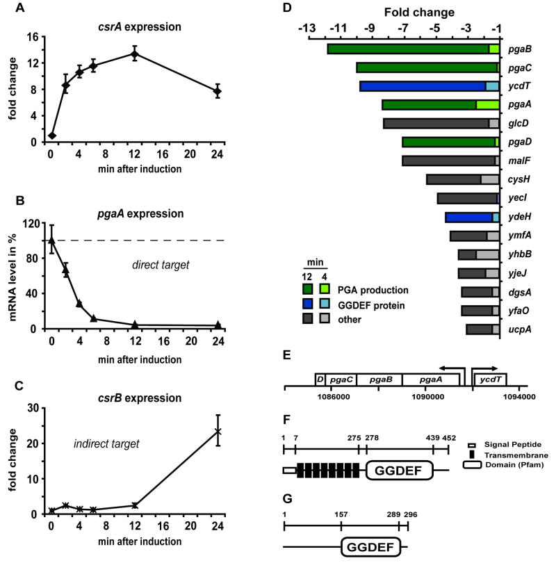
To screen for novel direct CsrA targets we decided to adopt a microarray based approach, which has previously been used to identify direct sRNA targets (Papenfort et al., 2006; Tjaden et al., 2006; Vogel and Wagner, 2007). Our strategy involved the pulse overexpression of csrA, followed by the immediate analysis of changes in whole genome expression patterns. The approach is based on the assumption that CsrA not only blocks the translation of many of its mRNA targets, but also secondarily destabilizes them. Hence, pulse overexpression of CsrA from an inducible vector is expected to lead to a rapid decrease in the transcript level of the directly regulated targets. Changes in the transcript levels of indirect CsrA targets are assumed to occur first at later time points after csrA induction. Such differential changes in the transcript level can be monitored over time by microarray analysis. To verify that our approach was working, we first monitored csrA expression as well as the expression of the known direct target pgaA and the known indirect target csrB in response to csrA overexpression by quantitative real-time reverse transcriptase PCR (RT-PCR). Our data show that addition of arabinose (at 0 minutes) to E. coli KJ157 (KSB837 csrA∷kan), carrying the arabinose inducible vector pBADcsrA, resulted in a strong upregulation of csrA expression within 2 minutes (Fig. 1A). Consistent with our prediction mRNA levels of the direct target pgaA dramatically decreased within less than 10 minutes (Fig. 1B). The expression of the sRNA CsrB, known to be indirectly and positively controlled by CsrA (Gudapaty et al., 2001), began to increase after a delay of ∼12 minutes (Fig. 1C). These data suggest that our approach was successful in discriminating between direct and indirect targets for CsrA.
Figure 1.
Identification of ycdT and ydeH as novel targets for CsrA. Plasmid encoded csrA (pBADcsrA) was expressed in KSB837 (csrA∷kan) upon induction with 0.1 % arabinose at OD600 1.5. (A) Increased csrA expression in response to arabinose addition (0 min) was measured by Real-time RT-PCR over time. CsrA is assumed to bind to its direct mRNA targets, to inhibit translation and thereby to destabilize the mRNAs. (B) mRNA levels of the known CsrA target pgaA rapidly decreased within a few minutes after pBADcsrA induction. (C) The indirect CsrA target csrB was changed in expression first after 12 minutes. (D) A genome-wide screen for genes, whose transcript levels decrease within 4 and 12 minutes (> 3 fold compared to 0 min) in response to CsrA overexpression, identified ycdT and ydeH as novel CsrA targets. (E) The ycdT gene is located adjacent to the pga operon and is divergently transcribed. (F) YcdT harbours 8 transmembrane regions and is predicted to contain a GGDEF motif. (G) YdeH contains a GGDEF motif and is predicted to be cytoplasmic.
In the next step we screened for novel direct CsrA targets by using an Affymetrix whole genome E. coli array. Since pgaA mRNA was downregulated within less than 12 minutes, we compared the transcriptional profiles 4 and 12 minutes after arabinose addition with the profile before arabinose induction (0 minutes). To eliminate genes downregulated in a CsrA-independent manner, we normalised the observed signal ratios against the signal ratios resulting from induction of the vector control pBAD28.
Four of the genes showing the strongest repression (> 7 fold) 12 minutes after pBADcsrA expression belonged to the pga operon (Fig. 1D). The mRNA levels of ycdT, followed the same kinetics upon CsrA overexpression as the pga mRNAs, suggesting that ycdT may be regulated by CsrA in a similar manner. Database search revealed that ycdT is located directly adjacent to the pga operon but on the reverse strand. ycdT encodes a transmembrane protein with a C-terminal GGDEF domain (Fig. 1E, F). Among the most downregulated genes in response to CsrA overexpression we found another GGDEF protein encoding ORF, ydeH (Fig. 1D, G). YdeH is predicted to encode a cytoplasmic protein, not containing any known domains involved in signalling (Fig. 1G).
The two GGDEF proteins YcdT and YdeH are regulated by CsrA
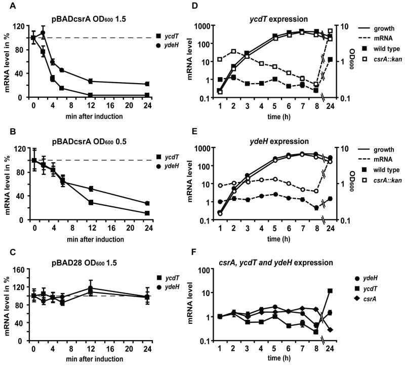
To confirm the effect of CsrA overexpression on ycdT and ydeH transcripts, we determined the kinetics of CsrA-dependent downregulation by RT-PCR. In accordance with our array data, ycdT and ydeH mRNA levels decreased strongly upon arabinose addition in late exponential phase (OD600 1.5) (Fig. 2A). The mRNA level of ycdT was halved within 4 minutes and reached a minimum of 3 % between 12 and 24 minutes. ydeH mRNA decreased to 50 % within five minutes and continued to decrease to approximately 22 % after 24 minutes. Similar results were observed when arabinose induction was performed earlier during growth at OD600 0.5 (Fig. 2B). In contrast, addition of arabinose to a strain carrying the empty vector pBAD28 did not affect the levels of ycdT and ydeH transcripts (Fig. 2C).
Figure 2.
CsrA dependent regulation of ycdT and ydeH expression measured by Real-Time RT-PCR. (A) Induction of pBADcsrA with 0.1 % arabinose (at 0 min) leads to a rapid decrease in ycdT (squares) and ydeH (circles) transcript levels during late (OD600 1.5) exponential growth. (B) Similar results were observed during early exponential growth (OD600 0.5). (C) Induction of pBAD28 had no effect on ycdT and ydeH mRNA levels. (D) In the csrA∷kan mutant TRMG1655 (open symbols), ycdT mRNA levels (dashed line) were strongly increased compared to the wild type MG1655 (filled symbols) over the entire growth cycle. (E) Likewise, ydeH expression (dashed line) was significantly higher in the csrA mutant. (F) Analysis of ycdT, ydeH and csrA (diamonds) expression over time in the wild type indicates that ycdT and ydeH are inversely regulated with respect to csrA.
To test the effect of a csrA mutation on ycdT and ydeH expression, we measured the mRNA levels of ycdT and ydeH by RT-PCR along the entire growth curve in the wild type and isogenic csrA mutant strains. In the wild type strain, expression of ycdT slightly decreased within the first 8 h of growth (Fig. 2D), whereas ydeH mRNAs remained at constant levels (Fig. 2E). Between 8 and 24 h the expression of both genes strongly increased. In the csrA mutant, ycdT and ydeH mRNA levels were significantly elevated. ycdT expression was more strongly upregulated (up to 30-fold) during exponential growth compared to later time points (Fig. 2D), whereas the transcript levels of ydeH were approximately 10 fold higher throughout the growth (Fig. 2E). Monitoring csrA transcript levels over time in the wild type strain shows that csrA expression rapidly decreased between 8 and 24 h (Fig. 2F), demonstrating that csrA is inversely regulated with ycdT and ydeH. However, the fact that CsrA activity is in large part under the control of CsrB and CsrC makes it difficult to correlate csrA mRNA levels with its activity. Nevertheless, these data confirm that CsrA is a negative regulator of ycdT and ydeH expression.
Effects of other components of the BarA-UvrY-Csr cascasde on ydeH and ycdT expression
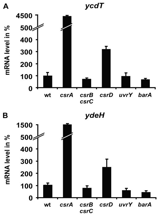
CsrA is antagonized by the CsrB and CsrC sRNAs. These sRNAs are transcriptionally activated by the BarA-UvrY two-component system and negatively controlled by CsrD at the level of RNA stability (Suzuki et al., 2002; Suzuki et al., 2006; Weilbacher et al., 2003). By using genetic mutants we tested the contribution of these components on ycdT and ydeH expression. Disruption of csrB and csrC resulted in a slight decrease in ycdT and ydeH mRNA levels (approx. 70 %) compared to the wild type (Fig. 3). Similar weak effects were observed in uvrY and barA mutants. These modest effects are consistent with the earlier finding that levels of CsrA protein normally exceeds the binding capacity of these small RNAs (Gudapaty et al., 2001). A more pronounced effect on ycdT and ydeH mRNAs was observed in a csrD mutant, in which the cellular levels of CsrB and CsrC are increased. Compared to the wild type the mRNA levels of ycdT and ydeH were approximately 3-fold increased (Fig. 3). All together, these results indicate that the entire Csr regulatory network is involved in the regulation of the expression of ycdT and ydeH.
Figure 3.
Effects of other components of the BarA-UvrY-Csr network on ycdT and ydeH expression. mRNA levels of ycdT (A) and ydeH (B) were measured in MG1655 (wt), TRMG (csrA), KJ230 (csrB csrC), KJ205 (csrD), AKP200 (uvrY) and AKP199 (barA) by Real-Time RT-PCR when the bacterial cultures had reached an OD600 of 1.5.
CsrA directly interacts with the ycdT and ydeH transcripts
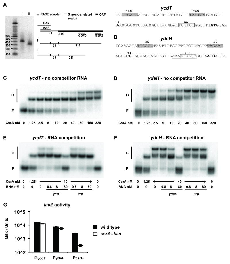
Previous studies have suggested that CsrA binds to the consensus sequence, ACA-GGAUG, with the GGA motif representing the most highly conserved nucleotides (Baker et al., 2002; Dubey et al., 2005). To make predictions about the binding of CsrA to the ycdT and ydeH mRNAs, we analysed the 5′ leader sequences of both transcripts for the existence of potential CsrA binding motifs. Since no information about the ycdT promoter was available in the data base, we determined the transcriptional initiation site of ycdT by Rapid Amplification of 5′ cDNA Ends (5′ RACE). A single band was observed for a PCR reaction, amplifying the 5′ non-translated region of the ycdT transript (Fig. 4A). Sequencing of the RACE PCR product identified the nucleotide A, 35 bp upstream of AUG, as the transcription start site. The -10 and -35 regions of ycdT [TATTAA (-10) and TTGACA (-35)], separated by a 19 bp spacing region, exhibited 4 and 6 bp of identity with respect to the consensus sequences for these promoter elements [TATAAT (-10) and TTGACA (-35)] (Hawley and McClure, 1983). We identified two potential CsrA binding sites with degenerate motifs in the 5′ non-translated region of the ycdT mRNA, one of them close to the transcription start and the other one overlapping the AUG translation initiation start codon (Fig. 4A). The ydeH transcript starts 29 nucleotides upstream of the initiation codon AUG (Yamamoto and Ishihama, 2006). Also in the 5′ non-translated region of the ydeH mRNA two potential CsrA binding sites binding sites were found, one of which overlaps the Shine-Dalgarno sequence and the other one is close to the 5′ end of the transcript (Fig. 4B).
Figure 4.
Physical interaction between CsrA and ycdT and ydeH transcripts. (A) The transcription start site for ycdT was determined by 5′RACE. An approximately 410 nt PCR product was detected after amplification of reverse transcribed cDNA with the gene specific primer GSP2 and the adapter primer AAP (lane 1 (I)). The product of a second nested PCR reaction with primers GSP3 and UAP was approximately 100 nt shorter (lane 2 (II) – as labelled in figure). Sequencing of the shorter fragment identified A (35 nt upstream of ATG) as transcription start site. Analysis of the 5′ leader of ycdT suggests two potential CsrA binding sites (underlined). (B) Sequence analysis of the intergenic region of ydeH suggests two potential CsrA binding sites (underlined), one of them overlapping the Shine-Dalgarno sequence (SD). (C, D) Gel mobility shift analyses of CsrA-ycdT and CsrA-ydeH interactions in the absence of RNA competitor. 5′ end-labelled ycdT or ydeH transcripts (80 pM) were incubated with CsrA at the indicated concentrations. The positions of free (F) and bound (B) RNA are shown. (E, F) Competition reactions using specific (ycdT, ydeH) or non-specific (trpL from B. subtilis) unlabelled RNA competitors. The concentration of competitor RNA is shown at the bottom of each lane. (G) β-galactosidase activity for plasmid encoded transcriptional ycdT-, ydeH- and csrB-lacZ fusions in wild type MG1655 and its csrA mutant. Bacteria containing pPYCDT, pPYDEH or pCBZ1, respectively, were grown until OD600 1.5. The mean values and the standard deviations were calculated for each strain from two parallel experiments.
To experimentally determine whether CsrA directly binds to the ycdT and ydeH transcripts, quantitative RNA gel mobility shift assays were performed with a ycdT transcript, consisting of a 36 nt leader and the first 20 nt of the coding region, and a ydeH transcript containing the 29 nt untranslated leader and the first 25 nt of the coding sequence. CsrA bound strongly to both ycdT and ydeH transcripts (Fig. 4C, D). For the ycdT transcript, two distinct complexes were observed at 2.5 nM CsrA, and essentially all of the starting RNA was shifted at 80 nM CsrA (Fig. 4C). For the ydeH transcript, two distinct shifted complexes were formed at 5 nM CsrA. However, complete binding was not seen until 320 nM CsrA, and at this concentration essentially all of the RNA was present in the upper complex (Fig. 4D). These gel shift patterns suggested that two CsrA proteins were bound to each transcript at higher CsrA concentrations, although the stoichiometry of binding was not experimentally determined. A nonlinear least-squares analysis of these data yielded an apparent equilibrium binding constant (Kd) of 2.6 ± 0.3 nM for ycdT and 2.3 ± 0.1 nM for ydeH.
The specificity of CsrA interaction with ycdT and ydeH transcripts was investigated by performing competition experiments with specific (ycdT or ydeH transcripts) and non-specific (Bacillus subtilis trp leader) unlabelled RNA competitors. Both ycdT and ydeH RNAs were able to compete for binding to CsrA while B. subtilis trp RNA did not effectively compete with the CsrA-ycdT or CsrA-ydeH interaction (Fig. 4E, F). These results establish that CsrA binds specifically to both ycdT and ydeH RNA.
In most cases CsrA downregulates its direct mRNA targets by binding to the leader, preventing translation and destabilizing the transcript (Baker et al., 2002; Dubey et al., 2003; Wang et al., 2005). However, in the case of hfq the binding of CsrA to the leader does not lead to mRNA destabilization, but to altered transcription (Baker et al., 2007). To test whether CsrA influences ycdT and ydeH mRNA levels by modulating promoter activity, we constructed plasmid-borne transcriptional ycdT- and ydeH-lacZ fusions, containing the upstream intergenic region and only 2 or 3 nucleotides of each transcript (ycdT -547 to +2; ydeH -222 to +3). Measuring of β-galactosidase activity of these reporter fusions revealed that both promoters were highly active in the wild type but were not altered by a csrA mutation (Fig. 4G). In contrast, lacZ expression from a control plasmid carrying the csrB promoter, which has earlier been reported to be regulated in a CsrA-dependent manner (Gudapaty et al., 2001), was clearly decreased in the csrA mutant (Fig. 4G). This demonstrates that CsrA does not change transcription of ycdT and ydeH, but rather modulates the stability of the messages.
YcdT and YdeH regulate motility
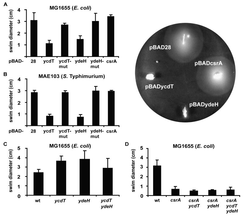
Proteins with GGDEF and EAL domains have been demonstrated to be involved in the regulation of bacterial physiology, including motility, biofilm formation, cell morphology and virulence [see reviews by (Cotter and Stibitz, 2007; D'Argenio and Miller, 2004; Jenal, 2004; Romling et al., 2005; Romling and Amikam, 2006)]. To characterise the phenotype of ycdT and ydeH in motility we analysed the swimming behaviour of strains, in which ycdT and ydeH were expressed from pBADycdT and pBADydeH, respectively, as well as respective knock-out mutants. Overexpression of both pBADycdT and pBADydeH led to a strong repression of swimming behaviour (Fig. 5A). The same effect was observed in a Salmonella enterica serovar Typhimurium (S. Typhimurium) background, although S. Typhimurium do not contain orthologs of these proteins (Fig. 5B). Mutations in ydeH and ycdT led to slightly increased swimming ability compared to the wild type. A ydeH ycdT double mutant was, however, not more motile than the wild type (Fig. 5C).
Figure 5.
The effects of YcdT and YdeH on motility. Motility was analysed by measuring the diameter of the swimming zone on 0.3 % agar plates, supplemented with 0.1 % arabinose, if necessary. (A) MG1655 (wt), carrying pBAD28, pBADycdT, pBADycdT-mut, pBADydeH, pBADydeH-mut or pBADcsrA. (B) Salmonella Typhimurium strain MAE103, carrying pBAD28, pBADycdT, pBADycdT-mut, pBADydeH, pBADydeH-mut or pBADcsrA. (C) MG1655 (wt) and mutants XWMGΔT (ycdT), KJ295 (ydeH) and the double mutant KJ311 (ycdT ydeH). (D) MG1655 and mutants TRMG (csrA), KJ331 (csrA ycdT), KJ330 (csrA ydeH) and KJ369 (csrA ycdT ydeH). A representative image of a motility agar plate for MAE103 carrying pBAD28, pBADycdT, pBADydeH or pBADcsrA is illustrated. All motility assays were repeated at least two times independently and the mean and the standard deviation were calculated.
Earlier studies have demonstrated that site-directed mutations in the GGDEF signature sequence of other proteins disrupt the function of this domain (Garcia et al., 2004; Paul et al., 2004; Simm et al., 2004). To test whether the repressing effect of ycdT and ydeH overexpression on motility was due to the activity of both proteins as diguanylate cyclases, we engineered mutants, in which the two first glycine residues of the respective GGEEF motifs were replaced by two alanine residues (GGEEF → AAEEF). The swimming behaviour of E. coli or Salmonella expressing the plasmids encoding these mutant YdeH and YcdT variants (pBADycdT-mut, pBADydeH-mut) was indistinguishable from the bacteria expressing the empty vector (Fig. 5A, B). This strongly suggests that the effect of YcdT and YdeH on motility is mediated by the second messenger c-di-GMP.
Previous results have demonstrated that a csrA mutant is strongly impaired in motility and that CsrA upregulates flhDC expression by binding to and stabilizing this mRNA (Wei et al., 2001). To test whether deletions in ycdT or ydeH can compensate for the swimming defect of the csrA mutant we constructed csrA ycdT and csrA ydeH double mutants as well as a csrA ycdT ydeH triple mutant. All three strains were not more motile than the csrA mutant (Fig. 5D), suggesting complex regulation of motility by pathways within the Csr network.
Beside their impact on motility, many proteins with GGDEF domains have been shown to regulate biofilm formation. In accordance, results of a parallel ongoing study show that YdeH significantly affects biofilm formation (C. Goller and T. Romeo, unpublished). Furthermore, the effect on biofilm formation seems to be mediated through increased synthesis of the biofilm polysaccharide PGA. YcdT has earlier been characterised regarding its phenotype in biofilm formation (Wang et al., 2005). However, neither biofilm formation nor pgaA-lacZ expression was influenced by YcdT under the given conditions (Wang et al., 2005).
In Enterobacteriaceae several GGDEF proteins have been shown to control biofilm formation by regulating the expression of curli fibres (Romling, 2005). Here, we analysed the expression of curli by analysing the colony morphology on Congo Red (CR) agar plates as well as the ability to form pellicles and to adhere to glass culture tubes at the air-liquid interface. However, we were not able to detect distinct ycdT or ydeH dependent phenotypes with respect to CR binding, pellicle formation or glass adherence at the air-liquid interface, suggesting that neither YcdT nor YdeH influence curli production under the conditions tested (data not shown).
AFM analysis of YcdT and YdeH mediated phenotypes
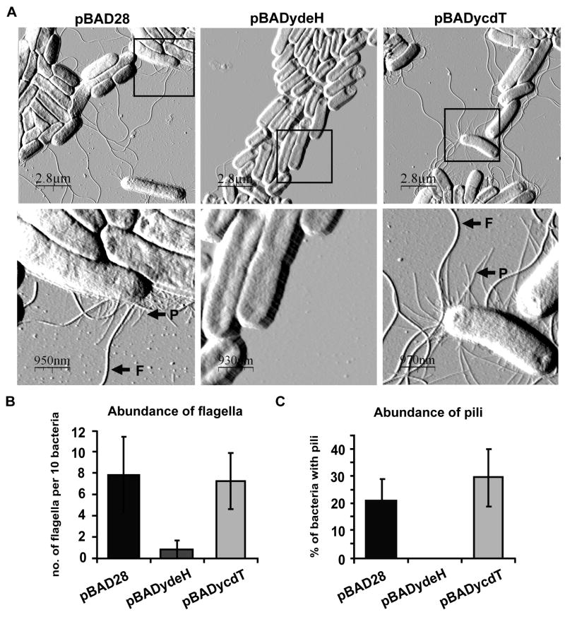
To further investigate the phenotypes mediated by YcdT and YdeH, we employed Atomic Force Microscopy (AFM), a technique recently shown to be a suitable tool for the study of bacterial morphology (Jonas et al., 2007). We allowed the bacteria to grow and to adhere to the substratum mica, which was submerged in the growth medium. For immobilization, the samples were air-dried at room temperature prior to AFM analysis. Images of the wild type strain carrying pBAD28 showed rod-shaped bacteria expressing flagella and pili-like structures (Fig. 6A). Overexpression of pBADydeH resulted in a clear reduction in the abundance of flagella (Fig. 6A, B), suggesting a role for YdeH in the regulation of flagellum biosynthesis. We also noted that YdeH overexpression completely abolished the appearance of the pili-like structures (Fig. 6A, C), indicating that flagella and pili synthesis might be co-regulated by YdeH. In contrast to ydeH, overexpression of ycdT did not affect the occurrence of flagella or pili or another distinct phenotype (Fig. 6), indicating that YcdT might have functions in the cell different from YdeH, which cannot be visualized by AFM under the conditions we have tested.
Figure 6.
High-resolution AFM analysis of cell morphology. Bacteria were grown on mica surfaces for 24 h at 28 °C in LB medium containing 0.1 % arabinose, but no salt. Afterwards the samples were air-dried and analysed with the AFM in contact mode. Representative images were chosen for presentation. (A) Lower (first row) and higher magnification (second row) AFM images of MG1655 (wt), carrying pBAD28, pBADydeH or pBADycdT. The arrows highlight the appearance of flagella (F) and pili (P). (B) Flagella expression was quantified by counting the number of flagella per total number of bacteria at five different locations on the microscope slide. The mean values and the standard deviations were calculated for each strain. (C) Pili expression was quantified by counting the number of bacteria expressing pili per total number of bacteria at five independent sites, from which the mean and standard deviation were determined.
YcdT and YdeH influence c-di-GMP levels in vivo
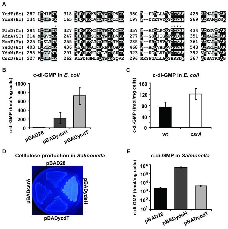
Both YcdT and YdeH contain GGDEF domains with consensus motifs, which are predicted to be dedicated to the synthesis of c-di-GMP (Ausmees et al., 2001; Paul et al., 2004; Schmidt et al., 2005; Simm et al., 2004). So far, proteins with a highly conserved active site motifs have been shown to possess DGC activity (Ausmees et al., 2001; Kirillina et al., 2004; Paul et al., 2004; Simm et al., 2004; Weber et al., 2006). In contrast, CsrD with a degenerate motif (HRSDF) failed to produce c-di-GMP (Suzuki et al., 2006). The amino acid sequences of the GGDEF domain of YcdT and YdeH perfectly match the conserved GG(D/E)EF motif as well as additional more extended conserved amino acid signatures of other enzymatically active proteins (Fig. 7A). Together with the finding that site-directed mutations of the GGDEF domains of YcdT and YdeH disrupted the effect on motility (Fig. 5), this strongly suggests that both proteins synthesize c-di-GMP. To prove this experimentally, we measured the c-di-GMP concentrations produced by E. coli MG1655 containing plasmid-encoded ydeH (pBADydeH), ycdT (pBADycdT) or the empty vector using HPLC and MALDI-TOF. Expression of ydeH resulted in clearly increased c-di-GMP levels (2215.3 fmol mg-1 cells) compared to the low levels, close to the limit of detection, in the control strain, carrying pBAD28 (94.6 fmol mg-1 cells). Even higher levels were detected when ycdT was overexpressed (7213.0 fmol mg-1 cells) (Fig. 7B). These data provide strong evidence that both of these proteins function as diguanylate cyclases (DGC) in vivo.
Figure 7.
Functional characterization of YcdT and YdeH. (A) The amino acid sequences of YcdT and YdeH were compared with other GGDEF domain proteins from Caulobacter crescentus (Cc), Salmonella Typhimurium (ST), Yersinia pestis (Yp) and E. coli (Ec), proven to synthesize c-di-GMP (PleD, AdrA, HmsT, YedQ and YdaM) or demonstrated not to be involved in c-di-GMP metabolism (CsrD). Dark background indicates a high level of similarity between the proteins. The stars (*) depict amino acid residues that have been demonstrated to be critical for substrate binding or catalysis (Chan et al., 2004). (B) c-di-GMP concentrations were determined in MG1655 carrying pBAD28, pBADydeH or pBADycdT grown to OD600 1.5 in LB medium with 0.1 % arabinose at 37 °C. (C) c-di-GMP concentrations of wt MG1655 and its isogenic csrA mutant TRMG, cultivated under equal conditions as (B), but without arabinose. (D) Expression of pBADydeH in the Salmonella adrA mutant MAE103 successfully restored cellulose production as visualized on calcofluor (CF), when grown for 20 h at 28 °C on LB agar with 0.1 % arabinose, but without salt. No dye binding was observed for MAE103 carrying pBADycdT, pBADcsrA or the control vector pBAD28. (E) c-di-GMP measurements in Salmonella MAE103, carrying pBAD28, pBADydeH and pBADycdT, grown for 20 h at 28 °C on LB agar with 0.1 % arabinose, but without salt.
The strong effect of CsrA on ycdT and ydeH transcript levels led us to analyse the overall effect of CsrA on the cellular c-di-GMP pool, by measuring the levels of the second messenger in the wild type strain MG1655 and its csrA mutant. We were able to consistently detect slightly elevated c-di-GMP levels in the csrA mutant (120.8 fmol mg-1 cells) compared to the wild type (74.5 fmol mg-1 cells) (Fig. 7C). This finding demonstrates a net effect of CsrA in the regulation of c-di-GMP turnover and is consistent with the previously documented negative effect of CsrA on biofilm formation (Jackson et al., 2002).
Several genes encoding GGDEF and EAL proteins have previously been shown to cross-complement phenotypes (Garcia et al., 2004; Simm et al., 2004), even between different species (Simm et al., 2005). In Salmonella, a mutation in the GGDEF gene adrA results in deficiency in cellulose synthesis due to decreased c-di-GMP levels. Overexpression of enzymatically active GGDEF proteins in such a Salmonella mutant leads to the restoration of cellulose production, which can be visualized on agar plates containing the dyes calcofluor (CF) or Congo red (CR). Thus, the ability to produce cellulose can be used as an indicator for DGC activity. We utilized this effect to study the enzymatic activities of YcdT and YdeH in S. Typhimurium. Strain MAE103, mutated in adrA and carrying pBADycdT, pBADydeH or the controls pBAD28 or pBADcsrA, was allowed to grow on CF and CR agar plates at 28 °C. Overexpression of pBADydeH resulted in a strongly fluorescent colony appearance of Salmonella on the CF plates (Fig. 7D) and as pink and rough colonies on the CR plates (data not shown), suggesting that cellulose was produced due to the elevated production of c-di-GMP. However, no dye binding could be observed for the strain carrying pBADycdT, demonstrating that ycdT fails to cross-complement an adrA mutation in Salmonella under the given conditions. In agreement with these data, only subtle changes (< 2-fold) in c-di-GMP levels were observed by HPLC and MALDI-TOF, when ycdT was overexpressed in Salmonella, grown at 28 °C on LB agar without salt, whereas plasmid encoded expression of ydeH in the same background strain resulted in strongly elevated c-di-GMP levels (> 200 fold) (Fig. 7E). Thus, in contrast to YdeH, which apparently possesses high DGC activity in plate grown Salmonella at 28 °C, YcdT appears to produce c-di-GMP at very low concentrations under the given conditions. A previous study has already demonstrated that in Salmonella most, but not all GGDEF proteins with conserved sequence signatures could restore cellulose production in an adrA mutant (Garcia et al., 2004) and that the cross-complementation ability strongly depended on the experimental conditions.
Global role of CsrA in the regulation of other GGDEF/EAL proteins
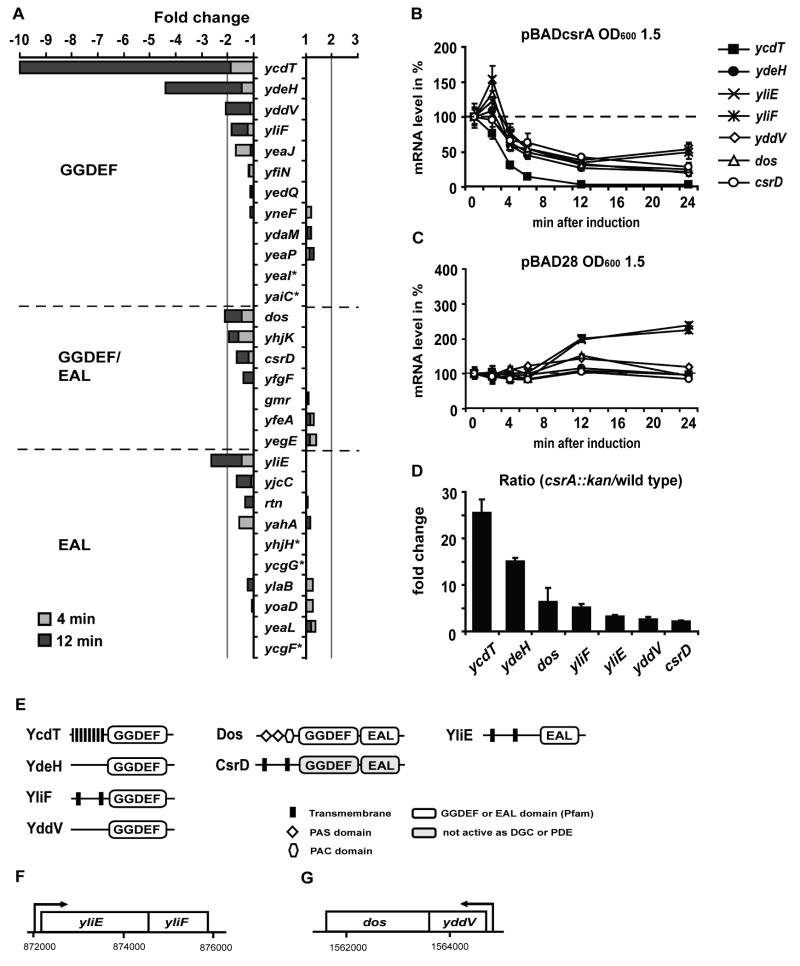
To test whether CsrA controls the expression of additional GGDEF/EAL proteins we analysed our array data for the expression patterns of all genes, annotated to contain a GGDEF and/or EAL domain, 4 and 12 minutes after CsrA pulse overproduction. For 4 of the 29 selected genes the signals were too low for reliable detection on the microarray (yeaI, yaiC, yhjH and ycgG). Most of the other GGDEF/EAL genes were relatively weakly expressed, but strongly enough for detection on the array. Interestingly, beside ycdT and ydeH several other genes showed changes in their transcript levels upon CsrA overproduction with an additive effect between 4 and 12 minutes: yddV (GGDEF), yliF (GGDEF), dos (GGDEF-EAL), yhjK (GGDEF-EAL), csrD (GGDEF-EAL), yliE (EAL) and yjcC (EAL) (Fig. 8A, E). CsrA dependent repression of these genes was, however, not as strong (1.5 to 2.5 fold) as repression of ycdT and ydeH. RT-PCR analysis of the kinetics of CsrA-dependent expression confirmed that mRNA levels of yliE, yliF, yddV, dos and csrD were indeed downregulated upon induction of pBADcsrA (Fig 8B), but remained constant or increased upon induction of the vector control (Fig. 8C). In the csrA mutant strain, expression of these genes was moderately increased (between 2 and 6 fold) compared to the wild type (Fig. 8D). Repression of yhjK and yjcC by CsrA overproduction could not be confirmed (data not shown). Noticeably, yddV and dos (Mendez-Ortiz et al., 2006) as well as yliE and yliF are organized as polycistronic units in operons (Fig. 8F, G). The expression patterns of yliE and yliF as well as yddV and dos followed almost identical kinetics (Fig 8B, C), indicating that these genes are co-regulated at the mRNA level by CsrA. The observation that CsrD is negatively regulated by CsrA agrees with the earlier finding that expression of a chromosomal csrD-lacZ translational fusion was modestly repressed (2-fold) by CsrA (Suzuki et al., 2006). These data confirm that CsrD is part of an additional autoregulatory loop within the Csr system. In summary, our data demonstrate that beside ycdT and ydeH, genes for several other GGDEF and GGDEF-EAL proteins as well as one EAL protein are negatively regulated by CsrA. This finding suggests a global role for CsrA in the regulation of c-di-GMP metabolism.
Figure 8.
The global effect of CsrA on GGDEF/EAL proteins. (A) All 29 E. coli genes encoding GGDEF, GGDEF-EAL or EAL proteins were analysed for CsrA-dependent changes in gene expression using the array data. Genes expressed at levels too low for microarray detection are indicated with a star (*). (B) The kinetics of CsrA-dependent downregulation of ycdT, ydeH, yliE, yliF, yddV, dos and csrD upon induction of pBADcsrA were confirmed by RT-PCR. (C) Expression of pBAD28 did not lead to a decrease in ycdT, ydeH, yliE, yliF, yddV, dos and csrD expression. (D) The ratio in mRNA levels between TRMG (csrA∷kan) and MG1655 (wt) was determined for ycdT, ydeH, yliE, yliF, yddV, dos and csrD, indicating that expression of these genes is increased in the csrA mutant. (E) Four of the CsrA regulated genes encode GGDEF proteins (ycdT, ydeH, yddV and yliF), two GGDEF-EAL proteins (dos, csrD) and one of them encodes an EAL protein (yliE). (F) yliE and yliF are organized as an operon. (G) Likewise, yddV and dos are present in an operon.
Discussion
Post-transcriptional regulation of GGDEF/EAL proteins by CsrA
The present study was initiated with a genome wide search for novel targets for the post-transcriptional regulator CsrA to better understand its role in bacterial adaptation and the cross-talk between the Csr system and other regulatory systems. Our search led to the finding that CsrA controls the expression of several GGDEF/EAL proteins, in particular the GGDEF proteins YcdT and YdeH, by physically binding to and changing their mRNA levels. To our knowledge this is the first example of GGDEF/EAL proteins being regulated at the mRNA level by a global post-transcriptional regulator. This supports the idea that c-di-GMP signalling is a multilayer process, including transcriptional, translational and post-translational levels. The array data also indicated that CsrA controls other mRNAs, some of which with unknown functions, but these effects need to be confirmed and were not the focus of this study.
With binding constants (Kd) of approximately 2.5 nM, CsrA binding to the ycdT and ydeH transcripts was remarkably strong. The affinities to the other known mRNA targets pgaA, glgC, cstA and hfq were approximately 10-fold lower (22 nM, 39 nM, 40 nM and 38 nM, respectively) (Baker et al., 2002; Baker et al., 2007; Dubey et al., 2003; Wang et al., 2005). Noteworthy, for each of the ycdT and ydeH transcripts only two GGA boxes were found in the 5′ leader sequences and binding of two CsrA proteins per transcript was observed at higher concentrations. For comparison, pgaA, glgC and cstA contain four to six potential CsrA binding sites. Moreover, in the case of ycdT, the sequence signature of both sites showed relative poor similarity to the consensus sequence. Thus, in addition to the primary sequence conservations, other determinants seem to largely influence the affinity of CsrA to its targets.
Our array revealed that beside YcdT and YdeH, two additional GGDEF proteins (YddV, YliF), two GGDEF-EAL proteins (Dos, CsrD) and one EAL protein (YliE) were regulated by CsrA. Together with the finding that increased overall levels of cellular c-di-GMP were measured in a csrA mutant, this implicates a global role for CsrA in the regulation of c-di-GMP metabolism. It is plausible that under other experimental conditions CsrA might control the expression of additional GGDEF/EAL proteins. For most of the GGDEF/EAL genes, relatively weak signals were detected on the microarray, suggesting that these genes require specific conditions for enhanced expression, different from the standard conditions used in our experiment. Since CsrA homologs are present in many different Gram negative bacteria (White et al., 1996) the role of CsrA in the regulation of GGDEF/EAL proteins might be a conserved feature. With the exception of CsrD, no other of the CsrA regulated GGDEF/EAL genes have homologous genes in S. Typhimurium. Therefore, CsrA might act on other GGDEF/EAL proteins in other bacteria. Furthermore, we cannot exclude that our microarray approach, which requires the destabilization of the CsrA mRNA targets upon its binding, failed to identify other important CsrA targets, in which translation is regulated without a corresponding alteration in mRNA stability, similar to the previous findings for hfq (Baker et al., 2007).
Interplay between Csr and c-di-GMP signalling
While c-di-GMP-mediated phenotypes and the molecular mechanisms governing c-di-GMP synthesis and turnover have received much attention, the role of the c-di-GMP network in signal transduction, including its linkage to external signals of specific adaptive responses and its interconnection with other global networks is relatively unexplored. Nevertheless, in E. coli genes encoding GGDEF/EAL domains were recently reported to be over-represented in the σS (RpoS) regulon, suggesting a role for c-di-GMP during the general stress response (Weber et al., 2006). In Vibrio cholerae quorum sensing signalling was recently demonstrated to be connected to c-di-GMP signalling through the action of the major quorum sensing regulator HapR (Waters et al., 2008). Furthermore, some GGDEF/EAL proteins, exemplified by the response regulator PleD (Aldridge and Jenal, 1999), contain phospho-receiver domains or other signalling domains, facilitating cross-talk and the integration into other signal cascades (Paul et al., 2008).
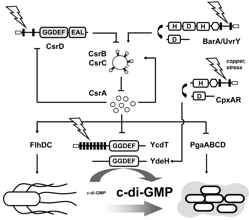
Our study revealed a direct link between the global Csr network and c-di-GMP signalling, placing both pathways in a broad cellular context. CsrA activity is controlled by the sRNAs CsrB and CsrC, whose expression levels are regulated by the BarA-UvrY two-component system and the probable inner membrane protein CsrD (Fig. 9). CsrA has previously been shown to control motility and biofilm formation by directly targeting the flhDC and pgaA mRNAs, respectively. Here, we show that in addition to the regulation of biosynthesis and global regulators CsrA regulates bacterial physiology in a c-di-GMP-dependent pathway by directly controlling the expression of ycdT and ydeH, which cause c-di-GMP accumulation and thereby favour the sessile life style. The combination of c-di-GMP dependent and c-di-GMP independent regulatory pathways allows CsrA to regulate biofilm related processes at various levels and thus to trigger the switch between a motile and a sessile life style. The CsrA and c-di-GMP specific adaptive responses are controlled by environmental signals, integrated at multiple sites within the signalling cascade. Although the nature of the signal sensed by BarA is not known, it is proposed to reflect the energy/growth status of the cell (Pernestig et al., 2003). In addition, BarA-UvrY signalling was recently demonstrated to be pH dependent (Mondragon et al., 2006). The prediction that CsrD and YcdT are membrane bound suggests that their activity is controlled from the outside. In addition, transcription of ydeH was previously demonstrated to depend on the CpxAR two-component system, responding to cell envelope stress and external copper (Yamamoto and Ishihama, 2005, 2006). A future challenge will be the identification of the nature of different input signals, controlling Csr and c-di-GMP signalling.
Figure 9.
Schematic view of the interconnection between Csr and c-di-GMP signalling in E. coli. The activity of the central player CsrA is controlled by the sRNAs CsrB and CsrC, which are regulated by the BarA-UvrY two-component system and CsrD, a GGDEF-EAL protein not involved in c-di-GMP metabolism. CsrA directly acts on motility and biofilm formation, by controlling mRNA levels of flhDC and pgaA, respectively. In addition, CsrA controls indirectly the switch between a motile and a sessile life style by regulating the levels of c-di-GMP through post-transcriptional regulation of the GGDEF proteins YcdT and YdeH and possibly additional proteins with GGDEF or EAL domains. Signals from the outside controlling the CsrA and c-di-GMP specific adaptive responses are integrated through the BarA-UvrY TCS and possibly through CsrD and YcdT. Transcription of ydeH was shown to be controlled by the CpxAR two-component system in response to cell envelope stress and external copper.
The roles of YcdT and YdeH in bacterial physiology
Numerous studies have shown that c-di-GMP controls bacterial behaviour [reviews by (D'Argenio and Miller, 2004; Jenal, 2004; Romling, 2005; Romling and Amikam, 2006)]. High levels of this second messenger favour sessility whereas low levels of c-di-GMP promote a motile life style. In accordance, YdeH and YcdT were found to repress swimming behaviour. YdeH seems to act at the level of flagellum synthesis while YcdT seems to modulate flagella function, raising the possibility that individual GGDEF proteins are dedicated to specific functions in the cell. We also observed that in the strain overexpressing YdeH the occurrence of pili was abolished. A recent study proposed a link between c-di-GMP signalling and type 1 pili and flagella expression in the Crohn-disease-associated adherent-invasive E. coli strain LF82 (Claret et al., 2007). While similar pathways might exist in E. coli K12, to this date we have no evidence for this hypothesis.
Our data show that overexpression of ydeH led to highly elevated c-di-GMP levels and to pronounced cellulose production in Salmonella. Consitent with these data, results from another parallel study suggest a significant role for YdeH in biofilm formation by regulating PGA synthesis (C. Goller and T. Romeo, unpublished). Although overexpression ycdT resulted in a strong accumulation of cellular c-di-GMP, we did not observe a distinct biofilm related phenotype neither in E. coli nor in S. Typhimurium. In addition, in an earlier study biofilm formation and pgaA-lacZ expression were not affected in the ycdT mutant XWMGΔT (Wang et al., 2005). Nevertheless, we suspect that not only YdeH, but also YcdT might have an impact on biofilm formation under other experimental growth conditions. The ycdT gene and the pga operon are divergently organized (Fig. 1E). A comprehensive bioinformatics study has recently demonstrated that chromosomal proximity indicates gene coregulation in prokaryotes independent of relative gene orientation and that adjacent bidirectionally transcribed genes with conserved gene orientation are strongly coregulated (Korbel et al., 2004). Furthermore, the ycdT homolog in Yersinia pestis, called HmsT, has been reported to be required for biofilm formation (Kirillina et al., 2004). Likewise, there is evidence that E. coli and Y. pestis produce the PGA polysaccharide as biofilm matrix component (Bobrov et al., 2008; Itoh et al., 2005). Synthesis of this polysaccharide was in a recent study shown to be positively regulated by HmsT, which was suggested to control c-di-GMP levels in close proximity to the glycosyltransferase HmsR, responsible for the production of the polysaccharide (Bobrov et al., 2008). Thus, regulation of PGA synthesis in Yersinia seems to occur in a c-di-GMP dependent fashion, similar to the production of the biofilm polysaccharide cellulose in Salmonella. Moreover, another recent study showed that the PEL biofilm polysaccharide synthesis in Pseudomonas aeruginosa is regulated by c-di-GMP. Here, the PelD protein serves as c-di-GMP receptor, activating the production of the PEL polysaccharide by a yet to be defined mechanism (Lee et al., 2007). These data suggest that there are related c-di-GMP dependent processes for controlling synthesis of the PGA exopolysaccharide in E. coli.
Noticeably, the pel genes in Pseudomonas, necessary for PEL synthesis, have been suggested to be regulated by the GacS-GacA-Rsm cascade, which is homologous to the BarA-UvrY-Csr pathway in E. coli (Goodman et al., 2004), further suggesting that the role of the Csr regulatory network in the regulation of biofilm components may be a conserved feature among γ-proteobacteria.
Experimental procedures
Bacterial strains and growth conditions
All strains used in this study are listed in Table 1. Chromosomal ydeH∷cat and csrB∷cat mutations were constructed using the Datsenko method (Datsenko & Wanner, 2000). The cat gene was amplified from pKD3 by PCR using primers ydeHKOFor2 and ydeHKORev2 or csrBKOFor and csrBKORev (Table 2), respectively, and introduced by electroporation into arabinose-treated BW25141 carrying pKD46. Transformants were selected on chloramphenicol plates, and their insertion sites were confirmed by PCR using the primer pairs ydeHKOtestFor/ydeHKOtestRev and csrBKOtestFor/csrBKOtestRev (Table 2). Mutations were transferred among strains by P1 transduction. For construction of the csrB csrC double mutant KJ230, the csrC∷tet allele from strain TWMG1655 was moved into the csrB mutant KJ227, from which the chloramphenicol cassette had been flipped out using the FLP recombinase. Strain KJ311 was generated by removing the chloramphenicol cassette from KJ295 by using the FLP recombinase and subsequent infection with a P1 lysate containing the ycdT∷cat mutation from XWMGΔT. For construction of strain KJ157, csrA∷kanr was moved from TRMG into KSB837. To generate the csrD mutant KJ205 the yhdA∷catr cassette from KJ27 was transduced into MG1655. To generate KJ331, KJ330 and KJ369, csrA∷kanr was transduced from TRMG into KJ295, XWMGΔT or KJ311, respectively. In most of the experiments, bacteria were grown in LB medium at 37 °C with shaking at 200 r.p.m. If necessary, antibiotics were added: ampicillin 100 μg ml−1, kanamycin 50 μg ml−1 and chloramphenicol 30 μg ml−1.
Table 1.
Bacterial strains and plasmids used in this study.
| Strain or plasmid | Description or genotype | Reference | ||
|---|---|---|---|---|
| Strains | ||||
| Escherichia coli | ||||
| MG1655 | F- λ- | Michael Cashel | ||
| BW25141 | lacIq rrnBT14 lacZWJ16 phoBR580 hsdR514 araBAD AH33 rhaBADLD78 galU95 endABT333 uidA (MluI)∷pir+ recA1 | (Datsenko and Wanner, 2000) | ||
| TRMG | MG1655 csrA∷kan | (Romeo et al., 1993) | ||
| KSB837 | CF7789 Δ(λatt-lom)∷bla φ (csrB-lacZ)1 (Hyb) ampr | (Suzuki et al., 2002) | ||
| KJ157 | KSB837 csrA∷kanr | This study | ||
| KJ227 | MG1655 csrB∷catr | This study | ||
| TWMG1655 | MG1655 csrC∷tetr | (Weilbacher et al., 2003) | ||
| KJ230 | MG1655 ΔcsrB csrC∷tetr | This study | ||
| KJ27 | KSB837 yhdA∷catr | (Jonas et al., 2006) | ||
| KJ205 | MG1655 yhdA∷catr | This study | ||
| AKP199 | MG1655 barA∷kanr | (Pernestig et al., 2003) | ||
| AKP200 | MG1655 uvrY∷catr | (Pernestig et al., 2003) | ||
| XWMGΔT | MG1655 ycdT∷catr | (Wang et al., 2005) | ||
| KJ295 | MG1655 ydeH∷catr | This study | ||
| KJ311 | MG1655 ΔydeHycdT∷catr | This study | ||
| KJ331 | KJ295 csrA∷kanr | This study | ||
| KJ330 | XWMGΔT csrA∷kanr | This study | ||
| KJ369 | KJ311 csrA∷kanr | This study | ||
| Salmonella enterica serovar Typhimurium | ||||
| MAE103 | ΔcsgBA102 adrA101∷MudJ | (Romling et al., 2000) | ||
| Plasmids | ||||
| pKD46 | temperature sensitive λ red recombinase expression vector | (Datsenko and Wanner, 2000) | ||
| pKD3 | template for mutant construction, carries chloramphenicol resistance cassette | (Datsenko and Wanner, 2000) | ||
| pBAD28 | pBAD expression plasmid | (Guzman et al., 1995) | ||
| pBADcsrA | CsrA expression plasmid, csrA is controlled by the plasmid-borne PBAD promoter | This study | ||
| pBADycdT | YcdT expression plasmid, ycdT is controlled by the plasmid-borne PBAD promoter | This study | ||
| pBADydeH | YdeH expression plasmid, ydeH is controlled by the plasmid-borne PBAD promoter | This study | ||
| pBADycdT-mut | mutagenised pBADycdT (G359A, G360A) | This study | ||
| pBADydeH-mut | mutagenised pBADydeH (G206A, G207A) | This study | ||
| pCBZ1 | pGE593, Φ(csrB-lacZ) | (Gudapaty et al., 2001) | ||
| pPYCDT | LacZ reporter plasmid, containing a ycdT-lacZ transcriptional fusion | This study | ||
| pPYDEH | LacZ reporter plasmid, containing a ydeH-lacZ transcriptional fusion | This study | ||
Table 2.
Primers used in this study.
| Primer name | Primer Sequence (5′ to 3′) |
|---|---|
| Genetic approaches | |
| ydeHKOFor2 | ATGGACTGTGCCAGTTTGGTCGGTGGATTGATCATCTGGGGCCACTCGTGTAGGCTGGAGCTGCTTC |
| ydeHKORev2 | CGGTTTGCTTACCCTCATACATTGCCCGGTCCGCTCTTCCAATGACCATATGAATATCCTCCTTAG |
| ydeHKOtestFor | ACAAGGAACTGTGAAAAAG |
| ydeHKOtestRev | ATCGTTGACACAGTAGCA |
| csrBKOFor | GAGTCAGACAACGAAGTGAACATCAGGATGATGACACTTCTGCGTAGGCTGGAGCTGCTTC |
| csrBKORev | AATAAAAAAAGGGAGCACTGTATTCACAGCGCTCCCGGTTCGTTTATATGAATATCCTCCTTAG |
| csrBKOtestFor | GTAGGAGATCGCCAGGAAAT |
| csrBKOtestRev | CACGCAGTAACGCTTCAAGC |
| CsrAForBAD | ACCTCTAGATCTTTCAAGGAGCAAAGAATG |
| CsrARevBAD | ACCAAGCTTGATGAGACGCGGAAAGATTA |
| pBADydeHFor | ACCTCTAGAGTGAAAAAGGAGTGGCAATG |
| pBADydeHRev | ACCAAGCTTTGAATGTTAAACGGAGCTTA |
| pBADycdTFor2 | ACCGAGCTCAGATTGGTGTAGCTTTATG |
| pBADycdTRev2 | ACCTCTAGAAGGATCAAAATGCCGCTTTA |
| YcdT-Mut-For | TAGCGCGCGTCGCCGCCGAAGAGTTTGGC |
| YcdT-Mut-Rev | GCCAAACTCTTCGGCGGCGACGCGCGCTA |
| YdeH-Mut-For | GAAACGGTTTATCGCTACGCGGCCGAAGAATTTATCATTATTG |
| YdeH-Mut-Rev | CAATAATGATAAATTCTTCGGCCGCGTAGCGATAAACCGTTTC |
| PydeHFor-EcoRI | ACCGAATTCTAAATTAGCCTGATGGCCTG |
| PydeHRev-BamHI | ACCGGATCCTGCGCGCTATTCTAACGAG |
| PycdTFor-EcoRI | ACCGAATTCTATTACTCCATGTATTGCC |
| PycdTRev-BamHI | ACCGGATCCTTCTATTATTAATAGATATAAG |
| Real-Time PCR | |
| RTrrnDFor | AGTTCCAGTGTGGCTGGTCAT |
| RTrrnDRev | GCTCACCAAGGCGACGAT |
| RTcsrAFor | TGGTGAGACCCTCATGATTGG |
| RTcsrARev | CGTACCTGGTTGCCCTTTACC |
| RTcsrBFor | CAAGGATGAGCAGGGAGCAA |
| RTcsrBRev | CGCTCCCGGTTCGTTTC |
| RTpgaAFor | TCGAACGTGAACCGCAAGA |
| RTpgaARev | ATGTACATCAACCGCACGTTTT |
| RTycdTFor | ACGCCTTATTGCGTCATGATT |
| RTycdTRev | CCCCAGGTGTCGTTGACTTT |
| RTydeHFor | AATAAGGCTATCGATGCCCACTAC |
| RTydeHRev | CGCGACCACGCTGTGA |
| RTyddVFor | TGCCCAGGTTGACGATGTC |
| RTyddVRev | ACTTCCGCGACGGTATGC |
| RTdosFor | CGCCGATGGCATTTTTTT |
| RTdosRev | ATTAACACCGCACCCATCATATT |
| RTyliEFor | TCGGTGGCTTCAGATGACTCT |
| RTyliERev | GGACGATCAAAGCAATTGTATGC |
| RTyliFFor | CCTGGACGACCTGACCAAA |
| RTyliFRev | GCGCTTTTAAATCTTCGTCAAAG |
| RTyhdAFor | GCCACGCTCACCGTTTAAGA |
| RTyhdARev | GCCGGGCAAGAATTGCT |
| RTyhjKFor | AGCCGGGAACACTGATTCTG |
| RTyhjKRev | GCATGAGGGTCGTCAATACGT |
| RTyjcCFor | GGCGCTGAAGCGTTGTTAC |
| RTyjcCRev | TCTGCCGGATTCATTATTTGC |
| 5′ RACE | |
| AAP | GGCCACGCGTCGACTAGTACGGGIIGGGIIGGGIIG |
| UAP | CUACUACUACUAGGCCACGCGTCGACTAGTAC |
| YcdTGSP1 | CTGACGAAACAAATAAT |
| YcdTGSP2HindIII | ACCAGGCTTGCTTGTCAAACGCTCCTCAATAA |
| YcdTGSP3HindIII | ACCAGGCTTATTGCCTACGGTCATAAATGAAAT |
| RNA Gel Mobility Shift Assays | |
| ycdT-T7 | TAATACGACTCACTATAGGGAAAGGGATCTACAACCTACAGATTGGTGTAGCTTTATGGAAAAAGACTATTTGAG |
| GC ycdT-T7 | CTCAAATAGTCTTTTTCCATAAAGCTACACCAATCTGTAGGTTGTAGATCCCTTTCCCTATAGTGAGTCGTATTA |
| ydeH-T7 | TAATACGACTCACTATAGGGCACAAGGAACTGTGAAAAAGGAGTGGCAATGATCAAGAAGACAACGGAAATTG |
| GC ydeH-T7 | CAATTTCCGTTGTCTTCTTGATCATTGCCACTCCTTTTTCACAGTTCCTTGTGCCCTATAGTGAGTCGTATTA |
Plasmid construction
All plasmids used in this study are listed in Table 1. For construction of pBADcsrA, pBADydeH and pBADycdT, the genes for csrA, ydeH and ycdT, were amplified from the MG1655 chromosome by PCR using the primer pairs CsrAForBAD/CsrARevBAD, pBADydeHFor/pBADydeHRev or pBADycdTFor2/pBADycdTRev2, respectively (Table 2). The PCR products of csrA and ydeH were cleaved with the enzymes HindIII and XbaI, while the product of ycdT was cut with SacI and XbaI. After cleavage of the pBAD28 vector at the corresponding sites followed by dephosphorylation (Shrimp Alkaline Phosphatase, Roche Diagnostics), the cleaved PCR fragments were inserted using the Rapid DNA Ligation Kit (Roche Diagnostics). For construction of pPYCDT and pPYDEH the upstream intergenic regions of the ycdT gene and the ydeH gene, including 2 or 3 nucleotides of the respective transcripts (ycdT -547 to +2; ydeH -222 to +3) were amplified using the primer pairs PycdTFor-EcoRI/ PycdTRev-BamHI or PydeHFor-EcoRI/ PydeHRev-BamHI (Table 2), respectively, and subsequently digested by BamHI and EcoRI. After removing the PcsrB insert from vector pCBZ1 (Gudapaty et al., 2001) by BamHI and EcoRI cleavage, the empty linearised vector was dephosphorylated and ligated with the respective ycdT or ydeH fragments to create pPYCDT and pPYDEH. Sequencing verified the integrity of all plasmid constructs.
Site-directed mutagenesis
To engineer the mutated ycdT and ydeH alleles, plasmids pBADycdT and pBADydeH were subjected to site-directed mutagenesis using the high-performance liquid chromatography-purified primer pairs YcdT-Mut-For/YcdT-Mut-Rev and YdeH-Mut-For/YdeH-Mut-Rev (Table 2) and the QuikChange II site-directed mutagenesis kit (Stratagene) to create plasmids pBADycdT-mut and pBADydeH-mut. Mutations introduced into ycdT and ydeH led to the replacement of the two glycines at positions 359 and 360 (ycdT) or 206 and 207 (ydeH) in the GGEEF motif by alanine (YcdT G359A, G360A; YdeH G206A, G207A). The mutations were confirmed by sequencing.
RNA extraction
Bacterial cultures were mixed with 2 vol of RNAprotect Bacterial Reagent (Qiagen) and incubated for 5 minutes at room temperature. Total cellular RNA was subsequently prepared by using the RNeasy Mini Kit with on-column DNA digestion (Qiagen). RNA concentrations were determined using the NanoDrop® ND-1000 UV-Vis Spectrophotometer (NanoDrop Technologies, Wilmington, DE). The quality of the RNA used for the microarray was assessed using the Agilent Bioanalyzer.
Microarray analysis
Microarray analysis was performed at the Bioinformatics and Expression Analysis Core Facility (BEA) at the Karolinska Institute (http://www.bea.ki.se) using the GeneChip E. coli Genome 2.0 Array (Affymetrix, P/N 900551, Santa Clara, CA). This array includes approximately 10,000 probe sets for all 20,366 genes present in four strains of E. coli. Affymetrix analysis was conducted according to the Affymetrix manual (www.affymetrix.com).The absolute signals from the samples, taken at 0 minutes (before arabinose induction), were compared with the signals from the 4 and 12 minutes samples. The signal ratios resulting from pBADcsrA overexpression were then normalised with the ratios resulting from overexpression of the empty vector pBAD28. Genes, whose expression levels were too low for reliable detection, or whose expression levels were decreased in response to induction of the empty vector pBAD28, were excluded from the analysis.
Quantitative Real-Time RT-PCR
500 ng of total RNA were used to synthesize cDNA using the High-Capacity cDNA Reverse Transcription Kit (Applied Biosystems). Primers were designed using the Primer Express Software v3.0 (Applied Biosystems). All RT primers used in this study are listed in Table 2. 0.1 ng template was used for the Real-Time PCR reaction using the Power SYBR Green PCR Master Mix (Applied Biosystems). Analysis was performed with an ABI 7500 Real Time PCR System (Applied Biosystems) using the standard run mode of the instrument. For detection of primer dimerization or other artifacts of amplification, a dissociation curve was run immediately after completion of the Real-Time PCR. Individual gene expression profiles were normalised against the rrnD gene, serving as an endogenous control. All results were analysed using the 7500 SDS Software v1.3.1 (Applied Biosystems) and further prepared using Excel (Microsoft). The data values presented in all figures represent the mean expression level of quadruplicates from one Real-Time PCR assay, relative to a calibrator value (time point 0 minutes or wild type). The error bars represent the standard error of the mean expression level calculated by the SDS software using the confidence value 95 %. Each experiment was repeated independently and representative data were chosen for presentation
5′ RACE
5′-ends of the ycdT transcripts were determined using the 5′ RACE System for Rapid Amplification of cDNA Ends (v2.0, Invitrogen). Three μg of total RNA were reverse transcribed using the primer YcdTGSP1 and the superscript II RT. cDNAs were purified, C-tailed with a terminal deoxynucleotidyl transferase and used as template in a PCR with an anchor primer (AAP), specific for the C-tail, and the gene specific primer YcdTGSP2HindIII (GSP2), complementary to a region upstream of the binding site of GSP1. To increase specificity, a nested PCR was carried out using the nested anchor primer UAP and the gene specific nested primer YcdTGSP3HindIII (GSP3). The PCR products were visualized on a 2 % agarose gel in TBE buffer and subsequently sequenced using the Big Dye Terminator Cycle Sequencing Kit (v3.1).
RNA Gel Mobility Shift Assays
Quantitative gel mobility shift assays followed a previously published procedure (Yakhnin et al., 2000). E. coli CsrA-His6 protein was purified as described previously (Mercante et al., 2006). DNA templates for generating ycdT and ydeH RNA transcripts were produced by annealing primers ycdT-T7 ((-36) − (+20)) and GC ycdT-T7 ((-36) − (+20)) and ydeH-T7 ((-29) − (+25)) and GC ydeH-T7 ((-29) − (+25)) in TES buffer (10 mM Tris-HCl pH 8.0, 1 mM EDTA, 100 mM NaCl). RNA was synthesized in vitro using the MEGAshortscript kit (Ambion) using the annealed DNA primers (for ydeH and ycdT) or linearized plasmid pPB77 (Babitzke et al., 1994) as templates. After gel purification, transcripts were 5′-end labeled using T4 polynucleotide kinase and [γ-32P]-ATP. Radiolabeled RNA was gel purified and resuspended in TE (10 mM Tris-HCl pH 8.0, 1 mM EDTA), heated to 85 °C and chilled on ice. Increasing concentrations of purified CsrA-His6 recombinant protein were combined with 80 pM radiolabeled RNA in 10 μl binding reactions (10 mM Tris-HCl pH 7.5, 10 mM MgCl2, 100 mM KCl, 3.25 ng total yeast RNA, 20 mM DTT, 7.5 % glycerol, 4 U SUPERasin (Ambion, Austin, TX)) for 30 minutes at 37 °C to allow for CsrA-RNA complex formation. Competition assays were performed in the absence or presence of unlabeled RNA specific and non-specific competitor. Binding reactions were separated using 12 % native polyacrylamide gels, and radioactive bands were visualized with a Molecular Dynamics phosphorimager. Free and bound RNA species were quantified with ImageQuant Software (Molecular Dynamics), and an apparent equilibrium binding constant (Kd) was calculated for CsrA-RNA complex formation according to a previously described cooperative binding equation (Mercante et al., 2006). The mean values and standard errors from two independent experiments were determined for each transcript. Graphpad Prism version 3.02 for Windows (San Diego, CA) software was used for calculations.
β-galactosidase assay
β-galactosidase activity was measured in 10 min reactions using the Miller protocol (1972). Twenty μl of bacterial culture, grown to an OD600 of 1.5, were used for each reaction. Each measurement was carried out independently at least two times.
Quantification of c-di-GMP
Nucleotide extracts were prepared essentially as previously described (Simm et al., 2004). For c-di-GMP extraction from liquid cultures, bacteria were grown in LB medium to OD600 1.5 at 37 °C, treated with formaldehyde (0.19 % final concentration) and pelleted by centrifugation. The pellet was resuspended in ice-cold water, heated to 95 °C for 10 minutes, before nucleotides were extracted by ethanol treatment. For c-di-GMP extraction from plate grown bacteria, approximately 100 mg of cells were harvested and immediately suspended in ice-cold 0.19 % formaldehyde, before being boiled for 10 minutes and treated with ethanol. Nucleotide extracts of 10 mg or 50 mg of cells (wet weight) were fractioned by high-performance liquid chromatography (HPLC) using a reversed-phase column (Hypersil ODS 5μ; Hypersil-Keystone). Runs were carried out with a multistep gradient using 0.1 M triethyl ammonium acetate (pH 6.0) at 1 ml min−1 with increasing concentrations of acetonitrile. Relevant fractions were collected, lyophilized, and resuspended in 10 μl water. Fractions containing c-di-GMP were pinpointed by matrix-assisted laser desorption ionization-time of flight (MALDI-TOF) analysis and pooled. Synthetic c-di-AMP was added to the pooled fractions at a suitable concentration to be used as an internal standard. A standard curve was established using fractions spiked with known amounts of c-di-GMP, using a fixed amount of synthetic c-di-AMP as internal control. The isotope areas of c-di-GMP and c-di-AMP were calculated, and the ratio was determined. Each c-di-GMP measurement was carried out independently at least two times.
AFM microscopy
Sample preparation and AFM imaging were performed as earlier described (Jonas et al., 2007). Bacteria were allowed to grow for 24 h at 28°C on mica slides Grade V-4 (SPI® Supplies, USA) submerged in Petri dishes containing 3 ml LB medium without NaCl. After incubation the mica slides were dipped 3 to 4 times into double distilled water, air-dried at room temperature in a dust-free environment for several hours and mounted onto glass microscope slides. Bacteria were imaged with the BioScope SZ (Veeco Instruments, Woodbury, NJ) operated in the contact mode using V-shaped silicon nitride nanoprobe cantilevers MLCT-AUHW (Veeco) with a spring constant of 0.05 N/m. Images were captured using NanoScope v6.13 (Veeco) and further analysed with the scanning probe software WSxM (Nanotec Electronica, Spain) (Horcas et al., 2007). To quantify flagella expression, the number of flagella and the number of bacteria was counted at five different locations on the microscope slide for each strain. The ratio of flagella per ten bacteria was calculated and the mean and the standard deviation determined. To quantify pili expression, the number of bacteria expressing pili per total number of bacteria was calculated at five different locations for each strain, from which the mean value and the standard deviation were calculated.
Phenotypic assays
To analyze the swimming behaviour of the bacteria, 0.3 % motility agar plates, if necessary supplemented with 0.1 % arabinose, were inoculated with 4 μl of overnight culture and incubated at 37 °C. The diameter of the swimming zone was measured over time. For analysis of colony morphology, bacteria from an overnight culture were streaked onto LB agar plates with or without NaCl supplemented with Congo Red (40 μg ml-1) and Coomassie brilliant blue (20 μg ml-1) or calcofluor (fluorescence brightener 28; 50 μg ml−1). Plates were incubated at 28 °C or 37 °C for 20 h or 24 h. The colony morphology and dye binding was analysed over time. Glass adherence was measured by culturing the bacteria in standing glass culture tubes containing LB medium with or without salt at 28 °C or 37 °C for 24 h. After analysing the formation of pellicles visually, the culture liquid was discarded by decanting and the bacteria, adherent to the glass tubes, were stained with crystal violet solution. The tubes were subsequently rinsed with water, allowed to air-dry in the upside-down position and adherence of the bacteria to the glass was analysed visually.
Bioinformatic analysis
The protein domain structures were analysed using Pfam (http://www.sanger.ac.uk) and UniProt (http://beta.uniprot.org/) and aligned using clustalw (http://www.ebi.ac.uk). The genomic context of the genes was analysed using EcoCyc (http://www.ecocyc.org).
Acknowledgments
We thank Dr. Jörg Vogel for helpful discussions and the Bioinformatics and Expression Analysis Core Facility (BEA) at the Karolinska Institute for carrying out the microarray experiment. This work was supported by a grant from the Marie Curie Early Stage Research Training Fellowship of the European Community's Sixth Framework Program under contract number MEST-CT-2004-8475 and by National Institutes of Health grants to T.R. GM059969, GM066794.
References
- Aldridge P, Jenal U. Cell cycle-dependent degradation of a flagellar motor component requires a novel-type response regulator. Mol Microbiol. 1999;32:379–391. doi: 10.1046/j.1365-2958.1999.01358.x. [DOI] [PubMed] [Google Scholar]
- Altier C, Suyemoto M, Lawhon SD. Regulation of Salmonella enterica serovar typhimurium invasion genes by csrA. Infect Immun. 2000;68:6790–6797. doi: 10.1128/iai.68.12.6790-6797.2000. [DOI] [PMC free article] [PubMed] [Google Scholar]
- Ausmees N, Mayer R, Weinhouse H, Volman G, Amikam D, Benziman M, Lindberg M. Genetic data indicate that proteins containing the GGDEF domain possess diguanylate cyclase activity. FEMS Microbiol Lett. 2001;204:163–167. doi: 10.1111/j.1574-6968.2001.tb10880.x. [DOI] [PubMed] [Google Scholar]
- Babitzke P, Stults JT, Shire SJ, Yanofsky C. TRAP, the trp RNA-binding attenuation protein of Bacillus subtilis, is a multisubunit complex that appears to recognize G/UAG repeats in the trpEDCFBA and trpG transcripts. J Biol Chem. 1994;269:16597–16604. [PubMed] [Google Scholar]
- Babitzke P, Romeo T. CsrB sRNA family: sequestration of RNA-binding regulatory proteins. Curr Opin Microbiol. 2007;10:156–163. doi: 10.1016/j.mib.2007.03.007. [DOI] [PubMed] [Google Scholar]
- Baker CS, Morozov I, Suzuki K, Romeo T, Babitzke P. CsrA regulates glycogen biosynthesis by preventing translation of glgC in Escherichia coli. Mol Microbiol. 2002;44:1599–1610. doi: 10.1046/j.1365-2958.2002.02982.x. [DOI] [PubMed] [Google Scholar]
- Baker CS, Eory LA, Yakhnin H, Mercante J, Romeo T, Babitzke P. CsrA inhibits translation initiation of Escherichia coli hfq by binding to a single site overlapping the Shine-Dalgarno sequence. J Bacteriol. 2007;189:5472–5481. doi: 10.1128/JB.00529-07. [DOI] [PMC free article] [PubMed] [Google Scholar]
- Barnard FM, Loughlin MF, Fainberg HP, Messenger MP, Ussery DW, Williams P, Jenks PJ. Global regulation of virulence and the stress response by CsrA in the highly adapted human gastric pathogen Helicobacter pylori. Mol Microbiol. 2004;51:15–32. doi: 10.1046/j.1365-2958.2003.03788.x. [DOI] [PubMed] [Google Scholar]
- Bobrov AG, Kirillina O, Forman S, Mack D, Perry RD. Insights into Yersinia pestis biofilm development: topology and co-interaction of Hms inner membrane proteins involved in exopolysaccharide production. Environ Microbiol. 2008 doi: 10.1111/j.1462-2920.2007.01554.x. [DOI] [PubMed] [Google Scholar]
- Chan C, Paul R, Samoray D, Amiot NC, Giese B, Jenal U, Schirmer T. Structural basis of activity and allosteric control of diguanylate cyclase. Proc Natl Acad Sci U S A. 2004;101:17084–17089. doi: 10.1073/pnas.0406134101. [DOI] [PMC free article] [PubMed] [Google Scholar]
- Claret L, Miquel S, Vieille N, Ryjenkov DA, Gomelsky M, Darfeuille-Michaud A. The flagellar sigma factor FliA regulates adhesion and invasion of Crohn disease-associated Escherichia coli via a cyclic dimeric GMP-dependent pathway. J Biol Chem. 2007;282:33275–33283. doi: 10.1074/jbc.M702800200. [DOI] [PubMed] [Google Scholar]
- Cotter PA, Stibitz S. c-di-GMP-mediated regulation of virulence and biofilm formation. Curr Opin Microbiol. 2007;10:17–23. doi: 10.1016/j.mib.2006.12.006. [DOI] [PubMed] [Google Scholar]
- D'Argenio DA, Miller SI. Cyclic di-GMP as a bacterial second messenger. Microbiology. 2004;150:2497–2502. doi: 10.1099/mic.0.27099-0. [DOI] [PubMed] [Google Scholar]
- Datsenko KA, Wanner BL. One-step inactivation of chromosomal genes in Escherichia coli K-12 using PCR products. Proc Natl Acad Sci U S A. 2000;97:6640–6645. doi: 10.1073/pnas.120163297. [DOI] [PMC free article] [PubMed] [Google Scholar]
- Dubey AK, Baker CS, Suzuki K, Jones AD, Pandit P, Romeo T, Babitzke P. CsrA regulates translation of the Escherichia coli carbon starvation gene, cstA, by blocking ribosome access to the cstA transcript. J Bacteriol. 2003;185:4450–4460. doi: 10.1128/JB.185.15.4450-4460.2003. [DOI] [PMC free article] [PubMed] [Google Scholar]
- Dubey AK, Baker CS, Romeo T, Babitzke P. RNA sequence and secondary structure participate in high-affinity CsrA-RNA interaction. Rna. 2005;11:1579–1587. doi: 10.1261/rna.2990205. [DOI] [PMC free article] [PubMed] [Google Scholar]
- Garcia B, Latasa C, Solano C, Garcia-del Portillo F, Gamazo C, Lasa I. Role of the GGDEF protein family in Salmonella cellulose biosynthesis and biofilm formation. Mol Microbiol. 2004;54:264–277. doi: 10.1111/j.1365-2958.2004.04269.x. [DOI] [PubMed] [Google Scholar]
- Goodman AL, Kulasekara B, Rietsch A, Boyd D, Smith RS, Lory S. A signaling network reciprocally regulates genes associated with acute infection and chronic persistence in Pseudomonas aeruginosa. Dev Cell. 2004;7:745–754. doi: 10.1016/j.devcel.2004.08.020. [DOI] [PubMed] [Google Scholar]
- Gudapaty S, Suzuki K, Wang X, Babitzke P, Romeo T. Regulatory interactions of Csr components: the RNA binding protein CsrA activates csrB transcription in Escherichia coli. J Bacteriol. 2001;183:6017–6027. doi: 10.1128/JB.183.20.6017-6027.2001. [DOI] [PMC free article] [PubMed] [Google Scholar]
- Guzman LM, Belin D, Carson MJ, Beckwith J. Tight regulation, modulation, and high-level expression by vectors containing the arabinose PBAD promoter. J Bacteriol. 1995;177:4121–4130. doi: 10.1128/jb.177.14.4121-4130.1995. [DOI] [PMC free article] [PubMed] [Google Scholar]
- Hawley DK, McClure WR. Compilation and analysis of Escherichia coli promoter DNA sequences. Nucleic Acids Res. 1983;11:2237–2255. doi: 10.1093/nar/11.8.2237. [DOI] [PMC free article] [PubMed] [Google Scholar]
- Heeb S, Haas D. Regulatory roles of the GacS/GacA two-component system in plant-associated and other gram-negative bacteria. Mol Plant Microbe Interact. 2001;14:1351–1363. doi: 10.1094/MPMI.2001.14.12.1351. [DOI] [PubMed] [Google Scholar]
- Heeb S, Valverde C, Gigot-Bonnefoy C, Haas D. Role of the stress sigma factor RpoS in GacA/RsmA-controlled secondary metabolism and resistance to oxidative stress in Pseudomonas fluorescens CHA0. FEMS Microbiol Lett. 2005;243:251–258. doi: 10.1016/j.femsle.2004.12.008. [DOI] [PubMed] [Google Scholar]
- Heurlier K, Williams F, Heeb S, Dormond C, Pessi G, Singer D, Camara M, Williams P, Haas D. Positive control of swarming, rhamnolipid synthesis, and lipase production by the posttranscriptional RsmA/RsmZ system in Pseudomonas aeruginosa PAO1. J Bacteriol. 2004;186:2936–2945. doi: 10.1128/JB.186.10.2936-2945.2004. [DOI] [PMC free article] [PubMed] [Google Scholar]
- Horcas I, Fernandez R, Gomez-Rodriguez JM, Colchero J, Gomez-Herrero J, Baro AM. WSXM: a software for scanning probe microscopy and a tool for nanotechnology. Rev Sci Instrum. 2007;78:013705. doi: 10.1063/1.2432410. [DOI] [PubMed] [Google Scholar]
- Itoh Y, Wang X, Hinnebusch BJ, Preston JF, 3rd, Romeo T. Depolymerization of beta-1,6-N-acetyl-D-glucosamine disrupts the integrity of diverse bacterial biofilms. J Bacteriol. 2005;187:382–387. doi: 10.1128/JB.187.1.382-387.2005. [DOI] [PMC free article] [PubMed] [Google Scholar]
- Jackson DW, Suzuki K, Oakford L, Simecka JW, Hart ME, Romeo T. Biofilm formation and dispersal under the influence of the global regulator CsrA of Escherichia coli. J Bacteriol. 2002;184:290–301. doi: 10.1128/JB.184.1.290-301.2002. [DOI] [PMC free article] [PubMed] [Google Scholar]
- Jenal U. Cyclic di-guanosine-monophosphate comes of age: a novel secondary messenger involved in modulating cell surface structures in bacteria? Curr Opin Microbiol. 2004;7:185–191. doi: 10.1016/j.mib.2004.02.007. [DOI] [PubMed] [Google Scholar]
- Jonas K, Tomenius H, Romling U, Georgellis D, Melefors O. Identification of YhdA as a regulator of the Escherichia coli carbon storage regulation system. FEMS Microbiol Lett. 2006;264:232–237. doi: 10.1111/j.1574-6968.2006.00457.x. [DOI] [PubMed] [Google Scholar]
- Jonas K, Tomenius H, Kader A, Normark S, Romling U, Belova LM, Melefors O. Roles of curli, cellulose and BapA in Salmonella biofilm morphology studied by atomic force microscopy. BMC Microbiol. 2007;7:70. doi: 10.1186/1471-2180-7-70. [DOI] [PMC free article] [PubMed] [Google Scholar]
- Kay E, Dubuis C, Haas D. Three small RNAs jointly ensure secondary metabolism and biocontrol in Pseudomonas fluorescens CHA0. Proc Natl Acad Sci U S A. 2005;102:17136–17141. doi: 10.1073/pnas.0505673102. [DOI] [PMC free article] [PubMed] [Google Scholar]
- Kirillina O, Fetherston JD, Bobrov AG, Abney J, Perry RD. HmsP, a putative phosphodiesterase, and HmsT, a putative diguanylate cyclase, control Hms-dependent biofilm formation in Yersinia pestis. Mol Microbiol. 2004;54:75–88. doi: 10.1111/j.1365-2958.2004.04253.x. [DOI] [PubMed] [Google Scholar]
- Korbel JO, Jensen LJ, von Mering C, Bork P. Analysis of genomic context: prediction of functional associations from conserved bidirectionally transcribed gene pairs. Nat Biotechnol. 2004;22:911–917. doi: 10.1038/nbt988. [DOI] [PubMed] [Google Scholar]
- Lee VT, Matewish JM, Kessler JL, Hyodo M, Hayakawa Y, Lory S. A cyclic-di-GMP receptor required for bacterial exopolysaccharide production. Mol Microbiol. 2007;65:1474–1484. doi: 10.1111/j.1365-2958.2007.05879.x. [DOI] [PMC free article] [PubMed] [Google Scholar]
- Lenz DH, Miller MB, Zhu J, Kulkarni RV, Bassler BL. CsrA and three redundant small RNAs regulate quorum sensing in Vibrio cholerae. Mol Microbiol. 2005;58:1186–1202. doi: 10.1111/j.1365-2958.2005.04902.x. [DOI] [PubMed] [Google Scholar]
- Liu MY, Gui G, Wei B, Preston JF, 3rd, Oakford L, Yuksel U, Giedroc DP, Romeo T. The RNA molecule CsrB binds to the global regulatory protein CsrA and antagonizes its activity in Escherichia coli. J Biol Chem. 1997;272:17502–17510. doi: 10.1074/jbc.272.28.17502. [DOI] [PubMed] [Google Scholar]
- Liu MY, Romeo T. The global regulator CsrA of Escherichia coli is a specific mRNA-binding protein. J Bacteriol. 1997;179:4639–4642. doi: 10.1128/jb.179.14.4639-4642.1997. [DOI] [PMC free article] [PubMed] [Google Scholar]
- Lucchetti-Miganeh C, Burrowes E, Baysse C, Ermel G. The post-transcriptional regulator CsrA plays a central role in the adaptation of bacterial pathogens to different stages of infection in animal hosts. Microbiology. 2008;154:16–29. doi: 10.1099/mic.0.2007/012286-0. [DOI] [PubMed] [Google Scholar]
- Majdalani N, Vanderpool CK, Gottesman S. Bacterial small RNA regulators. Crit Rev Biochem Mol Biol. 2005;40:93–113. doi: 10.1080/10409230590918702. [DOI] [PubMed] [Google Scholar]
- Mendez-Ortiz MM, Hyodo M, Hayakawa Y, Membrillo-Hernandez J. Genome-wide transcriptional profile of Escherichia coli in response to high levels of the second messenger 3′,5′-cyclic diguanylic acid. J Biol Chem. 2006;281:8090–8099. doi: 10.1074/jbc.M510701200. [DOI] [PubMed] [Google Scholar]
- Mercante J, Suzuki K, Cheng X, Babitzke P, Romeo T. Comprehensive alanine-scanning mutagenesis of Escherichia coli CsrA defines two subdomains of critical functional importance. J Biol Chem. 2006;281:31832–31842. doi: 10.1074/jbc.M606057200. [DOI] [PubMed] [Google Scholar]
- Mondragon V, Franco B, Jonas K, Suzuki K, Romeo T, Melefors O, Georgellis D. pH-dependent activation of the BarA-UvrY two-component system in Escherichia coli. J Bacteriol. 2006;188:8303–8306. doi: 10.1128/JB.01052-06. [DOI] [PMC free article] [PubMed] [Google Scholar]
- Papenfort K, Pfeiffer V, Mika F, Lucchini S, Hinton JC, Vogel J. SigmaE-dependent small RNAs of Salmonella respond to membrane stress by accelerating global omp mRNA decay. Mol Microbiol. 2006;62:1674–1688. doi: 10.1111/j.1365-2958.2006.05524.x. [DOI] [PMC free article] [PubMed] [Google Scholar]
- Paul R, Weiser S, Amiot NC, Chan C, Schirmer T, Giese B, Jenal U. Cell cycle-dependent dynamic localization of a bacterial response regulator with a novel diguanylate cyclase output domain. Genes Dev. 2004;18:715–727. doi: 10.1101/gad.289504. [DOI] [PMC free article] [PubMed] [Google Scholar]
- Paul R, Jaeger T, Abel S, Wiederkehr I, Folcher M, Biondi EG, Laub MT, Jenal U. Allosteric regulation of histidine kinases by their cognate response regulator determines cell fate. Cell. 2008;133:452–461. doi: 10.1016/j.cell.2008.02.045. [DOI] [PMC free article] [PubMed] [Google Scholar]
- Pernestig AK, Georgellis D, Romeo T, Suzuki K, Tomenius H, Normark S, Melefors O. The Escherichia coli BarA-UvrY two-component system is needed for efficient switching between glycolytic and gluconeogenic carbon sources. J Bacteriol. 2003;185:843–853. doi: 10.1128/JB.185.3.843-853.2003. [DOI] [PMC free article] [PubMed] [Google Scholar]
- Romby P, Vandenesch F, Wagner EG. The role of RNAs in the regulation of virulence-gene expression. Curr Opin Microbiol. 2006;9:229–236. doi: 10.1016/j.mib.2006.02.005. [DOI] [PubMed] [Google Scholar]
- Romeo T, Gong M, Liu MY, Brun-Zinkernagel AM. Identification and molecular characterization of csrA, a pleiotropic gene from Escherichia coli that affects glycogen biosynthesis, gluconeogenesis, cell size, and surface properties. J Bacteriol. 1993;175:4744–4755. doi: 10.1128/jb.175.15.4744-4755.1993. [DOI] [PMC free article] [PubMed] [Google Scholar]
- Romeo T. Global regulation by the small RNA-binding protein CsrA and the non-coding RNA molecule CsrB. Mol Microbiol. 1998;29:1321–1330. doi: 10.1046/j.1365-2958.1998.01021.x. [DOI] [PubMed] [Google Scholar]
- Romling U, Rohde M, Olsen A, Normark S, Reinkoster J. AgfD, the checkpoint of multicellular and aggregative behaviour in Salmonella typhimurium regulates at least two independent pathways. Mol Microbiol. 2000;36:10–23. doi: 10.1046/j.1365-2958.2000.01822.x. [DOI] [PubMed] [Google Scholar]
- Romling U. Characterization of the rdar morphotype, a multicellular behaviour in Enterobacteriaceae. Cell Mol Life Sci. 2005;62:1234–1246. doi: 10.1007/s00018-005-4557-x. [DOI] [PMC free article] [PubMed] [Google Scholar]
- Romling U, Gomelsky M, Galperin MY. C-di-GMP: the dawning of a novel bacterial signalling system. Mol Microbiol. 2005;57:629–639. doi: 10.1111/j.1365-2958.2005.04697.x. [DOI] [PubMed] [Google Scholar]
- Romling U, Amikam D. Cyclic di-GMP as a second messenger. Curr Opin Microbiol. 2006;9:218–228. doi: 10.1016/j.mib.2006.02.010. [DOI] [PubMed] [Google Scholar]
- Ryjenkov DA, Tarutina M, Moskvin OV, Gomelsky M. Cyclic diguanylate is a ubiquitous signaling molecule in bacteria: insights into biochemistry of the GGDEF protein domain. J Bacteriol. 2005;187:1792–1798. doi: 10.1128/JB.187.5.1792-1798.2005. [DOI] [PMC free article] [PubMed] [Google Scholar]
- Sabnis NA, Yang H, Romeo T. Pleiotropic regulation of central carbohydrate metabolism in Escherichia coli via the gene csrA. J Biol Chem. 1995;270:29096–29104. doi: 10.1074/jbc.270.49.29096. [DOI] [PubMed] [Google Scholar]
- Schmidt AJ, Ryjenkov DA, Gomelsky M. The ubiquitous protein domain EAL is a cyclic diguanylate-specific phosphodiesterase: enzymatically active and inactive EAL domains. J Bacteriol. 2005;187:4774–4781. doi: 10.1128/JB.187.14.4774-4781.2005. [DOI] [PMC free article] [PubMed] [Google Scholar]
- Schubert M, Lapouge K, Duss O, Oberstrass FC, Jelesarov I, Haas D, Allain FH. Molecular basis of messenger RNA recognition by the specific bacterial repressing clamp RsmA/CsrA. Nat Struct Mol Biol. 2007;14:807–813. doi: 10.1038/nsmb1285. [DOI] [PubMed] [Google Scholar]
- Simm R, Morr M, Kader A, Nimtz M, Romling U. GGDEF and EAL domains inversely regulate cyclic di-GMP levels and transition from sessility to motility. Mol Microbiol. 2004;53:1123–1134. doi: 10.1111/j.1365-2958.2004.04206.x. [DOI] [PubMed] [Google Scholar]
- Simm R, Fetherston JD, Kader A, Romling U, Perry RD. Phenotypic convergence mediated by GGDEF-domain-containing proteins. J Bacteriol. 2005;187:6816–6823. doi: 10.1128/JB.187.19.6816-6823.2005. [DOI] [PMC free article] [PubMed] [Google Scholar]
- Storz G, Altuvia S, Wassarman KM. An abundance of RNA regulators. Annu Rev Biochem. 2005;74:199–217. doi: 10.1146/annurev.biochem.74.082803.133136. [DOI] [PubMed] [Google Scholar]
- Suzuki K, Wang X, Weilbacher T, Pernestig AK, Melefors O, Georgellis D, Babitzke P, Romeo T. Regulatory circuitry of the CsrA/CsrB and BarA/UvrY systems of Escherichia coli. J Bacteriol. 2002;184:5130–5140. doi: 10.1128/JB.184.18.5130-5140.2002. [DOI] [PMC free article] [PubMed] [Google Scholar]
- Suzuki K, Babitzke P, Kushner SR, Romeo T. Identification of a novel regulatory protein (CsrD) that targets the global regulatory RNAs CsrB and CsrC for degradation by RNase E. Genes Dev. 2006;20:2605–2617. doi: 10.1101/gad.1461606. [DOI] [PMC free article] [PubMed] [Google Scholar]
- Tjaden B, Goodwin SS, Opdyke JA, Guillier M, Fu DX, Gottesman S, Storz G. Target prediction for small, noncoding RNAs in bacteria. Nucleic Acids Res. 2006;34:2791–2802. doi: 10.1093/nar/gkl356. [DOI] [PMC free article] [PubMed] [Google Scholar]
- Vogel J, Wagner EG. Target identification of small noncoding RNAs in bacteria. Curr Opin Microbiol. 2007;10:262–270. doi: 10.1016/j.mib.2007.06.001. [DOI] [PubMed] [Google Scholar]
- Wang X, Dubey AK, Suzuki K, Baker CS, Babitzke P, Romeo T. CsrA post-transcriptionally represses pgaABCD, responsible for synthesis of a biofilm polysaccharide adhesin of Escherichia coli. Mol Microbiol. 2005;56:1648–1663. doi: 10.1111/j.1365-2958.2005.04648.x. [DOI] [PubMed] [Google Scholar]
- Waters CM, Lu W, Rabinowitz JD, Bassler BL. Quorum sensing controls biofilm formation in Vibrio cholerae through modulation of cyclic di-GMP levels and repression of vpsT. J Bacteriol. 2008;190:2527–2536. doi: 10.1128/JB.01756-07. [DOI] [PMC free article] [PubMed] [Google Scholar]
- Weber H, Pesavento C, Possling A, Tischendorf G, Hengge R. Cyclic-di-GMP-mediated signalling within the sigma network of Escherichia coli. Mol Microbiol. 2006;62:1014–1034. doi: 10.1111/j.1365-2958.2006.05440.x. [DOI] [PubMed] [Google Scholar]
- Wei BL, Brun-Zinkernagel AM, Simecka JW, Pruss BM, Babitzke P, Romeo T. Positive regulation of motility and flhDC expression by the RNA-binding protein CsrA of Escherichia coli. Mol Microbiol. 2001;40:245–256. doi: 10.1046/j.1365-2958.2001.02380.x. [DOI] [PubMed] [Google Scholar]
- Weilbacher T, Suzuki K, Dubey AK, Wang X, Gudapaty S, Morozov I, Baker CS, Georgellis D, Babitzke P, Romeo T. A novel sRNA component of the carbon storage regulatory system of Escherichia coli. Mol Microbiol. 2003;48:657–670. doi: 10.1046/j.1365-2958.2003.03459.x. [DOI] [PubMed] [Google Scholar]
- White D, Hart ME, Romeo T. Phylogenetic distribution of the global regulatory gene csrA among eubacteria. Gene. 1996;182:221–223. doi: 10.1016/s0378-1119(96)00547-1. [DOI] [PubMed] [Google Scholar]
- Yakhnin AV, Trimble JJ, Chiaro CR, Babitzke P. Effects of mutations in the L-tryptophan binding pocket of the Trp RNA-binding attenuation protein of Bacillus subtilis. J Biol Chem. 2000;275:4519–4524. doi: 10.1074/jbc.275.6.4519. [DOI] [PubMed] [Google Scholar]
- Yakhnin H, Pandit P, Petty TJ, Baker CS, Romeo T, Babitzke P. CsrA of Bacillus subtilis regulates translation initiation of the gene encoding the flagellin protein (hag) by blocking ribosome binding. Mol Microbiol. 2007;64:1605–1620. doi: 10.1111/j.1365-2958.2007.05765.x. [DOI] [PubMed] [Google Scholar]
- Yamamoto K, Ishihama A. Transcriptional response of Escherichia coli to external copper. Mol Microbiol. 2005;56:215–227. doi: 10.1111/j.1365-2958.2005.04532.x. [DOI] [PubMed] [Google Scholar]
- Yamamoto K, Ishihama A. Characterization of copper-inducible promoters regulated by CpxA/CpxR in Escherichia coli. Biosci Biotechnol Biochem. 2006;70:1688–1695. doi: 10.1271/bbb.60024. [DOI] [PubMed] [Google Scholar]


