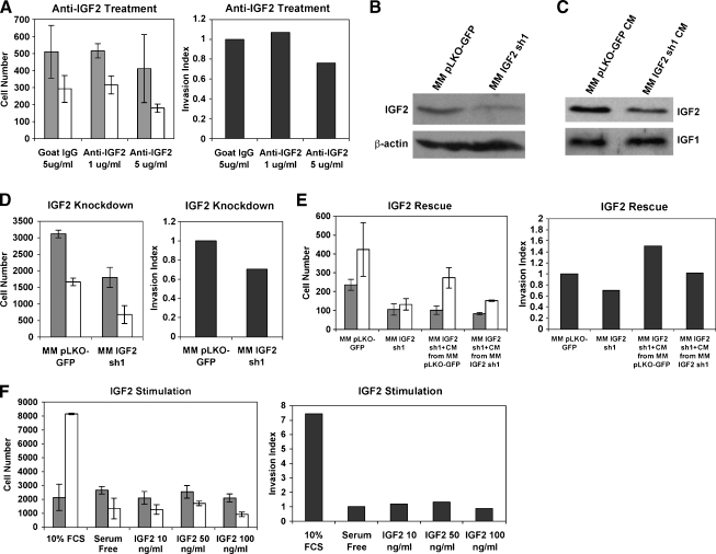
Figure 1.
IGF2 is required for HCC cell migration and invasion. (A) Migration (gray bars) and invasion (white bars) activities of MM189 HCC cells after inhibition of IGF2 using neutralizing antibodies. Error shown is SEM. The panel on the right displays the invasion index (see Materials and Methods). Data are from a representative experiment. (B) Immunoblot of whole-cell lysates confirming knockdown of IGF2 in MM189 HCC cells by a targeting shRNA. β-Actin serves as a loading control. (C) Immunoblot of conditioned serum-free medium collected from IGF2 knockdown cells and controls to determine levels of secreted IGF2 and IGF1. (D) Migration (gray bars) and invasion (white bars) activities of MM189 HCC cells after inhibition of IGF2 by shRNA. Error shown is SEM. The panel on the right displays the invasion index. Data are from a representative experiment. (E) Migration and invasion activities of MM189 cells with IGF2 knockdown or controls in the presence of serum-free medium (first two samples) or in the presence of serum-free conditioned medium collected from either control MM189 cells or MM189 cells expressing IGF2 shRNA (last two samples). Error shown is SEM. The panel on the right displays the invasion index. Data are from a representative experiment. (F) Migration (gray bars) and invasion (white bars) activities of MM189 HCC cells stimulated with recombinant IGF2. Error shown is SEM. The panel on the right displays the invasion index. IGF2 concentrations are shown on the x-axis. Data are from a representative experiment.

