Figure 3.
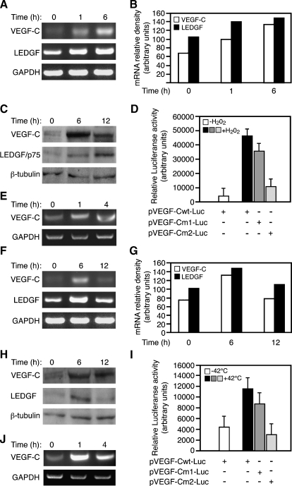
VEGF-C expression is induced by environmental stress. (A) VEGF-C and LEDGF-p75 mRNA expression was analyzed by RT-PCR analysis in H1299 human lung cancer cells exposed for 1 and 6 hours to 0.2 mM H2O2 relative to untreated cells. (B) Relative intensity of the bands normalized against GAPDH. (C) Immunoblot assay of VEGF-C, LEDGF, and β-tubulin in H1299 cells treated with 0.2 mM H2O2 for 6 or 12 hours or left untreated. (D) Luciferase reporter assay was used with a construct composed of a 468-bp VEGF-C wild-type gene fragment (pVEGF-Cwt-Luc) or mutation constructs carrying G-to-A substitutions in the LEDGF/p75 binding sites (pVEGF-Cm1-Luc and pVEGF-Cm2-Luc). H1299 cells transfected with the previously mentioned reporters were stimulated 24 hours after transfection with 0.2 mM H2O2 or were left untreated (mean±SD, n = 3). (E) LEDGF binding to VEGF-C gene sequences was analyzed by ChIP. Chromatin from cells that were exposed to 0.2 mM H2O2 for 1 and 4 hours or left untreated (0 hour) was immunoprecipitated (ChIP) with anti-LEDGF/p75-specific antibody and analyzed by PCR using primers spanning the 468-bp VEGF-C promoter (Figure 1A). Evaluation of total genomic DNA was carried out with GAPDH primers. (F) VEGF-C and LEDGF/p75 mRNA expression was analyzed by RT-PCR analysis either in H1299 lung cancer cells grown at 42°C for 6 hours, transferred for an additional 6-hour incubation at 37°C (12 hours) or left at 37°C (0 hour). (G) Relative intensity of the bands normalized to GAPDH. (H) Immunoblot assay of VEGF-C, LEDGF, and β-tubulin in H1299 cells treated as indicated in panel F. (I) Luciferase reporter assay carried out with pVEGF-Cwt-Luc or two mutated constructs pVEGF-Cm1-Luc and pVEGF-Cm2-Luc. H1299 cells transfected with the previously mentioned reporters were heat-activated (42°C) for 6 hours and then maintained for an additional 6 hours at 37°C (+ and - indicate presence and absence of each construct, respectively; mean ± SD n = 3). (J) ChIP analysis as was described in panel E, except that cells were heat-activated (42°C) for the indicated time.

