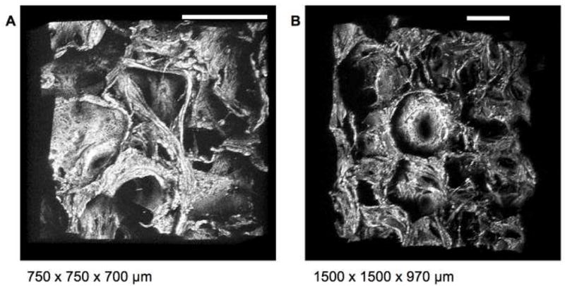
Figure 4. Three dimensional assessment of scaffold morphology.

Three dimensional reconstructions of aqueous scaffolds from TPEF optical sections acquired with 800nm excitation using a 455nm bandpass emission filter through an (A) 20× (0.7NA) and (B) 10× (0.3NA) objective allow for observation of scaffold architecture, pore size and interconnections at multiple angles. (Bar = 300 μm) (See corresponding animation Online)
