

Abstract
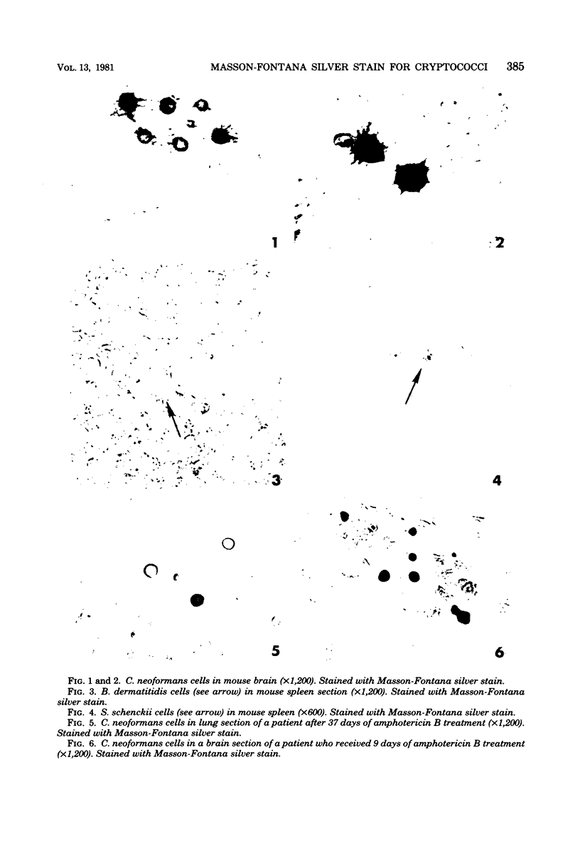
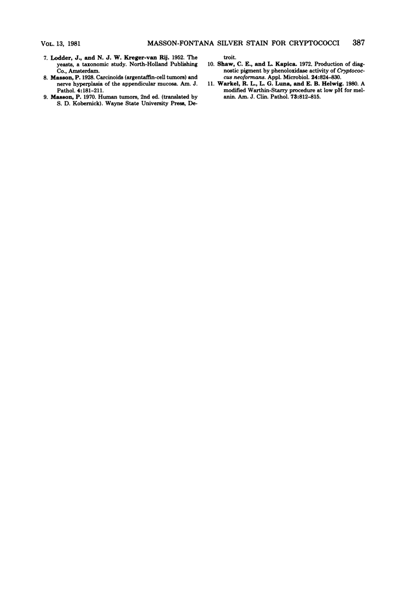
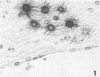
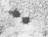
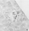
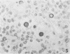
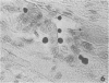
The Masson-Fontana silver stain for melanin was employed for the differentiation of pathogenic fungal species in human or mouse tissues. The fungi studied were Candida albicans, Candida tropicalis, Candida glabrata (Torulopsis glabrata), Cryptococcus neoformans, Cryptococcus bacillisporus, Coccidioides immitis, Blastomyces dermatitidis, Histoplasma capsulatum, Paracoccidioides brasiliensis, Sporothrix schenckii, Rhizopus rhizopodiformis, and Aspergillus fumigatus. The tissue sections stained with Masson-Fontana silver stain showed a dark brown to black color in the wall of cryptococci, whereas the walls of remaining fungal species were hyaline, except for those of S. schenckii. The yeastlike cells of S. schenckii showed faint brown pigment on the wall. Cultures of these fungi showed staining characteristics identical to those of the in vivo results. Cultures of four nonpathogenic Cryptococcus species, Cryptococcus uniguttulatus, Cryptococcus laurentii, Cryptococcus terreus, and Cryptococcus luteolus, were also tested for staining by the Masson-Fontana procedure. Of these, only C. laurentii stained positively, and the pigment on the cell wall was as intense as that of the cells of C. neoformans. These results indicate that the Masson-Fontana silver stain can be used as a specific stain in the histological diagnosis of cryptococcosis.
Full text
PDF
Images in this article
Selected References
These references are in PubMed. This may not be the complete list of references from this article.
- Kwon-Chung K. J. A new genus, filobasidiella, the perfect state of Cryptococcus neoformans. Mycologia. 1975 Nov-Dec;67(6):1197–1200. [PubMed] [Google Scholar]
- Kwon-Chung K. J. Gymnoascus demonbreunii Ajello & Cheng evidence that it is not the perfect state of Histoplasma capsulatum Darling. Sabouraudia. 1968 Feb;6(2):168–175. [PubMed] [Google Scholar]
- Masson P. Carcinoids (Argentaffin-Cell Tumors) and Nerve Hyperplasia of the Appendicular Mucosa. Am J Pathol. 1928 May;4(3):181–212.19. [PMC free article] [PubMed] [Google Scholar]
- Shaw C. E., Kapica L. Production of diagnostic pigment by phenoloxidase activity of cryptococcus neoformans. Appl Microbiol. 1972 Nov;24(5):824–830. doi: 10.1128/am.24.5.824-830.1972. [DOI] [PMC free article] [PubMed] [Google Scholar]
- Warkel R. L., Luna L. G., Helwig E. B. A modified Warthin-Starry procedure at low pH for melanin. Am J Clin Pathol. 1980 Jun;73(6):812–815. doi: 10.1093/ajcp/73.6.812. [DOI] [PubMed] [Google Scholar]