Abstract
The bacterial spore cortex is critical for spore stability and dormancy and must be hydrolyzed by germination-specific lytic enzymes (GSLEs), which allows complete germination and vegetative cell outgrowth. We created in-frame deletions of three genes that encode GSLEs that have been shown to be active in Bacillus anthracis germination: sleB, cwlJ1, and cwlJ2. Phenotypic analysis of individual null mutations showed that the removal of any one of these genes was not sufficient to disrupt spore germination in nutrient-rich media. This finding indicates that these genes have partially redundant functions. Double and triple deletions of these genes resulted in more significant defects. Although a small subset of ΔsleB ΔcwlJ1 spores germinate with wild-type kinetics, for the overall population there is a 3-order-of-magnitude decrease in the colony-forming efficiency compared with wild-type spores. ΔsleB ΔcwlJ1 ΔcwlJ2 spores are unable to complete germination in nutrient-rich conditions in vitro. Both ΔsleB ΔcwlJ1 and ΔsleB ΔcwlJ1 ΔcwlJ2 spores are significantly attenuated, but are not completely devoid of virulence, in a mouse model of inhalation anthrax. Although unable to germinate in standard nutrient-rich media, spores lacking SleB, CwlJ1, and CwlJ2 are able to germinate in whole blood and serum in vitro, which may explain the persistent low levels of virulence observed in mouse infections. This work contributes to our understanding of GSLE activation and function during germination. This information may result in identification of useful therapeutic targets for the disease anthrax, as well as provide insights into ways to induce the breakdown of the protective cortex layer, facilitating easier decontamination of resistant spores.
Bacillus anthracis, a gram-positive spore-forming bacterium, is the causative agent of anthrax. The dormant spore form is the infectious particle and produces three different forms of the disease depending on the route of entry into a suitable host (8). When spores enter through a skin lesion and when they are ingested, they cause cutaneous and gastrointestinal anthrax, respectively. Spores entering through the lungs cause the most severe form of the disease, inhalation anthrax, which is often fatal even with aggressive antibiotic therapy (1, 8, 34). Because true pneumonias are rarely seen in victims, it is believed that inhaled spores do not germinate in the lung but are phagocytosed by alveolar macrophages and germinate intracellularly en route to the mediastinal lymph nodes, which leads to dissemination, septicemia, toxemia, and often death (1, 34). It has been shown that the spores are able to germinate and the bacteria are able to multiply inside macrophages both in cell culture and in the lungs of challenged animals (7, 11, 28, 29).
Independent of the route of infection, spore germination inside a susceptible host is essential for disease. The highly stable spore form of the bacterium can remain viable under harsh environmental conditions for many decades (32). However, a spore can form a rapidly dividing vegetative cell upon entry into a host and recognition of specific chemical signals, or germinants, through specialized germinant receptors (32). The spore cortex, a thick layer of modified peptidoglycan (PG), contributes much of the spore's environmental resistance as it is necessary to maintain dehydration of the spore core (25). This protective barrier is broken down following the activation of germination-specific lytic enzymes (GSLEs), allowing full core rehydration and cell outgrowth (32). Experimentally, germination can also be triggered by nongerminant treatments, such as lysozyme treatment, high pressure, exogenous Ca2+-dipicolinic acid treatment, and treatment with cationic surfactants (32). Several of these treatments likely cause spore cortex hydrolysis, triggering spore germination. This indicates the importance of cortex degradation in the spore germination process.
Bacterial cell wall PG consists of polysaccharide chains of repeating N-acetylglucosamine and N-acetylmuramic acid, joined by β(1,4) glycosidic bonds (25). This basic structure is modified in several ways in spore cortex PG. In one major modification, 50% of the muramic acid residues (alternating every other residue) are converted to muramic-δ-lactam residues (25). This modification is essential for the specificity of GSLEs for degrading the cortex and prevents degradation of the bacterial cell wall during cortex hydrolysis (21).
Previous work on the role of GSLEs in Bacillus subtilis and, recently, in B. anthracis has shown that the enzymes SleB and CwlJ have partially redundant roles and are necessary together for full cortex hydrolysis and spore germination (6, 14). SleB is a lytic transglycosylase that, when activated by an unknown mechanism, hydrolyzes the bond between N-acetylmuramic acid and N-acetylglucosamine (5). In both B. subtilis and B. anthracis, the sleB gene is found in a bicistronic operon with ypeB. Although the function of YpeB is not known, deletion of ypeB prevents SleB activity in spore germination, and sleB and ypeB mutants have similar phenotypes (5). Expression of both gene products is necessary for the presence of SleB in the cortex and inner membrane of mature spores (2, 5).
Although no specific enzymatic activity has been attributed to CwlJ, it is required for full germination and it shares a homologous catalytic domain with SleB (20). In B. subtilis and Bacillus cereus, cwlJ is found in an operon with gerQ. Similar to the finding that ypeB is necessary for a functional SleB protein, gerQ is required for CwlJ activity (26). The B. anthracis genome contains two homologs of cwlJ (designated cwlJ1 and cwlJ2 [14]), whereas a single copy is present in B. subtilis and B. cereus. As it is in the related species, cwlJ1 is found in an operon with gerQ, but cwlJ2 is in a different locus and is not in an operon with a gerQ homolog (14). It has been shown that CwlJ is localized to the spore coat and that it is necessary for spore germination with exogenous Ca2+-dipicolinic acid treatment (3, 24).
GSLE activation represents a critical step in the complex process of germination. The relatively small number of genes involved and the apparent essential nature of their activity make them attractive targets for new therapeutics, as well as environmental decontamination compounds. The objective of this study was to test by using genetic analysis the role of the GSLE genes sleB, cwlJ1, and cwlJ2 in B. anthracis spore germination. Mutants lacking these three genes were tested to determine their effects on in vitro germination kinetics and colony-forming efficiency. Additionally, the virulence of these mutant strains was examined by comparing mutant and wild-type spores in an in vivo mouse model of inhalational anthrax.
MATERIALS AND METHODS
Bacterial strains and antibiotics.
Bacterial strains and plasmids used in this study are listed in Table 1. Vegetative B. anthracis cultures were grown in brain heart infusion media (BHI) (Difco), and Escherichia coli cultures were grown in Luria-Bertani medium (30). All media were supplemented with the following antibiotics to maintain selection as described for mutant construction below: ampicillin (100 μg/ml), erythromycin (400 μg/ml for E. coli and 5 μg/ml for B. anthracis), kanamycin (10 μg/ml), polymyxin B (60 U/ml), spectinomycin (100 μg/ml), and tetracycline (10 μg/ml).
TABLE 1.
B. anthracis strains and plasmids
| Strain or plasmid | Relevant characteristics | Reference |
|---|---|---|
| Strains | ||
| Sterne 34F2 | Wild type (pXO1+ pXO2−) | 33 |
| KC61 | 34F2 ΔsleB | This study |
| KC62 | 34F2 ΔypeB | This study |
| KC63 | 34F2 ΔsleB ΔypeB | This study |
| JG48 | 34F2 ΔcwlJ1 | This study |
| JG49 | 34F2 ΔcwlJ2 | This study |
| JG50 | 34F2 ΔsleB ΔypeB ΔcwlJ1 | This study |
| JG51 | 34F2 ΔsleB ΔypeB ΔcwlJ2 | This study |
| JG52 | 34F2 ΔcwlJ1 ΔcwlJ2 | This study |
| JG53 | 34F2 ΔsleB ΔypeB ΔcwlJ1 ΔcwlJ2 | This study |
| Plasmids | ||
| pBKJ236 | Allelic exchange vector, Emr | 16 |
| pBKJ258 | Allelic exchange vector, Kanr | 18 |
| pBKJ223 | Tetr, Pamy I-SceI | 16 |
| pJDG101 | ΔsleB construct cloned in pBKJ258 | This study |
| pJDG102 | ΔypeB construct cloned in pBKJ258 | This study |
| pJDG103 | ΔcwlJ1 construct cloned in pBKJ236 | This study |
| pJDG104 | ΔcwlJ2 construct cloned in pBKJ258 | This study |
B. anthracis spore stocks were prepared as previously described (7, 17). Briefly, vegetative cells were diluted 1:100 in modified G medium [0.2% yeast extract, 0.17 mM CaCl2, 2.87 mM K2HPO4, 0.81 mM MgSO4, 0.24 mM MnSO4, 17 μM ZnSO4, 20 μM CuSO4, 1.8 μM FeSO4, 15.5 mM (NH4)2SO4; pH 7.2] and incubated at 37°C and 300 rpm for 72 h. Following incubation, cultures were checked microscopically to verify the presence of phase-bright spores. Spores were purified as previously described, and the final stock contained >99% phase-bright spore particles (19).
Mutant construction.
Mutant bacterial strains were constructed using the allelic exchange method as previously described (16, 18). Allelic exchange plasmids were constructed as described previously for the ΔGBAA1941 construct (4). For each gene deleted, the initial 30 nucleotides were fused to the final 30 nucleotides, creating an in-frame deletion. In place of the deleted genetic material, three stop codons were inserted into each gene. cwlJ1 and cwlJ2 single mutants were constructed in a wild-type Sterne 34F2 background. The ΔsleB mutant was made by removal of the ΔsleB ΔypeB operon in a Sterne 34F2 background. ypeB is proposed to act in either stability or targeting of sleB. Both the SleB and YpeB proteins are required for SleB activity, and there is no phenotypic difference between ΔsleB and ΔypeB single mutants in the assays described here (data not shown). Double and triple mutants were created by deletion of additional genes in the single-mutant backgrounds. All oligonucleotide sequences used for construction and screening of mutant strains are available upon request.
Germination assays.
Spore germination was assayed by measuring the loss of heat resistance or the decrease in the optical density at 600 nm (OD600). To measure the loss of heat resistance, spore stocks were diluted to obtain a concentration of approximately 4 × 10⁵ spores/ml and heat activated by incubation at 65°C for 20 min. Germination was initiated by adding 10 μl of spores to 2.0 ml of germination medium. At each time point (2, 5, 10, 15, 20, and 30 min) 100-μl aliquots of this preparation were removed and incubated at 65°C for 20 min, and then 50-μl portions were plated on BHI agar and incubated overnight. The colonies that formed represented spores that failed to germinate and were therefore heat resistant. As a control, 50 μl of the germination reaction mixture at time zero was plated without heat treatment. The fraction of germinated spores for each time point was calculated using (1 − tn)/t0 (where tn is the time point of interest and t0 is time zero) and was expressed as a percentage. To determine the decrease in optical density, 5.0 ml of spores (OD600, 0.6) was heat activated by incubation at 65°C for 20 min. Germinants were then added to obtain a starting OD600 of approximately 0.3, and the reaction mixture was incubated at 37°C with shaking at 300 rpm. At 5-min intervals, 1.0-ml aliquots were removed, and the OD600 was measured using a Genesys 10UV spectrophotometer (Spectronic Unicam, Rochester, NY).
The germination media used in this study were BHI (Difco), whole bovine blood (HemoStat Laboratories), and bovine serum. Bovine serum was collected by pelleting whole blood at 4,000 rpm in an Eppendorf 5810 R swinging bucket centrifuge. Following centrifugation, the serum was filtered through a 0.2-μm syringe filter (Nalgene) and stored at 4°C. Heat-treated serum was incubated at 65°C for 30 min.
In this study, colony-forming efficiency is defined as the number of spore particles that represent 1 CFU. An improved Neubauer hemocytometer was used to determine the number of spore particles per ml for each spore stock assayed. The same spore stock was germinated in one of the germination media listed above for 10 min at 37°C. Germination reaction mixtures were then titrated on BHI plates to determine the number of CFU/ml.
Murine challenges.
Intratracheal infection of DBA/2J mice (Jackson Laboratories) was performed as previously described (13). Groups of eight mice were infected with mutant or wild-type strains using doses ranging from 1.5 × 10³ to 1.5 × 10⁷ spores per mouse. Mice were monitored for 14 days. B. anthracis bacteria were recovered from the blood, spleen, heart, and lungs of randomly selected individuals that died following infection. Strain identity was verified by using diagnostic PCR.
RESULTS
Effects of GSLE mutations on in vitro spore germination in BHI.
Germination appears to be a well-conserved process in spore-forming bacteria, including B. subtilis and B. cereus (2, 15, 22). Independently, and using assays different than those used in this study, Heffron et al. (14) showed that single-deletion mutations of the GSLE genes sleB, cwlJ1, and cwlJ2 in B. anthracis cause minor deficiencies in in vitro germination kinetics compared to those of wild-type spores and that the ΔsleB ΔcwlJ1 double mutant has severe germination deficiencies. We simultaneously constructed defined in-frame deletion mutations for each of the three GSLE genes individually and in combinations in order to better determine the contributions of these three genes to spore germination in vitro and to determine if these genes play a role in virulence in an in vivo mouse model of inhalation anthrax.
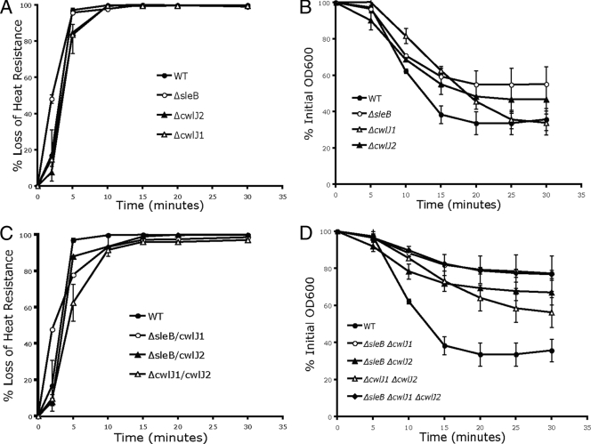
None of the strains created exhibited a gross defect in sporulation or the vegetative growth rate (data not shown). These strains were germinated in vitro using BHI as a germinant, and germination was monitored by determining the loss of heat resistance (9). All single mutants exhibited ∼100% germination in roughly the same time, 2 to 5 min (Fig. 1A). Small variations in the germination rates were observed from 0 to 5 min, including slight delays caused by the ΔcwlJ1 and ΔcwlJ2 mutations at the 5-min time point and a temporary increase in the germination kinetics of the ΔsleB mutant at the 2-min time point. Measuring spore germination by loss of heat resistance is limited since it can score only viable spores that are ultimately able to grow. Therefore, we also measured germination by monitoring the decrease in the OD600 following the addition of germinants. A 60% decrease in the initial optical density is considered to indicate 100% germination (23). The ΔsleB and ΔcwlJ2 mutants did not exhibit 100% germination (Fig. 1B). The ΔcwlJ1 mutant did exhibit 100% germination, but the germination was slightly delayed compared to that of the wild type (Fig. 1B). These results indicate that loss of any one GSLE caused only minor deficiencies in the germination kinetics compared to those of the wild type.
FIG. 1.
In vitro germination of spores in BHI. The loss of heat resistance (A and C) and the decrease in the OD600 (B and D) were measured as markers of spore germination. A 60% decrease in the OD600 represents approximately 100% germination (23). Spores were incubated in BHI at 37°C for 0 to 30 min. In all panels the symbols indicate the means for two or three independent experiments, and the error bars indicate standard errors. WT, wild type.
Germination of double mutants in BHI also revealed small fluctuations in germination kinetics when they were measured by using loss of heat resistance (Fig. 1C). Spores of the ΔsleB ΔcwlJ2 strain germinated with nearly wild-type kinetics. Spores of the ΔcwlJ1 ΔcwlJ2 strain had a delay in germination at the 5-min time point compared to wild-type spores. Despite this delay, the mutant spores still exhibited nearly 100% germination by the end of the 30-min time course. Similarly, we found only minor germination defects in ΔsleB ΔcwlJ1 spores compared to the wild type. Like the results for the ΔsleB single mutant, germination of the spores of this double mutant accelerated after 2 min, but otherwise the germination kinetics of spores in BHI were similar to the wild-type germination kinetics (Fig. 1C). However, as observed by Heffron et al. (14), we found severe defects in the germination of the ΔsleB ΔcwlJ1 mutant spores and also in the germination of the ΔsleB ΔcwlJ1 ΔcwlJ2 mutant spores when the germination was measured by the decrease in the optical density (Fig. 1D). Although the assay measuring the decrease in the optical density is better able to detect early events in spore germination, small subsets of spores are lost in the background of the population. We believe that this explains the different results for ΔsleB ΔcwlJ1 spore germination when it was measured by the two different methods. Although the ΔsleB ΔcwlJ1 spore population as a whole has major germination defects and cannot grow, the small number of spores that do grow germinate with wild-type kinetics (Fig. 1C and D).
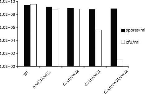
The germination kinetics of the ΔsleB ΔcwlJ1 ΔcwlJ2 triple mutant strain could not be assayed by measuring the loss of heat resistance because this strain was unable to germinate in response to BHI. However, this mutant had the same germination defect as the ΔsleB ΔcwlJ1 mutant as measured by the decrease in the optical density (Fig. 1D). To better characterize the nature of this dramatic defect and to determine if similar defects were present in the double-mutant strains, the colony-forming efficiency of each strain was measured. We tested the colony-forming efficiency by visually determining the number of spores in a sample using a hemocytometer and then inducing germination of the same spores with a variety of germinants. By comparing the difference between the concentration of a spore stock (spores/ml) determined with a hemocytometer and the number of CFU/ml of the same stock when it was titrated on BHI plates, we determined the colony-forming efficiency of all double and triple GSLE mutants in nutrient-rich media (Fig. 2). The colony-forming efficiency of the ΔsleB ΔcwlJ1 ΔcwlJ2 mutant was found to be 8.2 × 10⁷ spore particles per CFU, a 7-order-of-magnitude decrease compared to wild-type spores. Consistent with a recent report (14), the ΔsleB ΔcwlJ1 strain also exhibited a significant decrease in colony-forming efficiency (approximately 3 orders of magnitude) compared to the wild type. All other double mutants and wild-type strains exhibited a colony-forming efficiency of approximately 1 spore particle/CFU (Fig. 2). These data show that although the GSLEs do not play a major role in germination kinetics, they are necessary for spore outgrowth. Furthermore, it appears that the three genes have partially redundant functions as any one of them is sufficient for at least some level of colony formation. In order of increasing colony-forming efficiency, the ΔsleB ΔcwlJ1 ΔcwlJ2 triple mutant had the lowest colony-forming efficiency, followed by the ΔsleB ΔcwlJ1 mutant. The remaining strains, the ΔsleB ΔcwlJ2 and ΔcwlJ1 ΔcwlJ2 mutants, had the highest colony-forming efficiencies, which were comparable to the wild-type values. This indicates that the contributions of sleB and cwlJ1 have greater influence on colony-forming efficiency than the contributions of cwlJ2.
FIG. 2.
Colony-forming efficiency of GSLE mutants. The colony-forming efficiency was measured by visually determining the number of spores/ml (solid bars) of a given stock with an improved Neubauer hemocytometer and comparing the value obtained to the number of CFU/ml (open bars) for the same stock when it was plated on BHI. The data are representative of typical results. WT, wild type.
Virulence of GSLE mutant strains in mice.
Although the B. anthracis Sterne strain is attenuated in many mammalian species, certain strains of inbred mice, including DBA/2J, are susceptible to this bacterium, making this strain an excellent model system for virulence studies (12, 35). We used intratracheal injection of spores into the lungs as a model for inhalational anthrax (10). The virulence in DBA/2J mice of all double and triple mutants described above was compared to the virulence of wild-type bacteria. DBA/2J mice infected with wild-type spores had a median time to death of 2 days at a dose of 1.5 × 10⁶ spores/mouse, and the 50% lethal dose (LD50) was 1.5 × 10⁴ spores/mouse (Table 2).
TABLE 2.
DBA/2J mouse virulence
| Strain | Median time to death (days) with:
|
LD50 (spores/mouse)a | |
|---|---|---|---|
| 1.5 × 10⁶ spores/mouse | 1.5 × 10⁷ spores/mouse | ||
| 34F2 | 2 | NDb | 1.5 × 10⁴ |
| ΔsleB ΔcwlJ1 | 5.5 | 3.5 | 1.2 × 10⁶ |
| ΔsleB ΔcwlJ2 | 3.5 | ND | ND |
| ΔcwlJ1 ΔcwlJ2 | 3 | ND | ND |
| ΔsleB ΔcwlJ1 ΔcwlJ2 | Undefinedc | 4 | 8.3 × 10⁶ |
LD50s were determined by the method of Reed and Muench (27) and were based on survival data for four groups of mice (n = 8) with four different doses for each of the strains assayed.
ND, not determined.
Because only two of eight mice in the group died during the course of the experiment, the dose was not lethal enough to determine the median time to death.
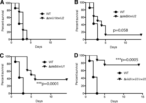
The ΔcwlJ1 ΔcwlJ2 double mutant, which exhibited a slight delay in germination kinetics in vitro (Fig. 1C and D), showed no attenuation of virulence compared to the wild type at a dose of 1.5 × 10⁶ spores/mouse (100 times the wild-type LD50) (Fig. 3A), and there was no significant increase in the median time to death (Table 2). The ΔsleB ΔcwlJ2 double mutant, which showed the least variation from wild-type germination kinetics in BHI, was slightly attenuated (Fig. 3B), and there was a slight increase in the median time to death (Table 2). Although the values were not significantly different from the wild-type values (P = 0.058), they did trend away from the values for wild-type spores. As expected from the in vitro colony-forming efficiency, the ΔsleB ΔcwlJ1 strain exhibited highly significant (P = 0.0001) attenuation compared to wild-type spores (Fig. 3C). The median time to death with this strain was 5.5 days, compared to 2 days with wild-type spores, and even at a 10-fold-higher dose (1.5 × 10⁷ spores/mouse) an increase in the median time to death was observed compared to the results for the lower dose of wild-type bacteria (2 days for the wild type and 3.5 days for the mutant) (Table 2).
FIG. 3.
Survival curves for GSLE mutants in mice. DBA/2J mice (eight mice per group) were challenged by intratracheal infection with wild-type (WT) and mutant spores at a dose of 1.5 × 10⁶ spores/mouse. The mice were monitored for 14 days, and then all surviving mice were euthanized. The survival curves for the ΔsleB ΔcwlJ1 (C) and ΔsleB ΔcwlJ1 ΔcwlJ2 (D) mutants were found to be significantly different from those for the wild type (P = 0.0001 and P = 0.0005, respectively, as determined by a log rank [Mantel-Cox] test). Although not meeting the significance threshold, the survival curve for the ΔsleB ΔcwlJ2 mutant (B) did show a trend away from the survival curve for the wild type (P = 0.058). The survival curve for the ΔcwlJ1 ΔcwlJ2 mutant was not significantly different from that of the wild type (A).
Next we tested the virulence of the ΔsleB ΔcwlJ1 ΔcwlJ2 triple mutant in our mouse model. The triple mutant was significantly attenuated (P = 0.0005) (Fig. 3D), but it did exhibit some virulence, killing 25% of the mice challenged in 4 days. This was somewhat surprising because the triple mutant was functionally unable to germinate in rich media. On BHI plates, only one spore per 82 million spores could germinate. As each mouse was given a dose of only 1.5 million spores, we did not expect to see any germination or subsequent mortality. Necropsies were performed on a subset of animals that succumbed to the infection, and mutant bacteria were isolated on BHI plates, confirming that mutant spores were able to geminate inside mice and multiply to nearly wild-type levels (data not shown). The median time to death with this strain could not be determined using the dose that was used for the strains described above (1.5 × 10⁶ spores/mouse) because it was not virulent enough to kill at least one-half of the mice challenged within the time course of the experiment (Table 2). At a 10-fold-higher dose, 1.5 × 10⁷ spores/mouse, the median time to death was 4 days. This was longer than the median time to death with the lower dose of wild-type spores (2 days) but was approximately the same as the median time to death with the high dose of ΔsleB ΔcwlJ1 double-mutant spores (3.5 days) (Table 2). Although both mutants are significantly attenuated compared to the wild type, the highest doses of the ΔsleB ΔcwlJ1 and ΔsleB ΔcwlJ1 ΔcwlJ2 strains tested (1.5 × 10⁷ spores/mouse and 8.4 × 10⁷ spores/mouse, respectively) did not produce significantly different survival curves and resulted in the same median time to death, 3.5 days (data not shown). This suggested that despite the higher number of mutant spores in the challenge dose, only a subset of the spores were able to germinate and cause disease.
In order to further define the virulence effects of the attenuated mutants, LD50s were determined for the wild-type, ΔsleB ΔcwlJ1, and ΔsleB ΔcwlJ1 ΔcwlJ2 strains. Groups of mice (n = 8) were infected by intratracheal injection with four doses of spores suspended in water. The parental strain Sterne 34F2 (wild type) had an LD50 of 1.5 × 10⁴ spores. The LD50s of the ΔsleB ΔcwlJ1 and ΔsleB ΔcwlJ1 ΔcwlJ2 strains were found to be 1.2 × 10⁶ spores and 8.3 × 10⁶ spores, respectively, which were 80-fold and 550-fold higher than the wild-type LD50, respectively (Table 2). Taken together, these data show that a lack of germination on nutrient-rich media in vitro does not indicate a block of germination in vivo and that another mechanism of spore germination may be active in the host.
Effects of GSLE mutations on in vitro germination in blood.
Although it was expected that the colony formation-deficient ΔsleB ΔcwlJ1 mutant would be at least somewhat virulent in mice, it did not seem likely that the extreme colony formation-deficient ΔsleB ΔcwlJ1 ΔcwlJ2 mutant would have any ability to cause disease. Based solely on the number of CFU on BHI, even the highest challenge dose of the ΔsleB ΔcwlJ1 ΔcwlJ2 mutant would be effectively 1 germinating spore/mouse, which is 10,000-fold lower than the wild-type LD50. These data led to the hypothesis that a host factor(s) was able to cause germination at levels higher than those calculated when BHI medium was used as a germinant and perhaps circumvent the requirement for GSLE activities.
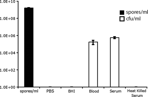
In order to test if such factors were present in blood, colony-forming efficiency assays were repeated with ΔsleB ΔcwlJ1 ΔcwlJ2 spores using defibrinated bovine whole blood as the germination medium. Spores were first incubated for 10 min in defibrinated bovine blood to initiate germination. Then the spores were plated on BHI and allowed to grow at 37°C. The colony-forming efficiency of the ΔsleB ΔcwlJ1 ΔcwlJ2 mutant increased from 8.2 × 10⁷ spores per CFU when it was germinated in BHI to 9.2 × 10³ spores per CFU when it was germinated in blood. This represents an almost 4-order-of-magnitude increase compared with the colony-forming efficiency in BHI (Fig. 4). The colony-forming efficiency of the triple mutant spores in blood was about 3 orders of magnitude less than that of wild-type spores germinated in blood (data not shown).
FIG. 4.
In vitro germination of ΔsleB ΔcwlJ1 ΔcwlJ2 spores in blood. The colony-forming efficiency of ΔsleB ΔcwlJ1 ΔcwlJ2 spores in blood and serum was examined. Spore dilutions were quantified visually with an improved Neubauer hemocytometer (spores/ml) and compared to the numbers of CFU/ml when spores were allowed to germinate in phosphate-buffered saline (PBS) (pH 7.5), BHI, whole bovine blood, bovine serum, or heat-treated bovine serum for 10 min before they were plated on BHI plates and incubated at 37°C overnight.
Next we wanted to further examine the source of the host germination factor by removing the contribution of whole cells in the blood. To do this, we used filtered bovine serum and heat-treated filtered bovine serum as germination media for triple-mutant spores. The mutant spores germinated in serum at a rate comparable to that in whole blood (2.9 × 10³ spores per CFU in bovine serum compared to 9.2 × 10³ spores per CFU in whole blood) (Fig. 4). This indicates that serum contains all the host components present in whole blood necessary to cause mutant spore germination and that whole cells in blood do not play a direct role. Bovine serum heat treated at 65°C for 30 min lost the ability to cause germination of ΔsleB ΔcwlJ1 ΔcwlJ2 spores (Fig. 4). This suggests that the host factor causing mutant spore germination is heat labile and may be an enzyme in serum.
DISCUSSION
GSLEs are expressed during B. anthracis sporulation and are active during germination (4, 14). Deletion of the genes that encode the enzymes SleB, CwlJ1, and CwlJ2 significantly disrupted spore germination and, in the case of the triple mutant, even led to a complete loss of germination in rich media in vitro. However, the germination-deficient spores exhibited some virulence in our mouse model, suggesting that there is an alternate mechanism of spore germination.
Individual deletions of the sleB, cwlJ1, and cwlJ2 genes did not result in major differences in germination kinetics or colony-forming efficiency (Fig. 1). The single mutants were not tested for virulence in mice. The ΔsleB ΔcwlJ2 and ΔcwlJ1 ΔcwlJ2 double mutants also had only minor effects on germination kinetics and had no effect on colony-forming efficiency (Fig. 1 to 2). However, the colony-forming efficiency of the ΔsleB ΔcwlJ1 double mutant on BHI was decreased by 3 orders of magnitude (Fig. 2). Despite this defect, the small percentage of the spore population that was able to grow germinated with kinetics very similar to those of wild-type spores (Fig. 1C). We believe that the assay that measured the decrease in the optical density was not sensitive enough to detect the germination kinetics of the subpopulation of spores that managed to germinate. Because only outgrowing spores can be detected by the loss of heat resistance assay, we were able to show that in fact the population defect was not uniform and that while the bulk of the spores are defective, a small number of spores germinate like wild-type spores. The ΔsleB ΔcwlJ1 ΔcwlJ2 triple mutant had a 7-order-of-magnitude decrease in colony-forming efficiency in BHI compared to the wild type (Fig. 2). Because of this severe defect in germination, the germination kinetics of growing spores could not be measured; however, the ΔsleB ΔcwlJ1 ΔcwlJ2 spore population exhibited the same severe defect as ΔsleB ΔcwlJ1 spores when it was assayed using the decrease in optical density (Fig. 1D).
B. subtilis and other related species of spore-forming bacteria contain one copy of the cwlJ gene, while the B. anthracis genome contains a second copy of this gene referred to as cwlJ2 (6, 14, 31). The single copy of cwlJ in other species and the cwlJ1 gene in B. anthracis are found in an operon with gerQ, a gene that has been shown to be required for proper targeting of cwlJ in the developing forespore (26). This conserved gene structure implies that cwlJ1 is more important than cwlJ2. Mutations in cwlJ2 result in no major defect in colony-forming efficiency (Fig. 2), suggesting that CwlJ2 is a less functional or redundant protein. Although CwlJ2 does not appear to be the primary protein involved, it is active during germination and can compensate in the absence of CwlJ1. The ΔcwlJ1 ΔcwlJ2 double mutant has a greater germination defect than either of the single mutants (Fig. 1). Also, the ΔsleB ΔcwlJ1 ΔcwlJ2 triple mutant has 100,000-fold-lower colony-forming ability than the ΔsleB ΔcwlJ1 double mutant in BHI (Fig. 2). These results show that SleB or CwlJ1 is sufficient for nearly wild-type levels of spore germination. CwlJ2 is sufficient for low-level germination, which can be observed when this protein is compensating for the absence of SleB and CwlJ1.
GSLEs play a critical role in the germination of all spore-forming bacteria, and it has long been thought that targeting the activity of these enzymes could provide useful therapies. The complete lack of in vitro germination of the ΔsleB ΔcwlJ1 ΔcwlJ2 mutant supports this theory, but the continued virulence in a mouse model indicates that there are alternate mechanisms of germination that can result in disease. We calculated that the LD50 of wild-type strain Sterne 34F2 is 1.5 × 10⁴ spores/mouse in the DBA/2J mouse model. The ΔsleB ΔcwlJ1 and ΔsleB ΔcwlJ1 ΔcwlJ2 mutant strains were attenuated and had LD50s that were 80-fold and 550-fold greater than that of the wild type, respectively (Table 2). Despite the increase in the LD50 and the large decrease in colony-forming efficiency for the ΔsleB ΔcwlJ1 ΔcwlJ2 mutant compared to the ΔsleB ΔcwlJ1 mutant, at no dose tested was there a significant difference in murine survival following infection with these two mutant strains (Fig. 3 and data not shown). This suggests that an alternate method of germination may involve limiting host factors in vivo. It is possible that these factors are host-specific germinants that activate unknown GSLEs. However, it is much more likely that a host factor is able to complement missing GSLE functions. Such potential factors are unknown and are currently under investigation. They may include host enzymes with PG-hydrolyzing activity, such as lysozyme. The ability of ΔsleB ΔcwlJ1 ΔcwlJ2 spores to germinate in bovine serum but not in heat-treated bovine serum suggests that a host enzyme has a role in germination and supports the hypothesis that there is a host factor complementation mechanism (Fig. 4). Interestingly, the colony-forming efficiency of the triple mutant in bovine serum is about 3 orders of magnitude less than that of the wild type (Fig. 4), which is consistent with the 550-fold difference in the LD50s between the triple mutant and the wild-type strain (Table 2).
It is generally believed that inhalation anthrax is the result of spore germination within alveolar macrophages (11, 29). Inhaled spores remain dormant in the lungs, possibly for many weeks, until they are engulfed by lung macrophages and trafficked to the nearest lymph node. It has been shown that B. anthracis spores engulfed by macrophages are not killed but germinate and replicate within the cells until they are ruptured (7). It would be interesting to know if the alternate germination mechanism provided by components of the serum is also active in alveolar macrophages.
The SleB, CwlJ1, and to a lesser extent CwlJ2 enzymes play important roles in the successful germination and outgrowth of B. anthracis spores. Removal of all three enzymes is sufficient to block in vitro germination under standard laboratory conditions. The continued virulence of mutant strains despite their lack of in vitro germination sheds new light on secondary, host-directed mechanisms of germination in vivo. Although it is clear that for disease therapies and spore decontamination there are many benefits to targeting germination-specific lytic enzymes, it is important to understand and control these secondary factors to fully combat disease.
Acknowledgments
We thank Brian Janes for his expertise and assistance with the preparation of the manuscript.
This work was supported in part by HHS contract N266200400059C/N01-AI-40059 and by the Great Lakes and the Southeast Regional Centers of Excellence for Biodefense and Emerging Infections. J.D.G. was supported by the University of Michigan Cellular Biotechnology Training Program.
Footnotes
Published ahead of print on 6 July 2009.
REFERENCES
- 1.Albrink, W. S. 1961. Pathogenesis of inhalation anthrax. Bacteriol. Rev. 25268-273. [DOI] [PMC free article] [PubMed] [Google Scholar]
- 2.Atrih, A., and S. J. Foster. 2001. In vivo roles of the germination-specific lytic enzymes of Bacillus subtilis 168. Microbiology 1472925-2932. [DOI] [PubMed] [Google Scholar]
- 3.Bagyan, I., and P. Setlow. 2002. Localization of the cortex lytic enzyme CwlJ in spores of Bacillus subtilis. J. Bacteriol. 1841219-1224. [DOI] [PMC free article] [PubMed] [Google Scholar]
- 4.Bergman, N. H., E. C. Anderson, E. E. Swenson, M. M. Niemeyer, A. D. Miyoshi, and P. C. Hanna. 2006. Transcriptional profiling of the Bacillus anthracis life cycle in vitro and an implied model for regulation of spore formation. J. Bacteriol. 1886092-6100. [DOI] [PMC free article] [PubMed] [Google Scholar]
- 5.Boland, F. M., A. Atrih, H. Chirakkal, S. J. Foster, and A. Moir. 2000. Complete spore-cortex hydrolysis during germination of Bacillus subtilis 168 requires SleB and YpeB. Microbiology 14657-64. [DOI] [PubMed] [Google Scholar]
- 6.Chirakkal, H., M. O'Rourke, A. Atrih, S. J. Foster, and A. Moir. 2002. Analysis of spore cortex lytic enzymes and related proteins in Bacillus subtilis endospore germination. Microbiology 1482383-2392. [DOI] [PubMed] [Google Scholar]
- 7.Dixon, T. C., A. A. Fadl, T. M. Koehler, J. A. Swanson, and P. C. Hanna. 2000. Early Bacillus anthracis-macrophage interactions: intracellular survival and escape. Cell. Microbiol. 2453-463. [DOI] [PubMed] [Google Scholar]
- 8.Dixon, T. C., M. Meselson, J. Guillemin, and P. C. Hanna. 1999. Anthrax. N. Engl. J. Med. 341815-826. [DOI] [PubMed] [Google Scholar]
- 9.Fisher, N., and P. Hanna. 2005. Characterization of Bacillus anthracis germinant receptors in vitro. J. Bacteriol. 1878055-8062. [DOI] [PMC free article] [PubMed] [Google Scholar]
- 10.Fisher, N., L. Shetron-Rama, A. Herring-Palmer, B. Heffernan, N. Bergman, and P. Hanna. 2006. The dltABCD operon of Bacillus anthracis Sterne is required for virulence and resistance to peptide, enzymatic, and cellular mediators of innate immunity. J. Bacteriol. 1881301-1309. [DOI] [PMC free article] [PubMed] [Google Scholar]
- 11.Guidi-Rontani, C., M. Weber-Levy, E. Labruyere, and M. Mock. 1999. Germination of Bacillus anthracis spores within alveolar macrophages. Mol. Microbiol. 319-17. [DOI] [PubMed] [Google Scholar]
- 12.Harvill, E. T., G. Lee, V. K. Grippe, and T. J. Merkel. 2005. Complement depletion renders C57BL/6 mice sensitive to the Bacillus anthracis Sterne strain. Infect. Immun. 734420-4422. [DOI] [PMC free article] [PubMed] [Google Scholar]
- 13.Heffernan, B. J., B. Thomason, A. Herring-Palmer, and P. Hanna. 2007. Bacillus anthracis anthrolysin O and three phospholipases C are functionally redundant in a murine model of inhalation anthrax. FEMS Microbiol. Lett. 27198-105. [DOI] [PubMed] [Google Scholar]
- 14.Heffron, J. D., B. Orsburn, and D. L. Popham. 2009. Roles of germination-specific lytic enzymes CwlJ and SleB in Bacillus anthracis. J. Bacteriol. 1912237-2247. [DOI] [PMC free article] [PubMed] [Google Scholar]
- 15.Ishikawa, S., K. Yamane, and J. Sekiguchi. 1998. Regulation and characterization of a newly deduced cell wall hydrolase gene (cwlJ) which affects germination of Bacillus subtilis spores. J. Bacteriol. 1801375-1380. [DOI] [PMC free article] [PubMed] [Google Scholar]
- 16.Janes, B. K., and S. Stibitz. 2006. Routine markerless gene replacement in Bacillus anthracis. Infect. Immun. 741949-1953. [DOI] [PMC free article] [PubMed] [Google Scholar]
- 17.Kim, H. U., and J. M. Goepfert. 1974. A sporulation medium for Bacillus anthracis. J. Appl. Bacteriol. 37265-267. [DOI] [PubMed] [Google Scholar]
- 18.Lee, J. Y., B. K. Janes, K. D. Passalacqua, B. F. Pfleger, N. H. Bergman, H. Liu, K. Hakansson, R. V. Somu, C. C. Aldrich, S. Cendrowski, P. C. Hanna, and D. H. Sherman. 2007. Biosynthetic analysis of the petrobactin siderophore pathway from Bacillus anthracis. J. Bacteriol. 1891698-1710. [DOI] [PMC free article] [PubMed] [Google Scholar]
- 19.Liu, H., N. H. Bergman, B. Thomason, S. Shallom, A. Hazen, J. Crossno, D. A. Rasko, J. Ravel, T. D. Read, S. N. Peterson, J. Yates III, and P. C. Hanna. 2004. Formation and composition of the Bacillus anthracis endospore. J. Bacteriol. 186164-178. [DOI] [PMC free article] [PubMed] [Google Scholar]
- 20.Moir, A. 2006. How do spores germinate? J. Appl. Microbiol. 101526-530. [DOI] [PubMed] [Google Scholar]
- 21.Moir, A., B. M. Corfe, and J. Behravan. 2002. Spore germination. Cell. Mol. Life Sci. 59403-409. [DOI] [PMC free article] [PubMed] [Google Scholar]
- 22.Moriyama, R., S. Kudoh, S. Miyata, S. Nonobe, A. Hattori, and S. Makino. 1996. A germination-specific spore cortex-lytic enzyme from Bacillus cereus spores: cloning and sequencing of the gene and molecular characterization of the enzyme. J. Bacteriol. 1785330-5332. [DOI] [PMC free article] [PubMed] [Google Scholar]
- 23.Nicholson, W. L., and P. Setlow. 1990. Sporulation, germination and outgrowth, p. 391-450. In C. R. Harwood and S. M. Cutting (ed.), Molecular biological methods for Bacillus. John Wiley & Sons Ltd., Chichester, United Kingdom.
- 24.Paidhungat, M., K. Ragkousi, and P. Setlow. 2001. Genetic requirements for induction of germination of spores of Bacillus subtilis by Ca2+-dipicolinate. J. Bacteriol. 1834886-4893. [DOI] [PMC free article] [PubMed] [Google Scholar]
- 25.Popham, D. L. 2002. Specialized peptidoglycan of the bacterial endospore: the inner wall of the lockbox. Cell. Mol. Life Sci. 59426-433. [DOI] [PMC free article] [PubMed] [Google Scholar]
- 26.Ragkousi, K., P. Eichenberger, C. van Ooij, and P. Setlow. 2003. Identification of a new gene essential for germination of Bacillus subtilis spores with Ca2+-dipicolinate. J. Bacteriol. 1852315-2329. [DOI] [PMC free article] [PubMed] [Google Scholar]
- 27.Reed, L. J., and H. Muench. 1938. A simple method of estimating fifty per cent endpoints. Am. J. Hyg. 27493-497. [Google Scholar]
- 28.Ross, J. M. 1955. On the histopathology of experimental anthrax in the guinea-pig. Br. J. Exp. Pathol. 36336-339. [PMC free article] [PubMed] [Google Scholar]
- 29.Ross, J. M. 1957. The pathogenesis of anthrax following the administration of spores by the respiratory route. J. Pathol Bacteriol. 73485-494. [Google Scholar]
- 30.Sambrook, J., and D. W. Russell. 2001. Molecular cloning: a laboratory manual, 3rd ed. Cold Spring Harbor Laboratory Press, Cold Spring Harbor, NY.
- 31.Setlow, B., L. Peng, C. A. Loshon, Y. Q. Li, G. Christie, and P. Setlow. 2009. Characterization of the germination of Bacillus megaterium spores lacking enzymes that degrade the spore cortex. J. Appl. Microbiol. 107318-328. [DOI] [PubMed] [Google Scholar]
- 32.Setlow, P. 2003. Spore germination. Curr. Opin. Microbiol. 6550-556. [DOI] [PubMed] [Google Scholar]
- 33.Sterne, M. 1939. The immunization of laboratory animals against anthrax. J. Vet. Sci. Anim. Ind. 13313-317. [Google Scholar]
- 34.Tournier, J. N., A. Quesnel-Hellmann, A. Cleret, and D. R. Vidal. 2007. Contribution of toxins to the pathogenesis of inhalational anthrax. Cell. Microbiol. 9555-565. [DOI] [PubMed] [Google Scholar]
- 35.Welkos, S. L., R. W. Trotter, D. M. Becker, and G. O. Nelson. 1989. Resistance to the Sterne strain of B. anthracis: phagocytic cell responses of resistant and susceptible mice. Microb. Pathog. 715-35. [DOI] [PubMed] [Google Scholar]


