Abstract
Neisseria gonorrhoeae, the etiologic agent of gonorrhea, is frequently asymptomatic in women, often leading to chronic infections. One factor contributing to this may be biofilm formation. N. gonorrhoeae can form biofilms on glass and plastic surfaces. There is also evidence that biofilm formation may occur during natural cervical infection. To further study the mechanism of gonococcal biofilm formation, we compared transcriptional profiles of N. gonorrhoeae biofilms to planktonic profiles. Biofilm RNA was extracted from N. gonorrhoeae 1291 grown for 48 h in continuous-flow chambers over glass. Planktonic RNA was extracted from the biofilm runoff. In comparing biofilm with planktonic growth, 3.8% of the genome was differentially regulated. Genes that were highly upregulated in biofilms included aniA, norB, and ccp. These genes encode enzymes that are central to anaerobic respiratory metabolism and stress tolerance. Downregulated genes included members of the nuo gene cluster, which encodes the proton-translocating NADH dehydrogenase. Furthermore, it was observed that aniA, ccp, and norB insertional mutants were attenuated for biofilm formation on glass and transformed human cervical epithelial cells. These data suggest that biofilm formation by the gonococcus may represent a response that is linked to the control of nitric oxide steady-state levels during infection of cervical epithelial cells.
Neisseria gonorrhoeae is the etiologic agent of gonorrhea, the second most commonly reported notifiable disease in the United States today (8). On average, 62 million new cases of gonorrhea are reported annually worldwide (24). Individuals infected with gonorrhea are at higher risk for contracting human immunodeficiency virus (HIV) (8, 22), as the presence of the gonococcus in the reproductive tract has been shown to increase local expression of HIV viral RNA (10). In addition, women are susceptible to chronic complications from undiagnosed gonorrhea infection, which is associated with the lack of noticeable symptoms in most women (30-32). Women infected with N. gonorrhoeae frequently develop upper genital tract infection, which leads to pelvic inflammatory disease in up to 45% of women with asymptomatic infection (31).
Antibiotic resistance in N. gonorrhoeae is an increasing problem in the treatment of gonococcal infection (7, 8, 30, 32, 63). Control strategies for gonococcal infection have traditionally relied on single-dose therapy to promptly clear infection and prevent transmission to others. However, antimicrobial resistance has often compromised these strategies (63). Attempts to design an effective vaccine for the prevention of gonococcal infection have been universally unsuccessful (30). In 2006, the number of cases of gonorrhea in the United States increased for the second consecutive year (8). Thus, it is becoming increasingly important to identify new strategies for treatment of this disease.
Many illnesses and infections in humans are exacerbated and/or caused by biofilms, including dental caries, otitis media, osteomyelitis, native valve endocarditis, and a number of nosocomial infections (13). N. gonorrhoeae is among those organisms that have recently been investigated for the ability to form biofilms. N. gonorrhoeae forms structures consistent with biofilms on abiotic surfaces (glass coverslips) under conditions of continuous fluid flow and over primary urethral and cervical epithelial cells under static culture conditions. These structures contain water channels and a continuous matrix, consistent with classically observed biofilm structures (25). Results from our laboratory indicate that N. gonorrhoeae also forms biofilms over human papillomavirus E6/E7-transformed human cervical epithelial cells (THCEC) under continuous-flow conditions, while biopsy evidence from patients with culture-proven gonorrhea indicates that biofilms are present during cervical infection (59). Altogether, these observations suggest that N. gonorrhoeae forms biofilm during natural cervical infection in women, which may contribute to persistent infection and could be associated with the apparent absence of symptoms in women.
Although the ability of N. gonorrhoeae to form biofilms has been established, little is currently known about the mechanism of biofilm formation or the signals that regulate biofilm production, architecture, and dispersal. Thus, we elected to compare the transcriptional profiles of N. gonorrhoeae biofilms to those of planktonic modes of growth in order to identify genetic pathways involved in biofilm formation and maintenance. We found that when biofilm growth was compared to planktonic growth, 3.8% of the genome was differentially regulated. Our study provides new insights into gonococcal metabolism during the interaction of the gonococcus with human epithelial cells.
MATERIALS AND METHODS
Bacteria.
N. gonorrhoeae strain 1291, a piliated clinical isolate that expresses Opa proteins, was used in this study. This strain was reconstituted from frozen stock cultures and propagated at 37°C with 5% CO2 on GC agar (Becton Dickinson, Franklin Lakes, NJ) supplemented with 1% IsoVitaleX (Becton Dickinson).
RNA isolation.
Overnight plate cultures were used to create cell suspensions of wild-type N. gonorrhoeae 1291 for inoculation of biofilm flow chambers. N. gonorrhoeae was grown in continuous-flow chambers in 1:10 GC broth (37) diluted in phosphate-buffered saline with 1% IsoVitaleX and 100 μM sodium nitrite. Nitrite was added to the biofilm medium, as biofilm formation is significantly enhanced by the addition of nitrite, allowing mature biofilms to form after 48 h of growth, although nitrite is not required for biofilm formation. Subsequently, nitrite was added to all biofilm media, unless otherwise noted. Cell suspensions (in biofilm medium) of 2 × 10⁸ CFU/ml were used to inoculate 37- by 5- by 5-mm (approximately 1-ml volume) flow cell chamber wells. These chambers were designed to reduce fluid sheer on biofilms (versus that in typical 1-mm-deep wells). Flow chambers were incubated under static conditions at 37°C for 1 h postinoculation. Chambers were then incubated for another 48 h at a flow rate of 150 μl/min. For the first 24 h of biofilm growth, the effluent was passed through a sterile glass wool filter for the removal of detached biofilm flocs, collected in a sterile waste flask, and cultured to assess culture purity. At this time, the waste flask was replaced with a second sterile flask containing 10 ml of RNAlater (Qiagen Corporation, Valencia, CA) and 100 μl of 10% sodium azide. For the final 24 h of biofilm growth, the filtered planktonic effluent was collected in this solution to preserve the RNA and to prevent transcriptional changes from occurring. Planktonic RNA was extracted from the collected effluent, and biofilm RNA was extracted directly from the biofilm chamber. Two flow chambers were filtered into a single planktonic collection flask, and each biofilm RNA sample was extracted from these two flow chambers, while each planktonic RNA sample was extracted from the combined effluent of these two flow cells. RNA was extracted using hot acid phenol extraction as follows. Samples were treated with 1 volume of acid phenol (prepared by adding an equal volume of 2 M citric acid, pH 4.3, to crystalline phenol and equilibrating it at room temperature for 24 h) with 0.1% RNase-free sodium dodecyl sulfate, shaken vigorously, and incubated in a water bath at 80°C for 10 min, with periodic shaking. The samples were cooled in an ice water bath until cold and then spun at 7,000 rpm for 20 min at 4°C. The supernatant was removed and extracted with 1 volume of phenol-chloroform (5:1; Ambion/Applied Biosystems, Austin, TX) and centrifuged for 15 min. The supernatant was then treated with 1 volume of chloroform and centrifuged for another 15 min. The supernatant was precipitated with 1 volume of isopropanol with 0.3 M sodium acetate overnight at −20°C. The RNA was pelleted and washed with 80% ethanol, and the pellet was dried at room temperature. The RNA was then digested with DNase I (New England Biolabs, Boston, MA) for 2 h at 37°C and purified using a Qiagen RNeasy MinElute kit. RNA purity was assessed on an Agilent 2100 bioanalyzer (Quantum Analytics, Foster City, CA), and only samples with RNA integrity numbers of 7.5 or greater were used for hybridization to microarrays.
Microarray analysis.
A total of two biofilm and two planktonic RNA samples were hybridized to custom MPAUT1a520274F Affymetrix microarrays (Affymetrix Inc., Santa Clara, CA). These RNAs were transcribed into labeled target cRNAs as described in the Affymetrix GeneChip manual and were hybridized to the MPAUT1a520274F arrays. Following hybridization, the chips were stained, scanned, and analyzed by Affymetrix GeneChip operating software as described by Affymetrix. ArrayAssist 5.5.1 software (Stratagene, an Agilent Technologies Company, La Jolla, CA) was used to identify significantly differentially regulated genes. All chips were normalized using the RMA algorithm. In comparing the mean log2 ratios for biofilm versus planktonic samples, genes with absolute changes of 2.0-fold or greater and P values of 0.05 or less were identified as differentially regulated.
Quantitative real-time PCR.
Array results were validated using SYBR green quantitative real-time PCR (qRT-PCR). For each target, qRT-PCR primer sets (Table 1) were selected using Primer Express software (Agilent Technologies) and obtained from Integrated DNA Technologies (Coralville, IA). RNA samples were converted into cDNAs as follows. Two microliters of random hexamer primers (Invitrogen Corp., Carlsbad, CA) was added to 2 μg total RNA in a total volume of 12 μl and incubated at room temperature for 10 min to allow primers to anneal and then transitioned to 70°C to relax RNA secondary structure and finally cooled on ice for 2 min. Next, 2 μl 0.1 M dithiothreitol, 4 μl SuperScript II 10× reaction buffer, 1 μl of a mixture containing a 10 mM concentration of each deoxynucleoside triphosphate, and 1 μl SuperScript II were added to each tube (all reagents were obtained from Invitrogen Corp.) and incubated at 42°C for 4 h. At this point, RNA was degraded with 3.5 μl of 0.5 M EDTA at 65°C for 15 min, and reactions were neutralized with 5 μl 1 M Tris and 21.5 μl Tris-EDTA (all reagents were obtained from Ambion/Applied Biosystems). cDNAs were purified using a Qiagen PCR cleanup kit, quantitated, diluted to 10 ng/μl, and used as templates for qRT-PCR. Relative RNA quantities were determined with a standard curve (fivefold dilutions of purified genomic DNA ranging from 100 ng/μl to 0.00032 ng/μl), and all values were normalized to the amount of outer membrane protein 3 (OMP3) RNA in each sample. Fifty-microliter reaction mixtures were set up in triplicate and contained 1× SYBR Green master mix (Ambion/Applied Biosystems) with 3.5 mM magnesium chloride, 10 ng of template or genomic DNA standard, and a 1 μM final concentration of each primer. RT-PCR was performed on an ABI Prism 7000 sequence detection system (Quantum Analytics). Each RT-PCR assay was performed a minimum of two times, and genes were considered validated if results were consistent for both assays and absolute changes were equivalent to or greater than the corresponding array changes.
TABLE 1.
Primers used in this study
| Primera | Target | Sequence (5′-3′) |
|---|---|---|
| qRT-PCR primers | ||
| MFRTF8 | omp3 | CGTCGGCATCGCTTTTG |
| MFRTR8 | omp3 | CAGGCTGTTCATGCGGTAGTC |
| MFRTF27 | secD | ACCAACCGACAAGCCATCAT |
| MFRTR27 | secD | CAATCGACCGCGACTATCCT |
| MFRTF28 | aniA | CAATCGACCGCGACTATCCT |
| MFRTR28 | aniA | GGTTTTTTCGACGGTTTCCA |
| MFRTF29 | ccp | GCGCAAGGTGTATTCCAACCT |
| MFRTR29 | ccp | AACGGACGGATTTTCTGCAT |
| MFRTF30 | fimT | GACAAAAACGGCAATAAGGAATATG |
| MFRTR30 | fimT | CGCTGCGGAGGAAAACAT |
| MFRTF31 | nifU | TTCCGCCATCGCTTCGT |
| MFRTR31 | nifU | TCATCCAGACTTTTGCCTTTG |
| MFRTF32 | nuoF | GCATTGCTTGAATCGTTGGAA |
| MFRTR32 | nuoF | GGGAACGGCGGTTTGAA |
| MFRTF33 | opaB | CAACCGCTCCAGGCAAAA |
| MFRTR33 | opaB | TGCGTACGGATGTTTCTGAAAT |
| MFRTF35 | tatC | AGACCCTGCTTGCCATTCC |
| MFRTR35 | tatC | AGCGTCCGAACCAAATGC |
| MFRTF36 | ngo0023 | TTGAGCCCGATTGCGAAT |
| MFRTR36 | ngo0023 | CCGCCGGTAATGACAAACTG |
| MFRTF37 | modA13 | CGAGCAAGGCGGTATCTCAA |
| MFRTR37 | modA13 | GGAAAGTTCGGCATAAACAAACTC |
| MFRTF38 | norB | CGAAAGCATCCTGCCTTACTATC |
| MFRTR38 | norB | GCGGGTGGTTTGCAACTT |
| MFRTF39 | secF | GCAGGGTGCGGATGTCA |
| MFRTR39 | secF | CACCCATTTTCAGCGTATCGA |
| MFRTF40 | tatB | TTTGGGCGAGCTGATTTTTG |
| MFRTR40 | tatB | GGCGTTCTGGACCAAGGA |
| MFRTF41 | narP | CCGCAGCAGGCAGTCATT |
| MFRTR41 | narP | CGGTAAGGTCGTCGCTGTCT |
| MFRTF42 | narQ | TGCAGGAGCGTGCCAAA |
| MFRTR42 | narQ | TTGTGCTTGGGAACGGATTT |
| MFRTF43 | mucD | GCCGGTATGGGCAGTATCAA |
| MFRTR43 | mucD | CCGATGAGTTTGGCGGTATATT |
| Cloning primers | ||
| aniA_F | ATGAAACGCCAAGCCTTAG | |
| aniA_R | TAAACGCTTTTTTCGGATGCAG | |
| aniA_5′Xho1F | ACCGCTCGAGGCCGTACTTCCACATTCA | |
| aniA_5′Xho1R | TGAATGTGGAAGTACGGCCTCGAGCGGT | |
| aniA_upstm | AACTACCCTGCCTTTGCCTGATT | |
| aniA_downstm | CCGAGGAAAAATAACCGGACATAC | |
| NorBF | CAGAGCAGGCAAAGGCAG | |
| NorBRev | GAACAGCCCTACCGCATC |
An F as the last letter of the name denotes a forward primer, while an R denotes a reverse primer. For the cloning primers, the primer name denotes the target.
Mutant construction.
An insertion mutant construct of the aniA gene was made via introduction of a kanamycin resistance cassette (pUC4Kan; Amersham Biosciences, Piscataway, NJ) into a suitable unique restriction site in the coding region of the aniA gene. The aniA gene from N. gonorrhoeae strain 1291 was amplified in two fragments, A and B, using primer set A (aniA_F and aniA5′XhoI_R) and primer set B (aniA_R and aniA5′XhoI_F), which contains the restriction site XhoI. The aniA gene containing the XhoI site (at bp 454 in the coding region) was amplified using fragments A and B and the primers aniA_F and aniA_R and then cloned into pGEM-T Easy (Promega, Madison, WI), generating pGEM-aniA(XhoI). The aniA insertional mutations were created by digesting pGEM-aniA(XhoI) with XhoI and ligating it with the isolated SalI kanamycin resistance cassette-containing fragment derived from pUC4Kan. The mutant construct was linearized with SacII, and N. gonorrhoeae strain 1291 was transformed with aniA::kan as described previously (36). Multiple independent mutant strains were isolated and confirmed by sequencing and PCR, using the primers aniA_upstm and aniA_downstm. An insertion mutant construct of the norB gene was also made via introduction of the kanamycin resistance cassette from pUC4Kan (Amersham) into a suitable unique restriction site in the coding region of the norB gene. The norB gene from N. gonorrhoeae strain 1291 was amplified using primers NorBF and NorBRev, and the resulting product was cloned into pGEM-T Easy (Promega). The norB insertional mutation was created by digesting this plasmid with BsaBI and ligating it with the isolated HincII kanamycin resistance cassette-containing fragment derived from pUC4Kan (Amersham). The mutant construct was linearized with NotI and transformed into N. gonorrhoeae strain 1291 as described previously (36). For each mutant construct, multiple independent mutant strains were isolated and confirmed by sequencing and PCR, using the same primers. See Table 1 for cloning primers and Table 2 for plasmids and strains.
TABLE 2.
Strains and plasmids used in this study
| Strain or plasmid | Relevant genotype or properties | Source or reference |
|---|---|---|
| Strains | ||
| N. gonorrhoeae 1291 | Wild type | |
| aniA::kan mutant | aniA::kan ΔaniA | This study |
| norB::kan mutant | norB::kan ΔnorB | This study |
| ccp::kan mutant | ccp::kan Δccp | 53 |
| Plasmids | ||
| pGEM-T Easy | Promega | |
| pUC4Kan | pUC4; Kanr | Amersham Biosciences |
| pGEM-aniA(XhoI) | pGEM-T Easy/aniA | This study |
| pGEM-aniA::kan | pGEM-T Easy/aniA::kan ΔaniA | This study |
| pGEM-norB | pGEM-T Easy/norB | This study |
| pGEM-norB::kan | pGEM-T Easy/norB::kan ΔnorB | This study |
| pGFP | pLES98/GFP; Chlr | 17 |
Biofilm growth in continuous-flow chambers over glass.
Wild-type N. gonorrhoeae strain 1291 and aniA::kan, ccp::kan, and norB::kan insertional mutants were assayed for the ability to form biofilms. A ccp::kan mutant was described in an earlier publication (53) and was transformed with pGFP for examination of biofilms via confocal microscopy. Strains were propagated from frozen stock cultures on GC agar with 1% IsoVitaleX (Becton Dickinson, Franklin Lakes, NJ) and incubated at 37°C and 5% CO2. Overnight plate cultures were used to create cell suspensions for inoculation of biofilm flow chambers. N. gonorrhoeae was grown in continuous-flow chambers over glass as described previously. Chloramphenicol was added to the medium at a final concentration of 5 μg/μl to maintain pGFP. After 48 h, the biofilm effluent was cultured to assure culture purity, and biofilm formation was assessed via confocal microscopy.
Growth curves under oxygen tension conditions present in biofilm medium.
A GEM Premier 3000 blood gas meter (Instrumentation Laboratory Company, Bedford, MA) was used to measure the dissolved oxygen content present in the biofilm medium collected from the medium reservoir and the biofilm outflow at 0, 24, and 48 h of biofilm growth. The medium was collected under mineral oil to prevent gas exchange during collection. Approximately 0.2 ml of each sample was run on the blood gas meter immediately following collection. Static growth curves for N. gonorrhoeae aniA::kan, ccp::kan, and norB::kan mutants and the wild-type parent strain were then performed under similar oxygen tension conditions in GC broth with 1% IsoVitalex and 100 μM sodium nitrite and assessed by measuring the dissolved oxygen content of the culture medium. Growth was monitored for 48 h.
THCEC culture.
Primary cervical cells were obtained from cervical biopsies performed at the University of Iowa Hospitals and Clinics, Iowa City, IA, and immortalized by the methodology developed by Klingelhutz et al. (38). The resulting cell line was designated THCEC. Our studies have confirmed the presence of complement receptor type III on the surfaces of these transformed cells and their ability to secrete complement components. These factors are important in gonococcal attachment to and invasion of cervical epithelial cells (18). THCEC were cultured in 100-mm tissue culture plates in serum-free keratinocyte growth medium (K-SFM) supplemented with 12.5 mg bovine pituitary extract, 0.08 μg epidermal growth factor per 500-ml bottle, and a final concentration of 1% penicillin-streptomycin (Gibco Cell Culture) at 37°C and 5% CO2. Once confluent, the cells were split onto collagen-coated coverslips, which were prepared by autoclaving 22- by 50-mm no. 1 coverslips in bovine tendon collagen type I (Worthington Biochemical Corp., Lakewood, NY), removing the coverslips from solution, and allowing them to dry for 30 min in 100-mm tissue culture plates at room temperature. Cells were split as follows. Two milliliters of 0.25% tryspin-1 mM EDTA (Gibco) was applied to a confluent 100-mm tissue culture plate for 4 min at room temperature and then aspirated, and the plate was incubated for an additional 5 min at 37°C and 5% CO2. Cells were collected in 5 ml K-SFM with 5% fetal bovine serum (Gibco) to neutralize trypsin-EDTA and then spun down and resuspended in a final volume of K-SFM to result in a 1:8 dilution per glass coverslip. A total of 0.5 ml of this cell suspension was applied to each collagen-coated coverslip, covering the entire surface. Coverslips were then incubated for 3 to 4 h at 37°C and 5% CO2 to allow cells to adhere, and then plates were flooded with K-SFM (10 ml). Cells were grown until confluent at 37°C and 5% CO2 (2 days) and were stained with Cell Tracker Orange (Molecular Probes) just prior to infection.
Biofilm growth in continuous-flow chambers over cells.
N. gonorrhoeae was grown in continuous-flow chambers adapted for tissue culture in 1:5 JEM medium (2 parts serum-free hybridoma medium, 1 part McCoy's 5A medium, and 1 part defined K-SFM; all available from Gibco) diluted in phosphate-buffered saline with 1% IsoVitaleX, 100 μM sodium nitrite, 0.5 g/liter sodium bicarbonate to buffer the medium, and 5 μg/μl chloramphenicol to maintain pGFP. Fifty- by 22- by 5-mm tissue culture biofilm chambers (approximately 3-ml volume) were assembled with confluent THCEC coverslips, which were placed between the top and bottom portions of the chamber, which was then sealed using a rubber gasket and screws that fasten the top and bottom portions together. Chambers were inoculated at a multiplicity of infection (MOI) of 100:1, using cell suspensions prepared in biofilm medium. Flow chambers were incubated under static conditions at 37°C and 5% CO2 for 1 h postinfection to allow attachment to THCEC. Chambers were then incubated for 48 h at 37°C under flow at 180 μl/min. After 48 h, the biofilm effluent was cultured to assure culture purity, and biofilm formation was assessed via confocal microscopy.
Confocal microscopy of continuous-flow chambers.
z-Series photomicrographs of flow chamber biofilms were taken with a Nikon PCM-2000 confocal microscope scanning system (Nikon, Melville, NY), using a modified stage for flow cell microscopy. Green fluorescent protein (GFP) was excited at 450 to 490 nm for biofilm imaging. Three-dimensional images of the biofilms were created from each z series, using Volocity high-performance three-dimensional imaging software (Improvision Inc., Lexington, MA). The images were adjusted to incorporate the pixel sizes for the x, y, and z axes of each image stack.
COMSTAT analysis of confocal z series.
Quantitative analysis of each z series was performed using COMSTAT (28), available from http://www.im.dtu.dk/comstat/. COMSTAT is a mathematical script written for MATLAB 5.3 (The Mathworks, Inc., Natick, MA) that quantifies three-dimensional biofilm structures by evaluating confocal image stacks so that pixels may be converted to relevant measurements of biofilm, including total biomass and average thickness. To complete COMSTAT analysis, an information file was created for each z series to adjust for the pixel sizes of the x, y, and z axes and the number of images in each z series. COMSTAT was then used to obtain threshold images to reduce the background. Biomass and the average and maximum thicknesses in each z series were calculated by COMSTAT, using the threshold images.
Treatment of biofilms with nitric oxide donor and inhibitor.
N. gonorrhoeae 1291 wild-type biofilms were treated with the nitric oxide donor sodium nitroprusside (SNP), either at the start of the biofilm or after 24 h of biofilm formation. When treating cells at the start of the biofilm, a final concentration of 500 nM SNP was added to the biofilm medium prior to initiating flow, and this medium was used to create suspensions for inoculation of the biofilm. When treating cells after 24 h of biofilm formation, a final concentration of 500 nM SNP was added to the medium reservoir after the biofilm had been allowed 24 h of growth time. To demonstrate that the effects of SNP were due to nitric oxide release, wild-type biofilms were also treated with the nitric oxide quencher 2-phenyl-4,4,5,5-tetramethylimidazoline-1-oxyl 3-oxide (PTIO). These biofilms were treated at the start of the biofilm with final concentrations of 500 nM SNP and 1 μM PTIO. All treated samples and untreated controls were run in quadruplicate for a minimum of four experiments. N. gonorrhoeae 1291 norB::kan mutant biofilms were also treated with PTIO (0.1 mM) and compared to untreated norB::kan mutant biofilms, which were run at the same time. Biofilm formation was evaluated by confocal microscopy and COMSTAT analysis as previously described.
Statistical analysis of COMSTAT results.
Statistical analysis was performed with Prism 4 software (GraphPad Software, Inc., La Jolla, CA). Paired t tests and nonparametric one-way analysis of variance with Dunn's multiple comparison posttest were used to compare the biomass, average thickness, and maximum thickness of aniA::kan, ccp::kan, and norB::kan insertional mutant biofilms to those of the wild-type biofilm or treated samples to the 48-h untreated biofilm sample for the NO studies. Values that met a P value cutoff of 0.05 were considered statistically different.
RESULTS
Microarray analysis of genes differentially expressed during growth as a biofilm.
Our working hypothesis was that physiological differences between biofilm and planktonic cells would be reflected in their transcriptional profiles. We compared the transcriptional profiles of cells grown as a biofilm to those of planktonic cells collected from the biofilm outflow, allowing for the identification of gene expression patterns that are specific to biofilm growth and development. Genes with an absolute change of 2.0-fold or greater and a P value of 0.05 or less were identified as differentially regulated. Under these analysis conditions, 83 genes were identified as differentially regulated, comparing biofilm to planktonic growth (see Tables S1 and S2 in the supplemental material). This represents approximately 3.8% of the genome.
Forty-eight hypothetical genes were identified, which accounts for 57.8% of the differentially regulated genes. However, the majority of the upregulated genes (11 of 16 genes) do have identified functions. Five of these genes had changes that were >2.5-fold, including three genes, aniA, ccp, and norB, which play critical roles in the anaerobic respiratory pathways of N. gonorrhoeae (54). AniA is an inducible nitrite reductase (46) that is required for anaerobic growth (33) with nitrite as an electron acceptor (39). NorB is a nitric oxide reductase that is also required for oxygen-limited growth and has been shown to play a key role in protection of cells against nitric oxide toxicity (34). Ccp is a cytochrome c peroxidase that is expressed only during anaerobic growth (42) and is responsible for protecting the gonococcus from hydrogen peroxide-mediated killing (61). The majority of the downregulated genes did not have identified functions, as 43 of the 67 genes are hypotheticals. Of the 24 genes with identified functions, 6 belong to the nuo operon, which is an NADH dehydrogenase (ND-1) involved in respiratory electron transfer (65). All genes in the nuo operon (nuoA to nuoN) were found to be downregulated via microarray, although the remaining eight genes met only a 1.5-fold cutoff.
Validation of microarray results.
To confirm our microarray results, we performed SYBR Green qRT-PCR. We selected a variety of highly up- and downregulated genes, including aniA, ccp, norB, and a representative of the nuo operon, nuoF. In addition, we selected some genes with changes that were below our 2-fold cutoff (those that met a 1.5-fold cutoff). Relative RNA quantities were determined by a standard curve, and all values were normalized to the amount of OMP3 RNA in each sample. Reactions were performed in triplicate, and each qRT-PCR assay was performed a minimum of two times. Expression profiles were validated for differentially regulated genes (absolute change of 2.0-fold or greater) when results were consistent for both assays and absolute change values were equivalent to or greater than the corresponding microarray change values.
Thirteen of 16 selected targets were validated via qRT-PCR (Table 3). The three genes that did not meet our criteria for validation are known or suspected to be phase variable, which might account for their inconsistent qRT-PCR profiles. The unverified genes included ngo0023 (encoding a periplasmic binding protein of an ABC transporter), ngo0641 (modA13 methylase gene), and ngo0080 (opaB).
TABLE 3.
qRT-PCR validation of microarray resultsa
| Gene (KEGG accession no.) | Gene name | Fold change in array assay (biofilm/planktonic growth) | Fold change by qRT-PCR (biofilm/ planktonic growth) (mean ± SD) |
|---|---|---|---|
| Upregulated genes meeting a 2.0-fold cutoff | |||
| NGO0641 | modA13 | 2.3 | 1.4 ± 0.2 |
| NGO1769 | ccp | 2.6 | 6.6 ± 1.6 |
| NGO0070 | opaB | 2.8 | 1.7 ± 1.3 |
| NGO1275 | norB | 2.8 | 6.7 ± 2.0 |
| NGO1276 | aniA | 5.4 | 17.9 ± 4.2 |
| Upregulated gene not meeting a 2.0-fold cutoff | |||
| NGO0452 | fimT | 1.8 | 1.3 ± 0.3 |
| Downregulated genes meeting a 2.0-fold cutoff | |||
| NGO1746 | nuoF | 2.1 | 3.5 ± 0.8 |
| NGO0182 | tatB | 2.2 | 2.6 ± 0.6 |
| NGO0752 | narL | 2.4 | 3.1 ± 0.6 |
| NGO0023 | 5.1 | 1.3 ± 0.3 | |
| Downregulated genes not meeting a 2.0-fold cutoff | |||
| NGO0189 | secD | 1.5 | 2.6 ± 1.3 |
| NGO0190 | secF | 1.5 | 3.0 ± 1.4 |
| NGO0138 | mucD | 1.6 | 3.5 ± 1.2 |
| NGO0181 | tatC | 1.7 | 1.9 ± 0.3 |
| NGO0633 | nifU | 1.7 | 4.7 ± 0.8 |
| NGO0753 | narX | 1.9 | 2.5 ± 0.3 |
Genes shown in bold did not meet our criteria for validation.
Expression profiles of highly differentially regulated genes in biofilms grown over host cells.
Microarray and qRT-PCR results indicated that aniA, ccp, norB, and the nuo operon are among the most highly differentially expressed genes between biofilm and planktonic growth states. This observation and the identified roles of aniA, ccp, and norB in anaerobic respiration made them attractive candidates for further study. However, before characterizing the roles of these genes in biofilms, and in view of the fact that N. gonorrhoeae is an obligate human pathogen (30), we elected to study the expression profiles of these genes in biofilms grown over THCEC. We did not perform microarray studies for biofilms grown over THCEC in order to avoid the problem of contaminating THCEC RNAs that may have skewed the microarray results.
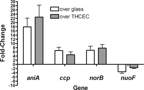
In order to study the expression profiles of aniA, ccp, norB, and nuoF in biofilms over cells, we grew biofilms over THCEC in flow cells for 48 h, confirmed the presence of a biofilm by confocal microscopy, and collected the planktonic effluent as described for the microarray studies. We collected duplicate planktonic and biofilm RNA samples from two independent experiments and used these RNAs as templates for qRT-PCR. We compared aniA, ccp, norB, and nuoF qRT-PCR expression profiles from this experiment to the qRT-PCR profiles for biofilms on glass (Fig. 1).
FIG. 1.
Relative RNA quantities were determined by a standard curve, and all values were normalized to the amount of OMP3 RNA in each sample. Reactions were performed in triplicate, and each qRT-PCR assay was performed a minimum of two times. qRT-PCR values for biofilms grown over THCEC are displayed by gray bars, and qRT-PCR values for biofilms grown over glass are displayed by white bars. The error bars represent 1 standard deviation from the mean for the averaged qRT-PCR data from duplicate assays.
We found that aniA, ccp, and norB were upregulated in biofilms grown over THCEC, similar to their expression in biofilms grown on glass. The nuoF gene was also downregulated in biofilms over THCEC, similar to the case for biofilms on glass. The similar expression of these genes in biofilms grown over cells and glass warranted pursuit of the role of anaerobic respiration in N. gonorrhoeae biofilms.
Characterization of the role of anaerobic metabolism genes in biofilms over glass.
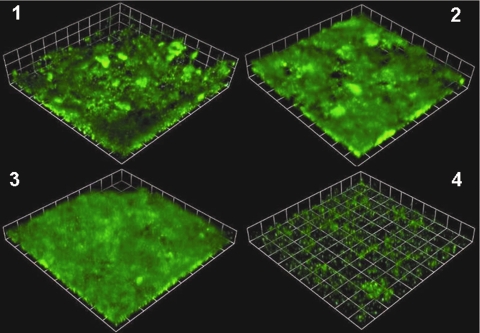
Wild-type N. gonorrhoeae clinical strain 1291 and isogenic aniA::kan, ccp::kan, and norB::kan mutants were assayed for the ability to form biofilms in flow cells over glass. These mutants were transformed with pGFP for examination of biofilms via confocal microscopy (Fig. 2). We found that there was no significant difference in the biomass of the aniA::kan or ccp::kan mutant when either was compared to the wild type. However, the aniA::kan and ccp::kan mutants did have significantly smaller average thicknesses (P ≤ 0.001). The norB::kan mutant had the most dramatic biofilm-deficient phenotype, with significantly less biomass and significantly smaller average thicknesses than the wild type (P ≤ 0.001) (Fig. 3). To confirm that the defect in biofilm formation was not due to a defect in growth under the oxygen tension conditions present in the medium in our biofilm system, we measured the dissolved oxygen content of medium entering and exiting the flow chambers at 0, 24, and 48 h. We found that the oxygen concentration (150 to 200 mm Hg) was much higher than concentrations that are typically considered to be anaerobic or microaerophilic (5 to 27 mm Hg) (15, 43). When we performed 48-hour growth curves under similar oxygen tension conditions, we found that there was no defect in the growth of aniA::kan, norB::kan, and ccp::kan insertion mutants compared to the wild type. This finding was the same for growth curves conducted under aerobic culture conditions (data not shown).
FIG. 2.
Biofilm masses over 2 days of growth for wild-type N. gonorrhoeae strain 1291 (1) and aniA::kan (2), ccp::kan (3), and norB::kan (4) insertion mutants. All four strains were visualized by GFP plasmid expression. The images are three-dimensional reconstructions of stacked z series taken at a magnification of ×200 and rendered by Volocity. These experiments were performed a minimum of three times, and representative results are shown.
FIG. 3.
COMSTAT and statistical analyses of biomass (A) and average thickness (B) for wild-type (checkered bars) and aniA::kan (black bars), ccp::kan (gray bars), and norB::kan (white bars) insertion mutant biofilms grown over glass for 2 days. Representative images of these biofilms are depicted in Fig. 2. All strains were run in duplicate in a minimum of three experiments, and at least four images of each biofilm chamber were used for COMSTAT analysis. The error bars represent 1 standard deviation from the mean. Statistical differences between mutants and the wild type, determined by Student's t test, are denoted by asterisks above the error bars. ***, P value of 0.001 or less.
Characterization of the role of anaerobic metabolism genes in biofilms over THCEC.
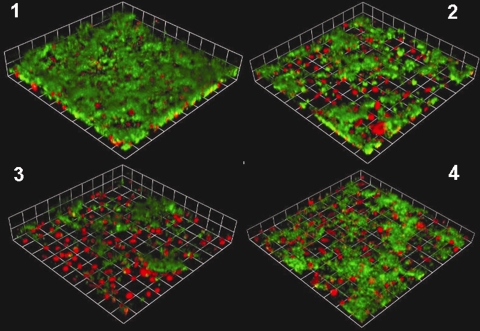
Wild-type N. gonorrhoeae clinical strain 1291 and aniA::kan, ccp::kan, and norB::kan insertional mutants were also assayed for the ability to form biofilms over THCEC, as all three anaerobic mutants were deficient in some aspect of biofilm formation over glass. We used confocal microscopy to evaluate whether the phenotypes of biofilms grown on glass were similar to or more exaggerated than those of biofilms grown over cells (Fig. 4). As the qRT-PCR results suggested, based on at least equivalent expression of these genes in biofilms grown over THCEC, these biofilms were more severely attenuated over THCEC than on glass. All three mutants had significantly less biomass and significantly smaller average thicknesses than the wild type did (P ≤ 0.001) (Fig. 5).
FIG. 4.
Biofilm masses over 2 days of growth for wild-type N. gonorrhoeae strain 1291 (1) and aniA::kan (2), ccp::kan (3), and norB::kan (4) insertion mutants. All four strains were visualized by GFP plasmid expression, while THCEC were visualized by Cell Tracker Orange (Molecular Probes, Invitrogen Corp., Carlsbad, CA) staining. The images are three-dimensional reconstructions of stacked z series taken at a magnification of ×200 and rendered by Volocity. These experiments were performed a minimum of three times, and representative results are shown.
FIG. 5.
COMSTAT and statistical analyses of biomass (A) and average thickness (B) for wild-type (checkered bars) and aniA::kan (black bars), ccp::kan (gray bars), and norB::kan (white bars) insertion mutant biofilms grown over THCEC for 2 days. Representative images of these biofilms are depicted in Fig. 4. All strains were run in duplicate in a minimum of three experiments, and at least four images of each biofilm chamber were used for COMSTAT analysis. The error bars represent 1 standard deviation from the mean. Statistical differences between mutants and the wild type, determined by Student's t test, are denoted by asterisks above the error bars. *, P value of 0.05 or less; **, P value of 0.01 or less.
Effect of nitric oxide on N. gonorrhoeae biofilm formation.
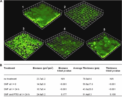
We observed that the norB mutant had a more pronounced biofilm-deficient phenotype than an aniA mutant when N. gonorrhoeae biofilms were grown over glass. One explanation for this phenotype is that NO accumulates in the norB mutant, and not in the aniA mutant, since AniA produces NO and this radical species is reduced by NorB to nitrous oxide. Findings for Pseudomonas aeruginosa (2) and Staphylococcus aureus (52) suggest that NO may be an important signaling molecule that causes biofilm detachment when present at sublethal concentrations within the biofilm. This prompted us to investigate the effect of NO on wild-type N. gonorrhoeae 1291 biofilms grown over glass. Wild-type biofilms treated with a sublethal concentration of NO (500 nM) either at the start of the biofilm or after 24 h of biofilm growth did have significantly less biomass and smaller average thicknesses than untreated biofilms. There was no apparent difference in biomass and average thickness for biofilms grown for 24 h without treatment and biofilms grown for 24 h and then treated with SNP for another 24 h. This suggests that the addition of SNP to a 24-hour biofilm halts development of the biofilm. Furthermore, biofilm formation was restored to wild-type levels when biofilms were simultaneously treated with NO and an NO quencher, suggesting that defects in biofilm formation were due to the accumulation of NO (Fig. 6). Treatment with the NO quencher (PTIO) alone did not alter biofilm formation (data not shown).
FIG. 6.
(A) Biofilm masses over 2 days of growth for wild-type N. gonorrhoeae strain 1291, either untreated (1), treated with SNP at the start of the biofilm (t = 0) (2), grown as a biofilm for only 24 h and left untreated (3), treated with SNP after 24 h of biofilm growth (t = 24 h) (4), or treated simultaneously with SNP and PTIO (5). All biofilms were visualized by GFP plasmid expression. The images are three-dimensional reconstructions of stacked z series taken at a magnification of ×200 and rendered by Volocity. These experiments were performed a minimum of four times, and representative results are shown. (B) Table showing results of COMSTAT analysis of biomass and average thickness. Statistical differences between treated samples and the untreated control (48-h biofilm) were determined via paired Student's t tests, and the corresponding P values are listed.
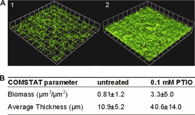
In order to directly assess the effect of NO accumulation on norB mutant biofilms, we also treated N. gonorrhoeae 1291 norB:kan biofilms with the NO quencher and compared these biofilms to untreated norB:kan biofilms. We found that treatment with the NO quencher enhanced norB mutant biofilm formation compared to untreated biofilm formation (Fig. 7). Moreover, we found that a higher concentration of NO quencher was required to restore the defect in biofilm formation by the norB mutant than that for wild-type biofilms artificially treated with an NO donor. This finding suggests that NO accumulates at concentrations in excess of 500 nM in norB mutant biofilms.
FIG. 7.
(A) Biofilm masses over 2 days of growth for N. gonorrhoeae strain 1291 norB::kan, either untreated (1) or treated with PTIO (2). Biofilms were visualized by GFP plasmid expression. The images are three-dimensional reconstructions of stacked z series taken at a magnification of ×200 and rendered by Volocity. These experiments were performed a minimum of three times, and representative results are shown. (B) Table showing results of COMSTAT analysis of biomass and average thicknesses. Statistical differences between treated samples and the untreated control were determined via paired Student's t tests, and the corresponding P values are listed.
DISCUSSION
Microarray analysis indicated that biofilms of N. gonorrhoeae possess a unique transcriptional profile distinguishing them from planktonic modes of growth. Specifically, genes required for anaerobic respiration (aniA, ccp, and norB) were more highly expressed during biofilm growth, while genes involved in respiration with NADH as an electron donor (nuo operon) were more highly expressed during planktonic growth. Mutants in which the aniA, ccp, or norB gene was interrupted by the insertion of a kanamycin cassette were attenuated for biofilm formation over glass and THCEC. Overall, these biofilms were less cohesive, were structurally instable, and often contained significantly less biomass or were thinner than wild-type biofilms. N. gonorrhoeae is frequently isolated from the genitourinary tract in the presence of obligate anaerobes (56), and AniA is a major antigen recognized in sera from patients with gonococcal disease (12). Thus, anaerobic respiration probably occurs naturally during infection, and since it also seems likely that N. gonorrhoeae forms biofilms during cervical infection (59), this suggests that biofilm formation is critical for successful infection of the female genitourinary tract.
N. gonorrhoeae was initially considered incapable of anaerobic growth (35), despite being isolated in the presence of obligate anaerobes (56). However, Short and coworkers demonstrated that N. gonorrhoeae is capable of survival under anaerobic conditions (55). Knapp and Clark later determined that anaerobic growth in N. gonorrhoeae is coupled to nitrite reduction and that supplementation with nitrite is required for growth under these conditions, explaining previous unsuccessful attempts to grow the organism anaerobically (39). Overton and coworkers went on to demonstrate that nitrous oxide is the end product of nitrite reduction, indicating that N. gonorrhoeae catalyzes partial denitrification, converting nitrite to nitrous oxide via nitric oxide (48).
AniA (formerly called Pan 1) was identified in a screen for anaerobically regulated genes, as one of three outer membrane proteins whose expression is induced under anaerobic growth conditions (11). Examination of sera from patients with complicated or uncomplicated gonococcal infection indicated that there is a strong antibody response to AniA, demonstrating that AniA is expressed in vivo and thus that anaerobic growth likely occurs during infection (12). AniA was later shown to be a lipoprotein (29) that functions as a nitrite reductase (46) and is subsequently required for anaerobic growth (33).
AniA is tightly regulated by oxygen availability and is virtually undetectable in aerobically cultured cells (33). Thus, elevated AniA expression in biofilms is a strong indicator that gonococcal biofilms grow anaerobically or microaerobically. Isolation of N. gonorrhoeae in the presence of obligate anaerobes suggests that oxygen is limited in the female genitourinary tract (56), and therefore biofilm formation may aid in the ability to respire under conditions of oxygen limitation in the host environment. With Neisseria meningitidis, Rock and Moir have shown that the addition of nitrite significantly enhances growth of this bacterium under oxygen-limited conditions (49), and thus it is possible that a combination of (micro)aerobic respiration and partial denitrification is optimal in gonococcal biofilms. Furthermore, if AniA is capable of modulating the immune response by binding complement regulatory proteins that can downregulate the immune response, as suggested by Cardinale and Clark (5), then biofilm growth may also convey an advantage to growth by aiding in immune evasion.
NO, the product of nitrite reduction by AniA in N. gonorrhoeae, has been shown to be an important signaling molecule and a modulator of cellular events (14, 41, 44, 47, 58, 60). Low basal levels of NO (nM scale) have been determined to be anti-inflammatory, while high levels of NO (μM scale) have been shown to be proinflammatory, stimulating the immune response (4, 14, 58). NO is also known to be toxic to some bacterial cells (14, 20, 44, 68) and is produced by nitric oxide synthases of polymorphonuclear neutrophils (6, 20, 44, 45). Thus, it has been postulated that NO production may be part of the innate immune response to bacterial infection, although this is currently a controversial topic (20).
NorB is a heme protein responsible for reducing AniA-generated NO to nitrous oxide (34). Unlike most NO reductases that are found in denitrifying organisms (68), such as Pseudomonas aeruginosa (1), NorB is composed of a single functional subunit (34). N. gonorrhoeae is capable of establishing a steady-state NO level during anaerobic growth and can reduce NO concentrations of >1 μM (proinflammatory) in the surrounding medium to approximately 100 nM (anti-inflammatory) in less than an hour (4). This suggests that N. gonorrhoeae is capable of rapidly reducing not only endogenously produced NO generated during anaerobic respiration but also environmentally produced NO from phagocytic cells. Cervical endothelial and epithelial cells have also been found to produce NO (40, 60), again implying that reduction of NO by the gonococcus may be critical during infection. During cervical infection, the gonococcus does not induce cytokine production in the host, suggesting that the organism either fails to induce the immune response or suppresses the response via an unknown mechanism (27, 50). Additionally, only low levels of antigonococcal antibody can be detected during uncomplicated infection in women (27), which cannot be attributed completely to the antigenic and phase variation of many surface-exposed gonococcal structures (30). The ability of the gonococcus to rapidly reduce host-produced NO from proinflammatory to anti-inflammatory levels may then help to account for the lack of a strong immune response in women with uncomplicated gonorrhea. Suppression of the immune response would likely result in the inability to clear the organism as well as in downregulation of the inflammatory response, resulting in the absence of noticeable symptoms. Thus, biofilm formation may be a special adaptation to growth in the cervical environment, enabling the gonococcus to persist in a chronic infection state.
N. gonorrhoeae is not the only organism that appears to catalyze anaerobic respiration during biofilm growth. P. aeruginosa, a well-studied biofilm-forming organism, has been shown to undergo anaerobic respiration during growth as a biofilm, and this is thought to be the primary mode of respiration during cystic fibrosis infection (21, 26, 62, 66). P. aeruginosa has been shown to form better biofilms under anaerobic than under aerobic growth conditions, and mutants with insertions in many of the anaerobic metabolism genes do not form biofilms as well as the wild type does (66). A recent study of Moraxella catarrhalis also determined that genes involved in anaerobic metabolism are highly upregulated during biofilm formation versus planktonic growth. Interestingly, M. catarrhalis is believed to be incapable of anaerobic growth. However, it is suggested that the ability to reduce NO may aid in the survival of M. catarrhalis in the nasopharynx (64). The observation that other organisms catalyze anaerobic respiration during biofilm growth suggests that biofilm formation could contribute to the ability to respire under oxygen-limited conditions. Our observation that a norB mutant had a more pronounced biofilm-deficient phenotype than an aniA mutant when biofilms were grown over glass bears similarity to findings for P. aeruginosa. In P. aeruginosa, a norBC (NO reductase) insertion mutant forms virtually no biofilm under anaerobic growth conditions, while a nir (nitrite reductase) mutant forms biofilms similar to those of the wild type (66). One explanation for this observation could be that NO accumulates under anaerobic growth conditions in the absence of an NO reductase, resulting in slow growth or cell death. An alternative explanation is that NO is an important signaling molecule in N. gonorrhoeae biofilms that elicits biofilm dispersal, which has been shown to be true for P. aeruginosa (2) and Staphylococcus aureus (52) biofilms. Inherent antimicrobial resistance in biofilms results in the inability to clear biofilm infection even with high doses of antibiotics (9, 16, 51), which makes biofilm dispersal an appealing target for the treatment of biofilm infection. NO treatment has been shown to augment the effects of antimicrobial treatment of P. aeruginosa biofilms (2). In agreement with these results, we found that treatment of N. gonorrhoeae biofilms with an NO donor either at the start of the biofilm or after 24 h of biofilm growth either hindered biofilm formation or elicited biofilm dispersal. Furthermore, addition of an NO quencher to norB mutant biofilms enhanced biofilm formation, suggesting that NO accumulation accounts for the severe biofilm-deficient phenotype observed with this mutant. We considered the possibility that production of NO in the norB::kan mutant could result in a growth defect by inhibition of cytochrome oxidase, subsequently impairing aerobic respiration. However, it has been shown that cytochrome cbb3 of Pseudomonas stutzeri, an isoenzyme in the family of cytochrome c oxidases, actually possesses nitric oxide reductase capability (23). The nitric oxide reductase activity of cytochrome cbb3, although considerably lower than the nitric oxide reductase activity in P. stutzeri, is the highest within the heme-copper oxidase superfamily (23). Furthermore, the norB::kan mutant showed no growth defect when grown under oxygen tension conditions that mimic those present in the medium of our biofilm apparatus, and cytochrome cbb3 is the only oxidase present in N. gonorrhoeae.
Cytochrome c peroxide, encoded by ccp, is a lipoprotein and is the sixth c-type cytochrome induced during anaerobic growth (61). Transcription of ccp is controlled by the fumarate and nitrate reductase regulator protein, which controls the expression of other important anaerobic metabolism genes in N. gonorrhoeae, such as aniA (42). Ccp plays an important role in oxidative stress defense by reducing hydrogen peroxide (H2O2) to water (H2O) (53). H2O2 is produced by polymorphonuclear neutrophils (6) and Lactobacillus species present in the female genitourinary tract (19). Specifically, H2O2-producing Lactobacillus strains are capable of inhibiting the growth of N. gonorrhoeae by acidification of the medium, H2O2 toxicity, and production of protein inhibitors (57, 67). Thus, upregulation of ccp during biofilm formation by N. gonorrhoeae may represent a critical adaptation for survival in the female genitourinary tract.
It is established from studies of the closely related mitochondrial complex 1 that NADH dehydrogenases of this type are highly susceptible to inactivation by nitric oxide, peroxynitrite, and S-nitrosothiols (3). The lower level of expression of nuo in biofilms suggests that the gonococcus is also able to alter expression of primary dehydrogenases in the respiratory chain.
Biofilm formation by the gonococcus may aid in oxidative stress tolerance during host infection by positively regulating factors, such as ccp and norB, which are required for the reduction of reactive oxygen and nitrogen species, respectively. Overall, anaerobic growth may be an important mechanism by which the gonococcus copes with the inherent oxidative stresses that are present during infection of human cervical tissues.
Supplementary Material
Acknowledgments
The M.A.A. lab is supported by NIH program grant R-01AI045728. The A.G.M. and M.P.J. labs are supported by NHMRC program grant 284214.
Editor: J. N. Weiser
Footnotes
Published ahead of print on 15 June 2009.
Supplemental material for this article may be found at http://iai.asm.org/.
REFERENCES
- 1.Arai, H., Y. Igarashi, and T. Kodama. 1995. The structural genes for nitric oxide reductase from Pseudomonas aeruginosa. Biochim. Biophys. Acta 1261279-284. [DOI] [PubMed] [Google Scholar]
- 2.Barraud, N., D. J. Hassett, S. H. Hwang, S. A. Rice, S. Kjelleberg, and J. S. Webb. 2006. Involvement of nitric oxide in biofilm dispersal of Pseudomonas aeruginosa. J. Bacteriol. 1887344-7353. [DOI] [PMC free article] [PubMed] [Google Scholar]
- 3.Brown, G. C., and V. Borutaite. 2004. Inhibition of mitochondrial respiratory complex I by nitric oxide, peroxynitrite and S-nitrosothiols. Biochim. Biophys. Acta 165844-49. [DOI] [PubMed] [Google Scholar]
- 4.Cardinale, J. A., and V. L. Clark. 2005. Determinants of nitric oxide steady-state levels during anaerobic respiration by Neisseria gonorrhoeae. Mol. Microbiol. 58177-188. [DOI] [PubMed] [Google Scholar]
- 5.Cardinale, J. A., and V. L. Clark. 2000. Expression of AniA, the major anaerobically induced outer membrane protein of Neisseria gonorrhoeae, provides protection against killing by normal human sera. Infect. Immun. 684368-4369. [DOI] [PMC free article] [PubMed] [Google Scholar]
- 6.Carreras, M. C., G. A. Pargament, S. D. Catz, J. J. Poderoso, and A. Boveris. 1994. Kinetics of nitric oxide and hydrogen peroxide production and formation of peroxynitrite during the respiratory burst of human neutrophils. FEBS Lett. 34165-68. [DOI] [PubMed] [Google Scholar]
- 7.Centers for Disease Control and Prevention. 2007. Update to CDC's sexually transmitted diseases treatment guidelines, 2006: fluoroquinolones no longer recommended for treatment of gonococcal infections. MMWR Morb. Mortal. Wkly. Rep. 56332-336. [PubMed] [Google Scholar]
- 8.Centers for Disease Control and Prevention. 2007. Sexually transmitted disease surveillance 2006. The Department of Health and Human Services, Atlanta, GA.
- 9.Ceri, H., M. E. Olson, C. Stremick, R. R. Read, D. Morck, and A. Buret. 1999. The Calgary biofilm device: new technology for rapid determination of antibiotic susceptibilities of bacterial biofilms. J. Clin. Microbiol. 371771-1776. [DOI] [PMC free article] [PubMed] [Google Scholar]
- 10.Chen, A., I. C. Boulton, J. Pongoski, A. Cochrane, and S. D. Gray-Owen. 2003. Induction of HIV-1 long terminal repeat-mediated transcription by Neisseria gonorrhoeae. AIDS 17625-628. [DOI] [PubMed] [Google Scholar]
- 11.Clark, V. L., L. A. Campbell, D. A. Palermo, T. M. Evans, and K. W. Klimpel. 1987. Induction and repression of outer membrane proteins by anaerobic growth of Neisseria gonorrhoeae. Infect. Immun. 551359-1364. [DOI] [PMC free article] [PubMed] [Google Scholar]
- 12.Clark, V. L., J. S. Knapp, S. Thompson, and K. W. Klimpel. 1988. Presence of antibodies to the major anaerobically induced gonococcal outer membrane protein in sera from patients with gonococcal infections. Microb. Pathog. 5381-390. [DOI] [PubMed] [Google Scholar]
- 13.Costerton, J. W., P. S. Stewart, and E. P. Greenberg. 1999. Bacterial biofilms: a common cause of persistent infections. Science 2841318-1322. [DOI] [PubMed] [Google Scholar]
- 14.Davis, K. L., E. Martin, I. V. Turko, and F. Murad. 2001. Novel effects of nitric oxide. Annu. Rev. Pharmacol. Toxicol. 41203-236. [DOI] [PubMed] [Google Scholar]
- 15.Dunn, G. M., R. A. Herbert, and C. M. Brown. 1979. Influence of oxygen tension on nitrate reduction by a Klebsiella sp. growing in chemostat culture. J. Gen. Microbiol. 112379-383. [DOI] [PubMed] [Google Scholar]
- 16.Dunne, W. M., Jr. 2002. Bacterial adhesion: seen any good biofilms lately? Clin. Microbiol. Rev. 15155-166. [DOI] [PMC free article] [PubMed] [Google Scholar]
- 17.Edwards, J. L., and M. A. Apicella. 2005. I-domain-containing integrins serve as pilus receptors for Neisseria gonorrhoeae adherence to human epithelial cells. Cell. Microbiol. 71197-1211. [DOI] [PubMed] [Google Scholar]
- 18.Edwards, J. L., E. J. Brown, K. A. Ault, and M. A. Apicella. 2001. The role of complement receptor 3 (CR3) in Neisseria gonorrhoeae infection of human cervical epithelia. Cell. Microbiol. 3611-622. [DOI] [PubMed] [Google Scholar]
- 19.Eschenbach, D. A., P. R. Davick, B. L. Williams, S. J. Klebanoff, K. Young-Smith, C. M. Critchlow, and K. K. Holmes. 1989. Prevalence of hydrogen peroxide-producing Lactobacillus species in normal women and women with bacterial vaginosis. J. Clin. Microbiol. 27251-256. [DOI] [PMC free article] [PubMed] [Google Scholar]
- 20.Fang, F. C. 1997. Perspectives series: host/pathogen interactions. Mechanisms of nitric oxide-related antimicrobial activity. J. Clin. Investig. 992818-2825. [DOI] [PMC free article] [PubMed] [Google Scholar]
- 21.Filiatrault, M. J., K. F. Picardo, H. Ngai, L. Passador, and B. H. Iglewski. 2006. Identification of Pseudomonas aeruginosa genes involved in virulence and anaerobic growth. Infect. Immun. 744237-4245. [DOI] [PMC free article] [PubMed] [Google Scholar]
- 22.Fleming, D. T., and J. N. Wasserheit. 1999. From epidemiological synergy to public health policy and practice: the contribution of other sexually transmitted diseases to sexual transmission of HIV infection. Sex. Transm. Infect. 753-17. [DOI] [PMC free article] [PubMed] [Google Scholar]
- 23.Forte, E., A. Urbani, M. Saraste, P. Sarti, M. Brunori, and A. Giuffre. 2001. The cytochrome cbb3 from Pseudomonas stutzeri displays nitric oxide reductase activity. Eur. J. Biochem. 2686486-6491. [DOI] [PubMed] [Google Scholar]
- 24.Gerbase, A. C., J. T. Rowley, D. H. Heymann, S. F. Berkley, and P. Piot. 1998. Global prevalence and incidence estimates of selected curable STDs. Sex. Transm. Infect. 74(Suppl. 1)S12-S16. [PubMed] [Google Scholar]
- 25.Greiner, L. L., J. L. Edwards, J. Shao, C. Rabinak, D. Entz, and M. A. Apicella. 2005. Biofilm formation by Neisseria gonorrhoeae. Infect. Immun. 731964-1970. [DOI] [PMC free article] [PubMed] [Google Scholar]
- 26.Hassett, D. J., J. Cuppoletti, B. Trapnell, S. V. Lymar, J. J. Rowe, S. S. Yoon, G. M. Hilliard, K. Parvatiyar, M. C. Kamani, D. J. Wozniak, S. H. Hwang, T. R. McDermott, and U. A. Ochsner. 2002. Anaerobic metabolism and quorum sensing by Pseudomonas aeruginosa biofilms in chronically infected cystic fibrosis airways: rethinking antibiotic treatment strategies and drug targets. Adv. Drug Deliv. Rev. 541425-1443. [DOI] [PubMed] [Google Scholar]
- 27.Hedges, S. R., D. A. Sibley, M. S. Mayo, E. W. Hook III, and M. W. Russell. 1998. Cytokine and antibody responses in women infected with Neisseria gonorrhoeae: effects of concomitant infections. J. Infect. Dis. 178742-751. [DOI] [PubMed] [Google Scholar]
- 28.Heydorn, A., A. T. Nielsen, M. Hentzer, C. Sternberg, M. Givskov, B. K. Ersboll, and S. Molin. 2000. Quantification of biofilm structures by the novel computer program COMSTAT. Microbiology 1462395-2407. [DOI] [PubMed] [Google Scholar]
- 29.Hoehn, G. T., and V. L. Clark. 1992. The major anaerobically induced outer membrane protein of Neisseria gonorrhoeae, Pan 1, is a lipoprotein. Infect. Immun. 604704-4708. [DOI] [PMC free article] [PubMed] [Google Scholar]
- 30.Hook, E. W., III, and H. H. Handsfield. 1999. Biology of Neisseria gonorrhoeae, p. 433-449. In K. K. Holmes, P. A. Mardh, P. F. Sparling, S. M. Lemon, W. E. Stamm, P. Piot, and J. N. Wasserjeot (ed.), Sexually transmitted diseases, 3rd ed. McGraw Hill, New York, NY.
- 31.Hook, E. W., III, and H. H. Handsfield. 1999. Gender perspectives and STDs, p. 117-127. In K. K. Holmes, P. A. Mardh, P. F. Sparling, S. M. Lemon, W. E. Stamm, P. Piot, and J. N. Wasserjeot (ed.), Sexually transmitted diseases, 3rd ed. McGraw Hill, New York, NY.
- 32.Hook, E. W., III, and H. H. Handsfield. 1999. Gonococcal infections in the adult, p. 451-466. In K. K. Holmes, P. A. Mardh, P. F. Sparling, S. M. Lemon, W. E. Stamm, P. Piot, and J. N. Wasserjeot (ed.), Sexually transmitted diseases, 3rd ed. McGraw Hill, New York, NY.
- 33.Householder, T. C., W. A. Belli, S. Lissenden, J. A. Cole, and V. L. Clark. 1999. cis- and trans-acting elements involved in regulation of aniA, the gene encoding the major anaerobically induced outer membrane protein in Neisseria gonorrhoeae. J. Bacteriol. 181541-551. [DOI] [PMC free article] [PubMed] [Google Scholar]
- 34.Householder, T. C., E. M. Fozo, J. A. Cardinale, and V. L. Clark. 2000. Gonococcal nitric oxide reductase is encoded by a single gene, norB, which is required for anaerobic growth and is induced by nitric oxide. Infect. Immun. 685241-5246. [DOI] [PMC free article] [PubMed] [Google Scholar]
- 35.James-Holmquest, A. N., R. D. Wende, R. L. Mudd, and R. P. Williams. 1973. Comparison of atmospheric conditions for culture of clinical specimens of Neisseria gonorrhoeae. Appl. Microbiol. 26466-469. [DOI] [PMC free article] [PubMed] [Google Scholar]
- 36.Jennings, M. P., M. Bisercic, K. L. Dunn, M. Virji, A. Martin, K. E. Wilks, J. C. Richards, and E. R. Moxon. 1995. Cloning and molecular analysis of the Isi1 (rfaF) gene of Neisseria meningitidis which encodes a heptosyl-2-transferase involved in LPS biosynthesis: evaluation of surface exposed carbohydrates in LPS mediated toxicity for human endothelial cells. Microb. Pathog. 19391-407. [DOI] [PubMed] [Google Scholar]
- 37.Kellogg, D. S., Jr., W. L. Peacock, Jr., W. E. Deacon, L. Brown, and D. I. Pirkle. 1963. Neisseria gonorrhoeae. I. Virulence genetically linked to clonal variation. J. Bacteriol. 851274-1279. [DOI] [PMC free article] [PubMed] [Google Scholar]
- 38.Klingelhutz, A. J., S. A. Barber, P. P. Smith, K. Dyer, and J. K. McDougall. 1994. Restoration of telomeres in human papillomavirus-immortalized human anogenital epithelial cells. Mol. Cell. Biol. 14961-969. [DOI] [PMC free article] [PubMed] [Google Scholar]
- 39.Knapp, J. S., and V. L. Clark. 1984. Anaerobic growth of Neisseria gonorrhoeae coupled to nitrite reduction. Infect. Immun. 46176-181. [DOI] [PMC free article] [PubMed] [Google Scholar]
- 40.Ledingham, M. A., A. J. Thomson, A. Young, L. M. Macara, I. A. Greer, and J. E. Norman. 2000. Changes in the expression of nitric oxide synthase in the human uterine cervix during pregnancy and parturition. Mol. Hum. Reprod. 61041-1048. [DOI] [PubMed] [Google Scholar]
- 41.Liaudet, L., F. G. Soriano, and C. Szabo. 2000. Biology of nitric oxide signaling. Crit. Care Med. 28N37-N52. [DOI] [PubMed] [Google Scholar]
- 42.Lissenden, S., S. Mohan, T. Overton, T. Regan, H. Crooke, J. A. Cardinale, T. C. Householder, P. Adams, C. D. O'Conner, V. L. Clark, H. Smith, and J. A. Cole. 2000. Identification of transcription activators that regulate gonococcal adaptation from aerobic to anaerobic or oxygen-limited growth. Mol. Microbiol. 37839-855. [DOI] [PubMed] [Google Scholar]
- 43.Loesche, W. J., F. Gusberti, G. Mettraux, T. Higgins, and S. Syed. 1983. Relationship between oxygen tension and subgingival bacterial flora in untreated human periodontal pockets. Infect. Immun. 42659-667. [DOI] [PMC free article] [PubMed] [Google Scholar]
- 44.MacMicking, J., Q. W. Xie, and C. Nathan. 1997. Nitric oxide and macrophage function. Annu. Rev. Immunol. 15323-350. [DOI] [PubMed] [Google Scholar]
- 45.McCall, T. B., N. K. Boughton-Smith, R. M. Palmer, B. J. Whittle, and S. Moncada. 1989. Synthesis of nitric oxide from l-arginine by neutrophils. Release and interaction with superoxide anion. Biochem. J. 261293-296. [DOI] [PMC free article] [PubMed] [Google Scholar]
- 46.Mellies, J., J. Jose, and T. F. Meyer. 1997. The Neisseria gonorrhoeae gene aniA encodes an inducible nitrite reductase. Mol. Gen. Genet. 256525-532. [DOI] [PubMed] [Google Scholar]
- 47.Ortega Mateo, A., and A. A. de Artiñano. 2000. Nitric oxide reactivity and mechanisms involved in its biological effects. Pharmacol. Res. 42421-427. [DOI] [PubMed] [Google Scholar]
- 48.Overton, T. W., R. Whitehead, Y. Li, L. A. Snyder, N. J. Saunders, H. Smith, and J. A. Cole. 2006. Coordinated regulation of the Neisseria gonorrhoeae-truncated denitrification pathway by the nitric oxide-sensitive repressor, NsrR, and nitrite-insensitive NarQ-NarP. J. Biol. Chem. 28133115-33126. [DOI] [PubMed] [Google Scholar]
- 49.Rock, J. D., and J. W. Moir. 2005. Microaerobic denitrification in Neisseria meningitidis. Biochem. Soc. Trans. 33134-136. [DOI] [PubMed] [Google Scholar]
- 50.Russell, M. W., S. R. Hedges, H. Y. Wu, E. W. Hook III, and J. Mestecky. 1999. Mucosal immunity in the genital tract: prospects for vaccines against sexually transmitted diseases—a review. Am. J. Reprod. Immunol. 4258-63. [DOI] [PubMed] [Google Scholar]
- 51.Schierholz, J. M., J. Beuth, D. Konig, A. Nurnberger, and G. Pulverer. 1999. Antimicrobial substances and effects on sessile bacteria. Zentralbl. Bakteriol. 289165-177. [DOI] [PubMed] [Google Scholar]
- 52.Schlag, S., C. Nerz, T. A. Birkenstock, F. Altenberend, and F. Gotz. 2007. Inhibition of staphylococcal biofilm formation by nitrite. J. Bacteriol. 1897911-7919. [DOI] [PMC free article] [PubMed] [Google Scholar]
- 53.Seib, K. L., H. J. Tseng, A. G. McEwan, M. A. Apicella, and M. P. Jennings. 2004. Defenses against oxidative stress in Neisseria gonorrhoeae and Neisseria meningitidis: distinctive systems for different lifestyles. J. Infect. Dis. 190136-147. [DOI] [PubMed] [Google Scholar]
- 54.Seib, K. L., H. J. Wu, S. P. Kidd, M. A. Apicella, M. P. Jennings, and A. G. McEwan. 2006. Defenses against oxidative stress in Neisseria gonorrhoeae: a system tailored for a challenging environment. Microbiol. Mol. Biol. Rev. 70344-361. [DOI] [PMC free article] [PubMed] [Google Scholar]
- 55.Short, H. B., V. L. Clark, D. S. Kellogg, Jr., and F. E. Young. 1982. Anaerobic survival of clinical isolates and laboratory strains of Neisseria gonorrhoeae: use in transfer and storage. J. Clin. Microbiol. 15915-919. [DOI] [PMC free article] [PubMed] [Google Scholar]
- 56.Smith, L. D. 1975. The pathogenic anaerobic bacteria. Charles C. Thomas, Springfield, IL.
- 57.St. Amant, D. C., I. E. Valentin-Bon, and A. E. Jerse. 2002. Inhibition of Neisseria gonorrhoeae by Lactobacillus species that are commonly isolated from the female genital tract. Infect. Immun. 707169-7171. [DOI] [PMC free article] [PubMed] [Google Scholar]
- 58.Stefano, G. B., Y. Goumon, T. V. Bilfinger, I. D. Welters, and P. Cadet. 2000. Basal nitric oxide limits immune, nervous and cardiovascular excitation: human endothelia express a mu opiate receptor. Prog. Neurobiol. 60513-530. [DOI] [PubMed] [Google Scholar]
- 59.Steichen, C. T., J. Q. Shao, M. R. Ketterer, and M. A. Apicella. 2008. Gonococcal cervicitis: a role for biofilm in pathogenesis. J. Infect. Dis. 198:1856-1861. [DOI] [PMC free article] [PubMed] [Google Scholar]
- 60.Tschugguel, W., C. Schneeberger, H. Lass, F. Stonek, M. B. Zaghlula, K. Czerwenka, C. Schatten, A. Kaider, P. Husslein, and J. C. Huber. 1999. Human cervical ripening is associated with an increase in cervical inducible nitric oxide synthase expression. Biol. Reprod. 601367-1372. [DOI] [PubMed] [Google Scholar]
- 61.Turner, S., E. Reid, H. Smith, and J. Cole. 2003. A novel cytochrome c peroxidase from Neisseria gonorrhoeae: a lipoprotein from a gram-negative bacterium. Biochem. J. 373865-873. [DOI] [PMC free article] [PubMed] [Google Scholar]
- 62.Van Alst, N. E., K. F. Picardo, B. H. Iglewski, and C. G. Haidaris. 2007. Nitrate sensing and metabolism modulate motility, biofilm formation, and virulence in Pseudomonas aeruginosa. Infect. Immun. 753780-3790. [DOI] [PMC free article] [PubMed] [Google Scholar]
- 63.Wang, S. A., A. B. Harvey, S. M. Conner, A. A. Zaidi, J. S. Knapp, W. L. Whittington, C. del Rio, F. N. Judson, and K. K. Holmes. 2007. Antimicrobial resistance for Neisseria gonorrhoeae in the United States, 1988 to 2003: the spread of fluoroquinolone resistance. Ann. Intern. Med. 14781-88. [DOI] [PubMed] [Google Scholar]
- 64.Wang, W., L. Reitzer, D. A. Rasko, M. M. Pearson, R. J. Blick, C. Laurence, and E. J. Hansen. 2007. Metabolic analysis of Moraxella catarrhalis and the effect of selected in vitro growth conditions on global gene expression. Infect. Immun. 754959-4971. [DOI] [PMC free article] [PubMed] [Google Scholar]
- 65.Yagi, T., S. Di Bernardo, E. Nakamuro-Ogiso, M. C. Kao, B. B. Seo, and A. Matsuno-Yagi. 2004. NADH dehydrogenases (NADH:quinone oxidoreductase), p. 15-40. In D. Zannoni (ed.), Respiration in archaea and bacteria, vol. 2. Kluwer Publishing, Dordrecht, The Netherlands. [Google Scholar]
- 66.Yoon, S. S., R. F. Hennigan, G. M. Hilliard, U. A. Ochsner, K. Parvatiyar, M. C. Kamani, H. L. Allen, T. R. DeKievit, P. R. Gardner, U. Schwab, J. J. Rowe, B. H. Iglewski, T. R. McDermott, R. P. Mason, D. J. Wozniak, R. E. Hancock, M. R. Parsek, T. L. Noah, R. C. Boucher, and D. J. Hassett. 2002. Pseudomonas aeruginosa anaerobic respiration in biofilms: relationships to cystic fibrosis pathogenesis. Dev. Cell 3593-603. [DOI] [PubMed] [Google Scholar]
- 67.Zheng, H. Y., T. M. Alcorn, and M. S. Cohen. 1994. Effects of H2O2-producing lactobacilli on Neisseria gonorrhoeae growth and catalase activity. J. Infect. Dis. 1701209-1215. [DOI] [PubMed] [Google Scholar]
- 68.Zumft, W. G. 1997. Cell biology and molecular basis of denitrification. Microbiol. Mol. Biol. Rev. 61533-616. [DOI] [PMC free article] [PubMed] [Google Scholar]
Associated Data
This section collects any data citations, data availability statements, or supplementary materials included in this article.


