Abstract
Many novel reassortant influenza viruses of the H9N2 genotype have emerged in aquatic birds in southern China since their initial isolation in this region in 1994. However, the genesis and evolution of H9N2 viruses in poultry in eastern China have not been investigated systematically. In the current study, H9N2 influenza viruses isolated from poultry in eastern China during the past 10 years were characterized genetically and antigenically. Phylogenetic analysis revealed that these H9N2 viruses have undergone extensive reassortment to generate multiple novel genotypes, including four genotypes (J, F, K, and L) that have never been recognized before. The major H9N2 influenza viruses represented by A/Chicken/Beijing/1/1994 (Ck/BJ/1/94)-like viruses circulating in poultry in eastern China before 1998 have been gradually replaced by A/Chicken/Shanghai/F/1998 (Ck/SH/F/98)-like viruses, which have a genotype different from that of viruses isolated in southern China. The similarity of the internal genes of these H9N2 viruses to those of the H5N1 influenza viruses isolated from 2001 onwards suggests that the Ck/SH/F/98-like virus may have been the donor of internal genes of human and poultry H5N1 influenza viruses circulating in Eurasia. Experimental studies showed that some of these H9N2 viruses could be efficiently transmitted by the respiratory tract in chicken flocks. Our study provides new insight into the genesis and evolution of H9N2 influenza viruses and supports the notion that some of these viruses may have been the donors of internal genes found in H5N1 viruses.
Wild birds, including wild waterfowls, gulls, and shorebirds, are the natural reservoirs for influenza A viruses, in which they are thought to be in evolutionary stasis (2, 33). However, when avian influenza viruses are transmitted to new hosts such as terrestrial poultry or mammals, they evolve rapidly and may cause occasional severe systemic infection with high morbidity (20, 29). Despite the fact that avian influenza virus infection occurs commonly in chickens, it is unable to persist for a long period of time due to control efforts and/or a failure of the virus to adapt to new hosts (29). In the past 20 years, greater numbers of outbreaks in poultry have occurred, suggesting that the avian influenza virus can infect and spread in aberrant hosts for an extended period of time (5, 14-16, 18, 32).
During the past 10 years, H9N2 influenza viruses have become panzootic in Eurasia and have been isolated from outbreaks in poultry worldwide (3, 5, 11, 14-16, 18, 24). A great deal of previous studies demonstrated that H9N2 influenza viruses have become established in terrestrial poultry in different Asian countries (5, 11, 13, 14, 18, 21, 24, 35). In 1994, H9N2 viruses were isolated from diseased chickens in Guangdong province, China, for the first time (4), and later in domestic poultry in other provinces in China (11, 16, 18, 35). Two distinct H9N2 virus lineages represented by A/Chicken/Beijing/1/94 (H9N2) and A/Quail/Hong Kong/G1/98 (H9N2), respectively, have been circulating in terrestrial poultry of southern China (9). Occasionally these viruses expand their host range to other mammals, including pigs and humans (6, 17, 22, 34). Increasing epidemiological and laboratory findings suggest that chickens may play an important role in expanding the host range for avian influenza virus. Our systematic surveillance of influenza viruses in chickens in China showed that H9N2 subtype influenza viruses continued to be prevalent in chickens in mainland China from 1994 to 2008 (18, 19, 36).
Eastern China contains one metropolitan city (Shanghai) and five provinces (Jiangsu, Zhejiang, Anhui, Shandong, and Jiangxi), where domestic poultry account for approximately 50% of the total poultry population in China. Since 1996, H9N2 influenza viruses have been isolated regularly from both chickens and other minor poultry species in our surveillance program in the eastern China region, but their genetic diversity and the interrelationships between H9N2 influenza viruses and different types of poultry have not been determined. Therefore, it is imperative to explore the evolution and properties of these viruses. The current report provides insight into the genesis and evolution of H9N2 influenza viruses in eastern China and presents new evidence for the potential crossover between H9N2 and H5N1 influenza viruses in this region.
MATERIALS AND METHODS
Virus isolation and identification.
Tracheal swabs and fecal samples were collected and put into 1.0 ml freezing medium (50% glycerol in phosphate saline buffer) containing antibiotics, as described previously (28, 36). Supernatants from processed samples were inoculated into the allantoic and the amniotic cavities of 9- to 10-day-old embryonated chicken eggs and incubated for 48 to 72 h at 35°C (28, 36). Allantoic fluids were harvested and tested for hemagglutinin (HA) activity and then frozen in aliquots at −70°C for viral RNA extraction and animal experiment. The HA of the viral isolates was subtyped by the hemagglutination inhibition (HI) test using the chickens' positive antiserum for avian influenza virus Ck/SH/F/98 (H9N2). The neuraminidase (NA) subtypes of the viruses were determined by conducting a BLAST search on viral nucleotide sequences available in GenBank from the National Center for Biotechnology Information, Bethesda, MD (http://www.ncbi.nlm.nih.gov/BLAST/). The viruses used in this study are listed in Table 1 (see also Table S1 in the supplemental material).
TABLE 1.
HI titers of antigenic analysis of H9N2 viruses with MAbs to Ck/SH/F/98 and Ck/SH/14/01 viruses
| Virus | Genotype | Virus HI titer determined with indicated MAb to strain:
|
|||||||||||
|---|---|---|---|---|---|---|---|---|---|---|---|---|---|
| Ck/SH/F/98
|
Ck/SH/14/01
|
||||||||||||
| 2A1 | 5D3 | 2H1 | 2B12 | 3C10 | 4A2 | 4E7 | 4G8 | 5B1 | 5C2 | 8E6 | 8E2 | ||
| Ck/AH/1/98 | A | <a | < | 5,120 | 5,120 | 640 | < | 10,240 | 40 | < | < | 2,560 | < |
| Ck/JS/1/98 | A | 640 | 80 | >10,240 | < | < | < | < | < | < | < | 2,560 | 160 |
| Ck/SD/1/99 | A | < | < | 5,120 | 5,120 | 320 | < | 10,240 | >10,240 | < | < | 2,560 | 40 |
| Ck/JS/1/99 | J | < | < | >10,240 | 5,120 | 10,240 | 5,120 | 10,240 | 10,240 | 10,240 | < | 5,120 | 160 |
| Ck/SD/1/00 | A | 320 | < | >10,240 | 10,240 | 2,560 | < | >10,240 | 160 | < | < | 5,120 | 40 |
| Ck/JS/2/01 | H | < | 1,280 | 640 | 2,560 | 5,120 | 2,560 | 5,120 | >10,240 | 10,240 | < | 5,120 | 160 |
| Ck/JS/1/02 | H | < | 5,120 | >10,240 | >10,240 | >10,240 | 10,240 | >10,240 | >10,240 | >10,240 | >10,240 | 10,240 | 1,280 |
| Dk/ZJ/3/02 | H | 10,240 | 10,240 | 10,240 | >10,240 | >10,240 | >10,240 | >10,240 | >10,240 | >10,240 | >10,240 | >10,240 | 640 |
| Ck/AH/2/02 | H | < | < | >10,240 | < | < | < | < | < | < | 2,560 | 1,280 | 40 |
| Dk/JS/1/02 | H | < | < | < | 10,240 | >10,240 | >10,240 | >10,240 | >10,240 | >10,240 | >10,240 | >10,240 | 640 |
| Ck/SD/1/03 | K | >10,240 | < | >10,240 | 5,120 | 10,240 | 5,120 | 10,240 | 10,240 | 640 | 10,240 | 10,240 | 5,120 |
| Ck/HN/2/04 | H | 1,280 | 1,280 | >10,240 | >10,240 | >10,240 | >10,240 | >10,240 | >10,240 | >10,240 | < | >10,240 | >10,240 |
| Dk/JS/3/05 | L | < | < | >10,240 | >10,240 | >10,240 | >10,240 | >10,240 | >10,240 | 10,240 | < | >10,240 | 640 |
<, titer of <10.
Phylogenetic and molecular analyses.
One or two isolates for each year from different provinces were selected for detailed characterization. Viral RNA extraction, reverse transcription-PCR, and gene sequencing were carried out as previously described (36). All eight gene segments from these viruses were partially sequenced and were phylogenetically analyzed with available virus sequence data from GenBank. The nucleotide sequences were analyzed with the Seqman module, and the nucleotide and deduced amino acid sequences were aligned and analyzed using the MegAlign module of the Lasergene sequence analysis package (DNAStar, Madison, WI) and MEGA version 4.0 (30). Evolutionary tree analyses were conducted using the neighbor-joining method (MEGA version 4.0) on the basis of the following gene sequences: nucleotides 88 to 1048 (960 bp) of HA1, 88 to 1369 or 1378 (1,282 or 1,291 bp, respectively) of NA, 589 to 1785 (1,197 bp) of PB2, 736 to 1888 (1,153 bp) of PB1, 784 to 2016 (1,233 bp) of PA, 76 to 1,483 (1,408 bp) of NP, 53 to 984 (932 bp) of M, and 66 to 838 (773 bp) of NS. Estimates of the phylogenies were calculated by performing 1,000 neighbor-joining bootstrap replicate assays.
Genotype definition.
Virus genotypes were defined by gene phylogeny, as previously described (35). Ck/BJ/94-like and Ck/SH/F/98-like viruses were designated genotypes A and H, respectively, as previously described (15).
Antigenic analysis.
Antigenic analysis was performed using a panel of monoclonal antibodies (MAbs). Two of them, 2H1 and 5D3, were kindly provided by X. A. Jiao and H. Y. Wang, without knowing detailed epitope specificity. Based on HI assay results against H9N2 isolates, these two MAbs have different binding epitopes. By using competitive binding enzyme-linked immunosorbent assay and Western blot analysis, a panel of MAbs was divided into four groups: group A (2B12, 3C10, 4E7, 4G8, 8E2, 8E6, and 2A1), group B (5B1), group C (4A11), and group D (5C7). Among these MAbs, 2A1 and 5C7 mainly recognize conformational epitopes while the other MAbs bind to the linear antigenic epitopes. Three MAbs (2A1, 5D3, and 2H1) are specific for A/Chicken/Shanghai/F/1998 (Ck/SH/F/1998), and nine MAbs (2B12, 3C10, 4A2, 4E7, 4G8, 5B1, 5C2, 8E6, and 8E2) are specific for A/Chicken/Shanghai/14/2001 (Ck/SH/14/01), as previously described (36).
Replication and transmission experiments in chickens.
Replication and transmission experiments were designed as previously described (36). Briefly, 10 or 11 5-week-old, specific-pathogen-free White Leghorn chickens were divided into (i) the direct inoculation group (three chickens), (ii) the physical contact group (three or four chickens), and (iii) the aerosol contact group (four chickens) for each of the viruses included. The physical contact group was raised in the same cage as chickens from the inoculated group at 30 min after the inoculation of the inoculated group. The aerosol contact group was placed in a cage directly adjacent to the infected group with a distance of 100 cm between cages. The direct inoculation group was inoculated intranasally or intratracheally with 107.0 50% egg infective doses (EID50)/ml of virus (equivalent to approximately 100 chicken infectious doses) (26, 36). One milliliter of virus inoculum was distributed to chickens as follows: 0.2 ml for intranasal inoculation and 0.8 ml for tracheal inoculations. Tracheal and cloacal swabs were collected 3, 5, and 7 days postinoculation, and viruses were titrated for infectivity in embryonated chicken eggs. Meanwhile, birds were observed daily for signs of disease during the 21-day study period.
Hemagglutination assays.
Erythrocytes from various animal species that differ in oligosaccharide composition were used. Agglutination of erythrocytes from cows, horses, and pigs requires NeuAcα2,3Gal or NeuGcα2,3Gal recognition, while agglutination of those from chickens, ducks, guinea pigs, and humans (type O) requires NeuAcα2,3Gal and NeuAcα2,6Gal recognition (7, 10, 12). To explore the receptor specificity of H9N2 viruses, hemagglutination assays were conducted with 50 μl 0.5% host red blood cells from each of the following: chicken, duck, goose, pigeon, quail, buffalo, donkey, goat, pig, dog, guinea pig, and human (type O) in phosphate buffer solution (pH 7.4) at 4°C for 45 min, as described previously (12).
Nucleotide sequence accession numbers.
The nucleotide sequences obtained in this study are available from GenBank under accession numbers EJ793281 to EJ793448.
RESULTS
Background.
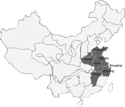
Since 1996, H9N2 viruses continue to be the most abundant influenza viruses isolated in eastern China. In late 1998, an H9N2 avian influenza outbreak occurred in chickens throughout eastern China. In order to explore the origin of H9N2 viruses that caused this outbreak, H9N2 influenza virus surveillance in poultry was first initiated in Shanghai in October 1998. Since 1999, this program has gradually expanded to include five other provinces in this region, including Jiangsu, Shandong, Zhejiang, Anhui, and Henan (Fig. 1). More than 100 H9N2 variants have been isolated from domestic poultry in eastern China during the past 10 years.
FIG. 1.
Geographical locations where H9N2 avian influenza viruses were isolated in eastern China.
Antigenic analysis.
To investigate the antigenic properties of the H9N2 viruses isolated from this region, we performed HI assays with a panel of MAbs raised against Ck/SH/F/98 and Ck/SH/14/01 viruses (Table 1; see also Table S1 in the supplemental material). It was very clear that a diverse set of reaction patterns existed among these H9N2 viruses. Patterns of reactions of MAbs 2B12, 3C10, 4E7, 4G8, 8E6, and 8E2 to these H9N2 viruses were very similar, suggesting that they could recognize the same antigenic epitopes. Both MAbs 4A2 and 5B1 reacted to these H9N2 isolates, but they could also bind another new antigenic epitope. The patterns of reactions of MAbs 5C2, 2A1, 5D3, and 2H1 with these H9N2 isolates were different, suggesting that they recognize four other antigenic epitopes. Based on the HI cross-reaction maps, the H9N2 viruses included in this study contained at least five antigenic epitopes with mutations. In addition, it is very interesting that three isolates, Ck/JS/1/98, Ck/AH/2/02, and Ck/HN/1/02, demonstrated a unique pattern by reacting only to five or six out of 12 MAbs included in this panel. Sequencing the HA genes of these three isolates revealed the presence of a potential N-linked carbohydrate site following a mutation from Ser to Asn at amino acid residue 145 of HA. Based on the presence or absence of this mutation, we determined that all of the viruses isolated from eastern China during the past 10 years could be separated into two groups, correlating well with the pattern of reactivity observed between the MAbs to Ck/SH/14/01. Ping et al. recently demonstrated that a single amino acid mutation from Ser to Asn at position 145 in the HA molecule could alter the recognition of the H9N2 influenza virus to a MAb by reverse genetics, suggesting that the new mutation of the glycosylation site at position 145 could play an important role in antigenic variation (25).
Phylogenetic analysis of surface genes.
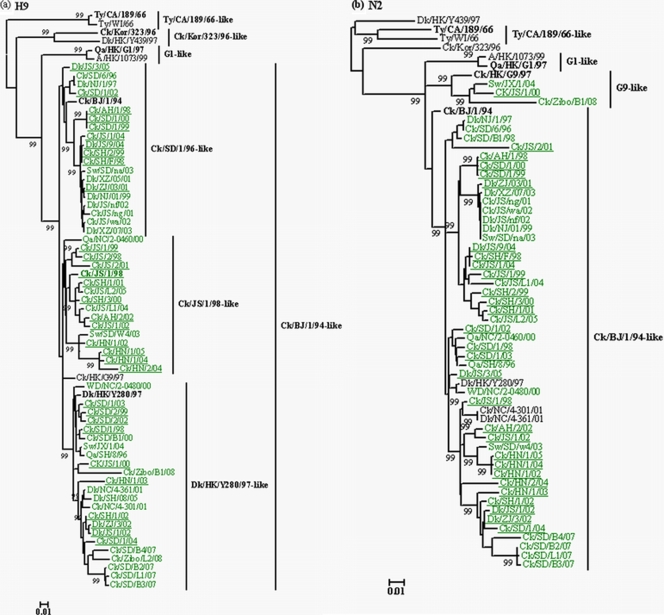
To better understand the evolution and ecology of H9N2 viruses isolated in eastern China, 27 representative viruses isolated in this region were genetically characterized. Phylogenetic analysis of the H9 HA genes showed that all of the H9N2 viruses isolated in poultry in eastern China during 1996 to 2008 belonged to the Ck/BJ/1/94-like lineage (Fig. 2a), which caused an outbreak of disease in chickens in southern China in 1994. Phylogenetic analysis further showed that these H9N2 viruses isolated from eastern China could be divided into three clusters: Ck/SD/1/96-like, Ck/JS/1/98-like, and Dk/HK/Y280/97-like. No Ck/Kor/323/96-like and Qa/HK/G1/97-like HA genes were found.
FIG. 2.
Phylogenetic trees for the HA (a) and NA (b) genes of representative influenza A viruses of the H9N2 subtype isolated in eastern China during the past 10 years. Trees were generated by the neighbor-joining method in the MEGA 4.0 program. Numbers above or below branches indicate neighbor-joining bootstrap values. Analysis was based on nucleotides 163 to 1048 of the HA gene and 88 to 1369 or 1378 of the NA gene. The HA and NA trees were rooted to A/Duck/Alberta/60/76 (H12N5) and A/Equine/Prague/1/56 (H7N7), respectively. The lengths of the horizontal lines are proportional to the minimum number of nucleotide differences required to join nodes. Vertical lines are for spacing and labeling. The viruses isolated in eastern China are highlighted in green. Viruses characterized in this study are underlined, and the names of the viruses and abbreviations can be found in Table 1 (see also Table S1 in the supplemental material). Genotypes characterized in this study are defined in Fig. 4. Abbreviations: Qa, quail; Gs, geese; G1, A/Quail/HongKong/G1/97; Ck, chicken; Dk, duck; Ty, Turkey; CA, California; WI, Wisconsin; HK, Hong Kong; Kor, Korea; BJ, Beijing; SD, Shandong; GD, Guangdong; SH, Shanghai; NC, Nanchang; YN, Yunnan; HB, Hubei; HN, Hunan; FJ, Fujian; ZJ, Zhengjiang.
Phylogenetic analysis of the NA gene showed an evolutionary pattern similar to that observed for the HA genes, wherein all viruses clustered within the Ck/BJ/1/94-like lineages, except for three viruses (Ck/JS/1/00, Ck/Zibo/B1/08, and Sw/JX/1/04), which clustered in the G9-like lineage (Fig. 2b). Taken together, our findings show that Ck/BJ/1/94-like viruses have been predominant in poultry in eastern China during the past 10 years.
Phylogenetic analysis of internal genes.
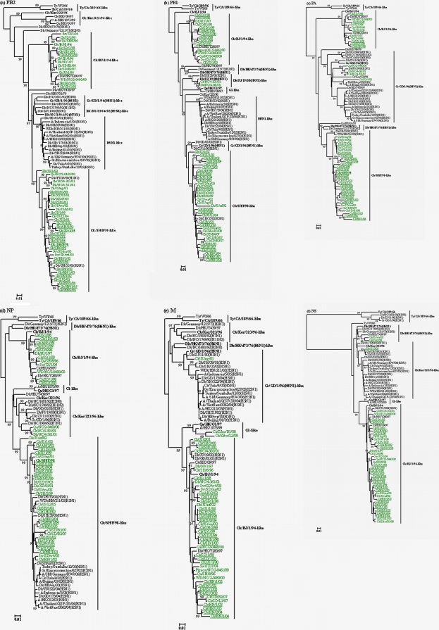
Phylogenetic analysis of the six internal genes revealed that H9N2 viruses from poultry in eastern China have undergone extensive reassortment during the past 10 years. The ribonucleoprotein (RNP) complex genes (PB2, PB1, PA, and NP genes) of all but one viral isolate prevailing in poultry in eastern China during the past 10 years clustered into two lineages, namely, the Ck/BJ/1/94-like lineage and the Ck/SH/F/98-like lineage (Fig. 3). One virus, Ck/Shandong/1/2003 (Ck/SD/1/03), is closely related to Gs/GD/1/96-like H5N1 viruses (Fig. 3c). It is notable that the RNP complex genes of the Ck/BJ/1/94-like lineage are completely distinct from those of the Ck/SH/F/98-like lineage, which are closely related to those of reassortant H5N1 variants isolated from domestic ducks since 2001, including the dominant H5N1 genotype Z prevailing in southeastern Asia. We noted that the RNP complex genes of a few H5N1 viruses derived from domestic ducks in this region also clustered into the Ck/SH/F/98-like (H9N2) lineage, indicating that reassortment events had occurred between these two virus subtypes. For example, the PB2 gene of H5N1 viruses Dk/FJ/19/00 and Dk/SH/35/02 (Fig. 3a); the PB1 gene of H5N1 virus Dk/SH/35/02 (Fig. 3b); the PA gene of H5N1 viruses WDk/HN/211/05, Dk/SH/38/01, and Dk/SH/35/02 (Fig. 3c); and the NP gene of eight H5N1 viruses including WDk/HN/211/05 and Dk/SH/35/02 (Fig. 3d) all clustered into the corresponding Ck/SH/F/98-like lineage. It is noteworthy that many human and poultry H5N1 viruses circulating in Eurasia since 2001 contain an NP gene segment with high homology to that of Ck/SH/F/98, including A/Beijing/01/03, A/Indonesia/5/05, A/HK/212/03, A/Thailand/2(SP-33)/04, and A/Vietnam/3062/04, suggesting that the Ck/SH/F/98-like virus may have been the donor of internal genes of the human and poultry H5N1 influenza viruses. Furthermore, the four RNP genes of some H9N2 viruses isolated from domestic ducks in eastern China since 2002, such as Dk/ZJ/3/02, Dk/JS/1/02 and Dk/JS/9/04, share very high homology with those of Ck/SH/F/98-like variants and clustered into the same lineage, indicating that interspecies transmission between different of types of poultry occurred in this region. Taken together, these findings suggest that the direction of interspecies transmission of Ck/SH/F/98-like viruses may be from chickens to domestic ducks and Ck/SH/F/98-like viruses may have become a possible provider of internal genes to H5N1 viruses identified since 2001.
FIG. 3.
Phylogenetic trees for the PB2 (a), PB1 (b), PA (c), NP (d), M (e), and NS (f) genes of representative influenza A viruses isolated in eastern China during the past 10 years. Trees were generated by the neighbor-joining method in the MEGA 4.0 program. Numbers above or below branches indicate neighbor-joining bootstrap values. Analysis was based on the following nucleotides: PB2, 589 to 1785; PB1, 736 to 1888; PA, 784 to 2016; NP, 76 to 1483; M, 78 to 947; and NS, 57 to 680. The phylogenetic trees of PB2, NP, M, and NS are rooted to A/Equine/Prague/1/56 (H7N7). The phylogenetic trees of PB1 and PA are rooted to A/Equine/London/1416/73 (H7N7). The lengths of the horizontal lines are proportional to the minimum number of nucleotide differences required to join nodes. Vertical lines are for spacing and labeling. The viruses isolated in eastern China are highlighted in green. Viruses characterized in this study are underlined, and the names of the viruses and abbreviations can be found in Table 1 (see also Table S1 in the supplemental material). Other virus names and abbreviations can be found in the legend to Fig. 2.
Similar to the HA and NA genes, the matrix (M) and nonstructural (NS) genes showed much less diversity than the other internal genes and belonged to the Ck/BJ/1/94-like lineage (Fig. 3e and f), except for two viruses, Ck/JS/1/99 and Dk/JS/1/05, whose NS genes clustered with those of viruses similar to Dk/HK/d73/76 (H6N1) and Ck/Kor/323/96 (Fig. 3f). However, phylogenetic analysis of the M and NS genes showed that some of the H5N1 isolates from ducks were closely related to these H9N2 variants, suggesting that a reassortment event had occurred in this region. Our findings further support the view that interspecies transmission between different types of poultry exists in southern China.
Molecular characterization.
The deduced amino acid sequences of these viruses were aligned and compared with those of other representative H9N2 viruses in this region. As shown in Table 2, except for seven viruses isolated since 2001, the HAs of all viruses characterized had an avian-like motif 226-Gln (H3 numbering), while those of the other seven viruses had a human-like motif 226-Leu at the receptor binding site (Table 2). In addition, all of these Ck/BJ/1/94-like viruses had a receptor-binding-site-related Ala-to-Val/Thr mutation at position 190 (H3 numbering). Interestingly, in two Ck/Bj/1/94-like viruses, a never-before-documented amino acid change from His to Ser at position 183 (H3 numbering) was observed. Most of the H9N2 viruses tested in this study had the same R-S-S-R amino acid motif at the connecting peptide, but a few of the viruses had an S→L and R→K mutation at positions −3 and −4, respectively, of the HA1 protein, giving an R-L-S-R and K-S-S-R motif at the connecting peptide (Table 2). Seven potential glycosylation sites, five in HA1 (29, 141, 200, 282, and 305) and two in HA2 (154 and 213), were highly conserved. However, a few of the viruses, for example, Ck/JS/1/98, Ck/AH/2/02, and Ck/HN/1/02, which exhibited unique antigenic patterns in the above antigenic analysis, have an additional potential glycosylation site at residue 145 due to the change of Ser to Asn. All of the Ck/BJ/1/94-like H9N2 viruses analyzed had the same “marking” deletion of three amino acids (positions 62 to 64) at the NA stalk region, as previously described (9, 11, 15). It is notable that Val27Ala and Ser31Asn mutations in the M2 protein, which is associated with amantadine resistance of influenza virus (1), had also been observed in a few of the H9N2 viruses isolated in this region (Table 2). Three viruses isolated from chickens since 2003 (Ck/HN/1/03, Ck/HN/1/04, and Ck/HN/2/04) contained a Ser31Ile mutation which had not been previously observed. All of the viruses had a G1-like NS1 motif, 92-Glu, which is reportedly associated with increased pathogenicity of H5N1 influenza virus in pigs (27).
TABLE 2.
Comparison of amino acid sequences of HA, NA, and M2 genes of representative viruses from eastern China
| Virus | Genotype | Residue at RBSa position:
|
NA deletion (aa) | Connecting peptideb | M2 residue at amantadine resistance mutation position
|
||||
|---|---|---|---|---|---|---|---|---|---|
| 183 | 190 | 226 | 228 | 27 | 31 | ||||
| Qa/HK/G1/97 | H | E | L | G | 38-39 | R-S-S-R | V | S | |
| Dk/HK/Y439/97 | H | E | Q | G | A-S-N-R | V | S | ||
| Ck/Kor/323/96 | H | E | Q | G | A-S-Y-R | V | S | ||
| Ck/HK/G9/97 | N | A | L | G | R-S-S-R | V | S | ||
| Ck/BJ/1/94 | A | N | V | Q | G | R-S-S-R | V | S | |
| Ck/SD/1/98 | A | N | T | Q | G | 62-64 | R-S-S-R | V | S |
| Ck/AH/1/98 | A | N | V | Q | G | 62-64 | R-S-S-R | V | S |
| Ck/JS/1/98 | A | N | V | Q | G | 62-64 | R-S-S-R | V | S |
| Ck/SH/F/98 | H | N | A | Q | G | 62-64 | R-S-S-R | V | N |
| Ck/SD/1/99 | A | N | V | Q | G | 62-64 | R-S-S-R | V | S |
| Ck/JS/1/99 | J | N | T | Q | G | 62-64 | K-S-S-R | V | S |
| Ck/SH/2/99 | H | N | A | Q | G | 62-64 | R-S-S-R | V | S |
| Ck/SD/1/00 | A | N | V | Q | G | 62-64 | R-S-S-R | V | N |
| Ck/SH/3/00 | H | N | T | Q | G | 62-64 | R-S-S-R | V | S |
| Ck/SH/1/01 | H | N | T | L | G | 62-64 | R-S-S-R | V | S |
| Ck/JS/2/01 | H | N | T | Q | G | 62-64 | K-S-S-R | V | S |
| Ck/AH/2/02 | H | N | V | Q | G | 62-64 | R-S-S-R | V | N |
| Ck/JS/1/02 | H | N | A | Q | G | 62-64 | R-S-S-R | V | S |
| Dk/ZJ/1/02 | H | N | A | L | G | 62-64 | R-S-S-R | V | S |
| DK/JS/1/02 | H | N | A | Q | G | 62-64 | R-S-S-R | V | S |
| Ck/HN/1/02 | H | N | V | Q | G | 62-64 | R-L-S-R | V | S |
| Ck/SH/1/02 | H | N | A | L | G | 62-64 | R-S-S-R | V | S |
| Ck/SD/1/03 | K | N | V | L | G | 62-64 | R-S-S-R | V | S |
| Ck/HN/1/03 | H | S | V | Q | G | 62-64 | R-S-S-R | V | I |
| Ck/JS/1/04 | H | N | A | Q | G | 62-64 | K-S-S-R | V | N |
| Ck/SD/1/04 | H | N | T | Q | G | 62-64 | R-S-S-R | A | S |
| Dk/JS/9/04 | H | N | A | Q | G | 62-64 | R-S-S-R | V | N |
| Ck/HN/1/04 | H | N | A | L | G | 62-64 | R-L-S-R | V | I |
| Ck/HN/2/04 | H | S | V | L | G | 62-64 | R-L-S-R | V | I |
| Dk/JS/3/05 | L | N | V | Q | G | 62-64 | R-S-S-R | V | S |
| Ck/HN/1/05 | H | N | A | L | G | 62-64 | R-L-S-R | A | S |
RBS, receptor binding site.
Connecting peptide from positions −4 to −1 of HA1. Boldface indicates the mutation amino acid motif at the cleavage site.
Genotyping.
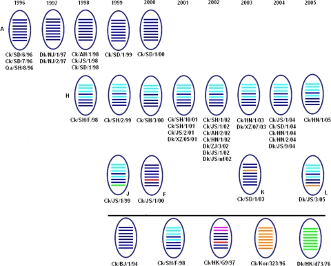
Here we assigned H9N2 virus genotypes, as previously described (15). A total of six H9N2 influenza virus genotypes (A, H, J, F, K, and L) were identified from different types of poultry under our surveillance, including four novel genotypes that have not been recognized in previous studies and were designated genotypes J, F, K, and L (Fig. 4). These novel genotypes all had undergone reassortment two or three times, with gene segments from Ck/BJ/1/94-like, G9-like, Ck/SH/F/98-like, Ck/Kor/323/96-like, and Dk/HK/d73/76(H6N1)-like viruses. H9N2 genotype A viruses were initially detected in quails and chickens in eastern China in 1996 and appeared in domestic ducks in 1997, suggesting that the interspecies transmission of this genotype occurred in eastern China. Although H9N2 genotype A viruses were a dominant genotype in poultry from 1996 onwards, the number of isolates detected from chickens decreased gradually and the isolates failed to be detected in poultry in 2000, which might correlate with the H9N2 homologous vaccine being widely used in chickens. Since 1998, an H9N2 genotype H virus emerged and became predominant in chickens from 2001 onwards, suggesting that this virus subtype established itself in chickens. However, it is notable that genotype H viruses have been detected in domestic ducks from 2002 onwards, indicating that the interspecies transmission from chickens to domestic ducks occurred in this region. Although the remaining and novel genotypes J, K, F, and L were only occasionally and transiently detected in chickens or domestic ducks, the number of genotypes being detected from chickens has increased since 1999, suggesting that H9N2 influenza viruses have become more and more diversified in this region. Two of the four novel genotypes, K and L, were novel reassortants containing gene segments similar to Ck/Kor/323/96-like viruses or Dk/HK/Y439/97-like viruses, showing that reassortment events occurred in this region.
FIG. 4.
Genotypes of H9N2 influenza viruses isolated from poultry in eastern China. The order of the eight gene segments from the top (horizontal bars) is PB2, PB1, PA, HA, NP, NA, M, and NS. Each color represents a virus lineage. Genotype definitions are described in Materials and Methods.
Replication and transmission of H9N2 influenza virus isolates in chickens.
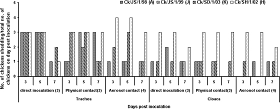
Because these H9N2 viruses had caused severe morbidity and mortality in chickens in the field during the past 10 years, we examined their replication and transmission in chickens. Most of these H9N2 viruses replicated in the inoculated chickens and were transmitted to contact birds, but none of the H9N2 viruses caused death during the 3-week observation period (Fig. 5; see also Table S2 in the supplemental material). Most of the H9N2 viruses were shed mainly from the respiratory tract with titers in the range of 4.0 to 7.0 log10 EID50/0.1 ml in tracheal swabs on day 5 postinfection (p.i.). The maximal virus shedding was observed between the 3rd and 5th day postinoculation. Viruses were detected in tracheal swabs of the contact birds from day 3 postcontact. Virus was mostly detected in the cloacal swabs of directly inoculated birds on day 3 p.i. Lower virus titers were detected in the cloacal swabs than in the tracheal swabs from both inoculated and contact birds. It is notable that biological differences in H9N2 viruses of different genotypes were obvious. For example, most genotype A viruses replicated mainly in inoculated chickens but failed to be transmitted efficiently to the aerosol contact group. In contrast, most genotype H and genotype K viruses from 2001 onwards not only replicated efficiently in infected chickens but also were transmitted efficiently among aerosol contact chickens (Fig. 5; see also Table S2 in the supplemental material).
FIG. 5.
Respiratory transmission of different genotypic H9N2 viruses. Directly inoculated chickens were inoculated with 107 EID50 (equivalent to approximately 100 chicken infectious doses) of different genotypic H9N2 viruses. Physical contact chickens and aerosol contact chickens were introduced at 30 min p.i. Tracheal and cloacal swabs were collected 3, 5, and 7 days postinoculation, and viruses were titrated for infectivity in embryonated chicken eggs.
Hemagglutination activity with erythrocytes from different animals.
Erythrocytes from different animal species differ in their oligosaccharide composition. For example, agglutination of erythrocytes from cows, horses, and pigs requires NeuAcα2,3Gal or NeuGcα2,3Gal recognition, while agglutination of those from chickens, ducks, guinea pigs, and humans (type O) requires NeuAcα2,3Gal and NeuAcα2,6Gal recognition (7, 10, 12). Therefore, receptor specificity of influenza A viruses correlates with the agglutination of erythrocytes from different animal species (7, 10, 12). In order to explore the receptor specificity of the H9N2 viruses, the agglutination patterns of these H9N2 variants were systematically analyzed with erythrocytes from different animal species (Table 3). All of these H9N2 variants agglutinate erythrocytes from chickens, ducks, geese, pigeons, quails, guinea pigs, dogs, and humans (type O), whereas most did not agglutinate donkey erythrocytes. Based on differences of patterns of hemagglutination to buffalo and goat erythrocytes, most of these H9N2 variants can be further differentiated into two groups. In addition, a number of H9N2 viruses also could agglutinate pig erythrocytes. These results clearly show that the difference lies in the receptor binding site of these H9N2 variants. Therefore, direct analysis of receptor specificity of these H9N2 variants, using defined glycoconjugates, is needed in order to understand the differences in agglutination patterns of these H9N2 variants. However, whether these H9N2 variants possess a human-like motif at the receptor site and have an ability to infect humans and other mammals requires further study.
TABLE 3.
Hemagglutination of H9N2 avian influenza virus with erythrocytes from different animal species
| Virus | Hemagglutination with erythrocytes froma:
|
|||||||||||
|---|---|---|---|---|---|---|---|---|---|---|---|---|
| Chicken | Duck | Goose | Pigeon | Quail | Buffalo | Donkey | Goat | Pig | Dog | Guinea pig | Human (type O) | |
| Ck/SD/1/98 | + | + | + | + | + | + | − | + | − | + | + | + |
| Ck/AH/1/98 | + | + | + | + | + | + | − | + | + | + | + | + |
| Ck/JS/1/98 | + | + | + | + | + | + | − | + | + | + | + | + |
| Ck/SH/F/98b | + | + | + | + | + | − | − | − | − | + | + | + |
| Ck/SD/1/99 | + | + | + | + | + | + | − | + | − | + | + | + |
| Ck/JS/1/99 | + | + | + | + | + | + | − | + | + | + | + | + |
| Ck/SH/1/99b | + | + | + | + | + | + | − | + | − | + | + | + |
| Ck/SD/1/00 | + | + | + | + | + | + | − | + | + | + | + | + |
| Ck/SH/1/00b | + | + | + | + | + | − | − | − | − | + | + | + |
| Ck/SH/1/01b | + | + | + | + | + | − | − | − | − | + | + | + |
| Ck/JS/2/01 | + | + | + | + | + | + | + | + | + | + | + | + |
| Ck/AH/2/02 | + | + | + | + | + | + | − | + | − | + | + | + |
| Ck/JS/1/02 | + | + | + | + | + | − | − | − | − | + | + | + |
| Dk/ZJ/3/02 | + | + | + | + | + | + | − | + | − | + | + | + |
| DK/JS/1/02 | + | + | + | + | + | − | − | − | − | + | + | + |
| Ck/HN/1/02 | + | + | + | + | + | + | − | + | − | + | + | + |
| Ck/SH/1/02b | + | + | + | + | + | − | − | − | − | + | + | + |
| Ck/SD/1/03 | + | + | + | + | + | + | − | + | − | + | + | + |
| Ck/HN/1/03 | + | + | + | + | + | + | − | + | + | + | + | + |
| Ck/JS/1/04 | + | + | + | + | + | + | − | + | − | + | + | + |
| Ck/SD/1/04 | + | + | + | + | + | − | − | − | − | + | + | + |
| Dk/JS/9/04 | + | + | + | + | + | − | − | − | − | + | + | + |
| Ck/HN/1/04 | + | + | + | + | + | − | − | − | − | + | + | + |
| Ck/HN/2/04 | + | + | + | + | + | + | − | + | − | + | + | + |
| Dk/JS/3/05 | + | + | + | + | + | + | − | + | − | + | + | + |
| Ck/HN/1/05 | + | + | + | + | + | − | − | − | − | + | + | + |
+, agglutination with virus-containing allantoic fluid; −, no agglutination with viruses containing >128 hemagglutination units/ml, as determined with red blood cells from different hosts in a microtiter assay.
Data from reference 36.
DISCUSSION
The first H9N2 avian influenza outbreak caused by Ck/BJ/1/94-like viruses was reported in eastern China in 1994. In late 1998, most chicken farms throughout eastern China suffered serious economic losses due to H9N2 avian influenza viruses. In the present study, our findings reveal that H9N2 influenza A viruses isolated during 1996 to 2008 in eastern China have evolved continuously and have undergone wide reassortment with aquatic avian influenza A viruses, generating six genotypes in eastern China during the past 10 years. Furthermore, it is noteworthy that all six virus genotypes identified in this study have different phenotypic characteristics. Most of the H9N2 viruses isolated before 2000 can replicate mainly in inoculated chickens but have failed to be transmitted efficiently to aerosol contact groups. In contrast, most of the viruses isolated from 2001 onwards not only replicated efficiently in the infected chickens but also were transmitted efficiently among aerosol contact chickens. Therefore, the H9N2 viruses circulating in eastern China appear to have undergone phenotypic changes.
Chickens are generally considered aberrant hosts of influenza viruses because the mutation rates of many chicken viruses are higher than those for viruses isolated from aquatic birds (29). However, previous studies have shown that H9N2 influenza viruses have circulated widely among chicken flocks for more than 10 years since they were first detected in mainland China in 1994 (4). Furthermore, it is worth noting that chickens not only are able to harbor genotype A (Ck/BJ/1/94-like) viruses but may have also carried genotype H (Ck/SH/F/98-like) viruses over the past 10 years. Zhang et al. (36) recently demonstrated that Ck/SH/F/98-like H9N2 influenza viruses have been circulating in chicken flocks from the same integrated chicken operation for at least 5 years. Ito et al. (12) also demonstrated that chickens have molecular characterizations classifying them as potential intermediate hosts for transmission of avian influenza viruses to humans. Furthermore, the recent transmission of avian H5N1 and H9N2 influenza viruses from chickens and/or quails to humans indicates that avian influenza viruses can directly infect humans without an intermediate host (17, 22). All available evidence suggests that H9N2 influenza viruses have adapted to chickens. Furthermore, chickens have played an important role in the evolution of influenza viruses by acting as mixing vessels or disseminators of avian/mammalian reassortant influenza A viruses.
Although many novel genotypes have been generated in eastern China during the past 10 years, only two genotypes (genotypes A and H) have been well adapted to chickens and have been circulating in chickens for some time. Genetic and antigenic studies demonstrated that genotype A was the predominant genotype circulating in poultry before 2000. However, from 2000 onward, the genotype A viruses were replaced by a novel genotype, genotype H (Ck/SH/F/98-like), which has become the predominant genotype in poultry in eastern China. Furthermore, the genotype H viruses appear to be better adapted to poultry and more easily reassorted with other subtype avian influenza A viruses. The other H9N2 virus genotypes were very short-lived reassortants, none of which had become established in poultry, suggesting that those viruses were not well adapted to chickens. This finding provides potentially useful information regarding key molecular determinants for such selection.
Because pigs contain both Neu5Acα2-3Gal and Neu5Acα2-6Gal linkages to support replication of both avian and human influenza viruses, pigs have been recognized as “mixing vessels” for avian and human viruses (12). During the past 5 years, H9N2 influenza viruses have been isolated repeatedly from pigs in eastern China (6, 34). However, phylogenetic analysis of the internal genes of those swine H9N2 viruses showed that almost all swine H9N2 viruses recently isolated from eastern China also possess Ck/SH/F/98-like lineage RNP genes (6, 34). Furthermore, the mutation Glu (Q)→Leu (L) at position 226 found in human H2 and H3 isolates occurred in those of genotype H H9N2 viruses. A key amino acid mutation from His to Ser at position 183 (H3 numbering) in the HA receptor binding site has never before been documented. These phenomena increased the chances of generating new influenza viruses with pandemic potential in this region. Therefore, surveillance of H9N2 viruses in this region for the next possible influenza pandemic should be given a high priority.
Donors of internal genes present in H5N1 variants circulating throughout the world have not been well established. However, based on the phylogenetic relationships of NP genes, the present study demonstrated that Ck/SH/F/-98-like H9N2 viruses may be a possible donor of H5N1/01-like internal genes to currently circulating viruses. Furthermore, based on the sequence of reassortment events, Ck/SH/F/98-like internal genes were first incorporated into H5N1/01-like viruses in 2001 or before. In addition, it was noted that Ck/BJ/1/94-like viruses and Ck/SH/F/98-like viruses first isolated in chickens have also been recognized in domestic ducks in this region. This raises the possibility of bidirectional transmission between chickens and domestic ducks. However, the possibility of gene flow from chickens to ducks could not be excluded in Ck/BJ/1/94-like and Ck/SH/F/98-like H9N2 viruses. Domestic ducks may serve as mixing vessels to facilitate reassortment events for H9N2 and H5N1 viruses and the emergence of novel genotypes in this region.
The receptor specificity of influenza viruses is one factor that allows avian influenza viruses to cross the species barrier. Some H9N2 viruses isolated since 2001 contained G1-like receptor HA signatures described by Gambarian et al. (8). Wan and Perez (31) also demonstrated that the Glu-to-Leu mutation at amino acid position 226 in HA allows H9N2 viruses to replicate more efficiently in human airway epithelial cells cultured in vitro. Substitution of Gln for Leu at residue 226 and the change of Ala to Thr at position 190 that occurred in the receptor binding site of these H9N2 variants have been previously reported to be involved in binding specificity to receptors in the host cell (23, 32). In this study, we found that H9N2 viruses isolated during the past 10 years differed from each other in their ability to agglutinate erythrocytes from goat and buffalo, suggesting differences in receptor specificities in H9N2 influenza viruses. Ito et al. (12) demonstrated that the α2,3 linkage and NeuGc, but not NeuAc, recognition appear essential for agglutination of bovine and equine erythrocytes. Therefore, additional studies are needed to understand the role of mutations in the receptor binding site in restricting the host range of these H9N2 variants.
The current study demonstrates the continued circulation of H9N2 influenza viruses from poultry in eastern China and shows that the genotypes of H9N2 viruses circulating in eastern China are different from those isolated in southern China. The Ck/SH/F/98-like lineage has replaced the Ck/BJ/1/94-like lineage and has become a predominant genotype circulating in poultry in eastern China. Furthermore, reassortment between H9N2 and H5N1 subtype viruses has contributed to the generation of novel genotypes of both subtypes. It is clear that Ck/SH/F/98-like viruses could provide internal genes for H5N1 viruses currently circulating throughout the world. Therefore, it is imperative that particular attention be paid to H9N2 viruses of avian origin to avert any future pandemic in humans in this region.
Supplementary Material
Acknowledgments
This study was supported in part by the National Key Technologies R & D Program of China (grant 2006BAD06A01), by the National High-Tech Research and Development Program of China (grant 2006AA10A205) (X. Liu), and by Jiangsu Nature & Science Fund grants BK2007581 (to National Drug Screening Lab, Jiangsu New Drug Screening Center, China Pharmaceutical University) and BK2006728 (to China-US Vaccine Research Center, The First Affiliated Hospital of Nanjing Medical University).
We are grateful to Xinan Jiao and Haiyan Wang for kindly providing Ck/SH/F/98-specific MAbs and to Guijie Hao, Min Gu, Xusheng Qiu, and Feng Xue for their help with animal experiments.
Footnotes
Published ahead of print on 24 June 2009.
Supplemental material for this article may be found at http://jvi.asm.org/.
REFERENCES
- 1.Aamir, U. B., U. Wernery, N. Ilyushina, and R. G. Webster. 2007. Characterization of avian H9N2 influenza viruses from United Arab Emirates 2000 to 2003. Virology 36145-55. [DOI] [PMC free article] [PubMed] [Google Scholar]
- 2.Alexander, D. J. 2000. A review of avian influenza in different bird species. Vet. Microbiol. 743-13. [DOI] [PubMed] [Google Scholar]
- 3.Cameron, K. R., V. Gregory, J. Banks, I. H. Brown, D. J. Alexander, A. J. Hay, and Y. P. Lin. 2000. H9N2 subtype influenza A viruses in poultry in Pakistan are closely related to the H9N2 viruses responsible for human infection in Hong Kong. Virology 27836-41. [DOI] [PubMed] [Google Scholar]
- 4.Chen, B. L., Z. J. Zhang, and W. B. Chen. 1994. Isolation and preliminary serological characterization of type A influenza viruses from chickens. J. Vet. Med. 223-5. (In Chinese.) [Google Scholar]
- 5.Choi, Y. K., H. Ozaki, R. J. Webby, R. G. Webster, J. S. Peiris, L. Poon, C. Butt, Y. H. C. Leung, and Y. Guan. 2004. Continuing evolution of H9N2 influenza viruses in southeastern China. J. Virol. 788609-8614. [DOI] [PMC free article] [PubMed] [Google Scholar]
- 6.Cong, Y. L., J. Pu, Q. F. Liu, S. Wang, G. Z. Zhang, X. L. Zhang, W. X. Fan, E. G. Brown, and J. H. Liu. 2007. Antigenic and genetic characterization of H9N2 swine influenza viruses in China. J. Gen. Virol. 882035-2041. [DOI] [PubMed] [Google Scholar]
- 7.Connor, R. J., Y. Kawaoka, R. G. Webster, and J. C. Paulson. 1994. Receptor specificity in human, avian, and equine H2 and H3 influenza virus isolates. Virology 20517-23. [DOI] [PubMed] [Google Scholar]
- 8.Gambarian, A. S., S. S. Iamnikova, D. K. L'vov, J. S. Robertson, R. G. Webster, and M. N. Matrosovich. 2002. Differences in receptor specificity between the influenza A viruses isolated from the duck, chicken, and human. Mol. Biol. (Moscow) 36542-549. [PubMed] [Google Scholar]
- 9.Guan, Y., K. F. Shortridge, S. Krauss, and R. G. Webster. 1999. Molecular characterization of H9N2 influenza viruses: were they the donors of the “internal” genes of H5N1 viruses in Hong Kong? Proc. Natl. Acad. Sci. USA 969363-9367. [DOI] [PMC free article] [PubMed] [Google Scholar]
- 10.Guo, C. T., N. Takahashi, H. Yagi, K. Kato, T. Takahashi, S. Q. Yi, Y. Chen, T. Ito, K. Otsuki, H. Kida, Y. Kawaoka, I. P. J. H. Kazuya, M. Daisei, S. Takashi, and S. Yasuo. 2007. The quail and chicken intestine have sialyl-galactose sugar chains responsible for the binding of influenza A viruses to human type receptors. Glycobiology 17713-724. [DOI] [PubMed] [Google Scholar]
- 11.Guo, Y. J., S. Krauss, D. A. Senne, I. P. Mo, K. S. Lo, X. P. Xiong, M. Norwood, K. F. Shortridge, R. G. Webster, and Y. Guan. 2000. Characterization of the pathogenicity of members of the newly established H9N2 influenza virus lineages in Asia. Virology 267279-288. [DOI] [PubMed] [Google Scholar]
- 12.Ito, T., Y. Suzuki, L. Mitanaul, A. Vines, H. Kida, and Y. Kawaoka. 1997. Receptor specificity of influenza A viruses correlates with the agglutination of erythrocytes from different animal species. Virology 227493-499. [DOI] [PubMed] [Google Scholar]
- 13.Kim, J. A., S. H. Cho, H. S. Kim, and S. H. Seo. 2006. H9N2 influenza viruses isolated from poultry in Korean live bird markets continuously evolve and cause the severe clinical signs in layers. Vet. Microbiol. 118169-176. [DOI] [PubMed] [Google Scholar]
- 14.Lee, Y. J., J. Y. Shin, M. S. Song, Y. M. Lee, J. G. Choi, E. K. Lee, O. M. Jeong, H. W. Sung, J. H. Kim, Y. K. Kwon, J. H. Kwon, C. J. Kim, R. J. Webby, R. G. Webster, and Y. K. Choi. 2007. Continuing evolution of H9 influenza viruses in Korean poultry. Virology 359313-323. [DOI] [PubMed] [Google Scholar]
- 15.Li, C., K. Yu, G. Tian, D. Yu, L. Liu, B. Jing, J. Ping, and H. Chen. 2005. Evolution of H9N2 influenza viruses from domestic poultry in Mainland China. Virology 34070-83. [DOI] [PubMed] [Google Scholar]
- 16.Li, K. S., K. M. Xu, J. S. Peiris, L. L. Poon, K. Z. Yu, K. Y. Yuen, K. F. Shortridge, R. G. Webster, and Y. Guan. 2003. Characterization of H9 subtype influenza viruses from the ducks of southern China: a candidate for the next influenza pandemic in humans? J. Virol. 776988-6994. [DOI] [PMC free article] [PubMed] [Google Scholar]
- 17.Lin, Y. P., M. Shaw, V. Gregory, K. Cameron, W. Lim, A. Klimov, K. Subbarao, Y. Guan, S. Krauss, K. Shortridge, R. Webster, N. Cox, and A. Hay. 2000. Avian-to-human transmission of H9N2 subtype influenza A viruses: relationship between H9N2 and H5N1 human isolates. Proc. Natl. Acad. Sci. USA 979654-9658. [DOI] [PMC free article] [PubMed] [Google Scholar]
- 18.Liu, H., X. Liu, J. Cheng, D. Peng, L. Jia, and Y. Huang. 2003. Phylogenetic analysis of the hemagglutinin genes of twenty-six avian influenza viruses of subtype H9N2 isolated from chickens in China during 1996-2001. Avian Dis. 47116-127. [DOI] [PubMed] [Google Scholar]
- 19.Lu, J. H., X. F. Liu, W. X. Shao, Y. L. Liu, D. P. Wei, and H. Q. Liu. 2005. Phylogenetic analysis of eight genes of H9N2 subtype influenza virus: a mainland China strain possessing early isolates genes that have been circulating. Virus Genes 31163-169. [DOI] [PubMed] [Google Scholar]
- 20.Ludwig, S., L. Stitz, O. Planz, H. Van, W. M. Fitch, and C. Scholtissek. 1995. European swine virus as a possible source for the next influenza pandemic? Virology 212555-561. [DOI] [PubMed] [Google Scholar]
- 21.Naeem, K., A. Ullah, R. J. Manvell, and D. J. Alexander. 1999. Avian influenza A subtype H9N2 in poultry in Pakistan. Vet. Rec. 145560-565. [DOI] [PubMed] [Google Scholar]
- 22.Peiris, M., K. Y. Yuen, C. W. Leung, K. H. Chan, P. L. Ip, R. W. Lai, W. K. Orr, and K. F. Shortridge. 1999. Human infection with influenza H9N2. Lancet 354916-917. [DOI] [PubMed] [Google Scholar]
- 23.Perez, D. R., W. Lim, J. P. Seiler, Y. Guan, M. Peiris, K. F. Shortridge, and R. G. Webster. 2003. Role of quail in the interspecies transmission of H9 influenza A viruses: molecular changes on HA that correspond to adaptation from ducks to chickens. J. Virol. 773148-3156. [DOI] [PMC free article] [PubMed] [Google Scholar]
- 24.Perk, S., A. Panshin, E. Shihmanter, I. Gissin, S. Pokamunski, M. Pirak, and M. Lipkind. 2006. Ecology and molecular epidemiology of H9N2 avian influenza viruses isolated in Israel during 2000-2004 epizootic. Dev. Biol. (Basel) 124201-209. [PubMed] [Google Scholar]
- 25.Ping, J. H., C. J. Li, D. H. Deng, Y. P. Jiang, G. B. Tian, S. X. Zhang, Z. G. Bu, and H. L. Chen. 2008. Single-amino-acid mutation in the HA alters the recognition of H9N2 influenza virus by a monoclonal antibody. Biochem. Biophys. Res. Commun. 371168-171. [DOI] [PubMed] [Google Scholar]
- 26.Reed, L. D., and H. Muench. 1938. A simple method for estimating fifty percent endpoints. Am. J. Hyg. 27493-497. [Google Scholar]
- 27.Seo, S. H., E. Hoffmann, and R. G. Webster. 2002. Lethal H5N1 influenza viruses escape host anti-viral cytokine responses. Nat. Med. 8950-954. [DOI] [PubMed] [Google Scholar]
- 28.Shortridge, K. F., N. N. Zhou, Y. Guan, P. Gao, T. Ito, Y. Kawaoka, S. Kodihalli, S. Krauss, D. Markwell, K. G. Murti, M. Norwood, D. Senne, L. Sims, A. Takada, and R. G. Webster. 1998. Characterization of avian H5N1 influenza viruses from poultry in Hong Kong. Virology 252331-342. [DOI] [PubMed] [Google Scholar]
- 29.Suarez, D. L. 2000. Evolution of avian influenza viruses. Vet. Microbiol. 7415-27. [DOI] [PubMed] [Google Scholar]
- 30.Tamura, K., J. Dudley, M. Nei, and S. Kumar. 2007. MEGA4: molecular evolutionary genetics analysis (MEGA) software version 4.0. Mol. Biol. Evol. 241596-1599. [DOI] [PubMed] [Google Scholar]
- 31.Wan, H. Q., and D. R. Perez. 2007. Amino acid 226 in the hemagglutinin of H9N2 influenza viruses determines cell tropism and replication in human airway epithelial cells. J. Virol. 815181-5191. [DOI] [PMC free article] [PubMed] [Google Scholar]
- 32.Webby, R. J., P. R. Woolcock, S. L. Krauss, D. B. Walker, P. S. Chin, K. F. Shortridge, and R. G. Webster. 2003. Multiple genotypes of nonpathogenic H6N2 influenza viruses isolated from chickens in California. Avian Dis. 47905-910. [DOI] [PubMed] [Google Scholar]
- 33.Webster, R. G., W. J. Bean, O. T. Gorman, T. M. Chambers, and Y. Kawaoka. 1992. Evolution and ecology of influenza A viruses. Microbiol. Rev. 56152-179. [DOI] [PMC free article] [PubMed] [Google Scholar]
- 34.Xu, C., W. Fan, R. Wei, and H. Zhao. 2004. Isolation and identification of swine influenza recombinant A/Swine/Shandong/1/2003(H9N2) virus. Microbes Infect. 6919-925. [DOI] [PubMed] [Google Scholar]
- 35.Xu, K. M., K. S. Li, G. J. D. Smith, J. W. Li, H. Tai, J. X. Zhang, R. G. Webster, J. S. M. Peiris, H. Chen, and Y. Guan. 2007. Evolution and molecular epidemiology of H9N2 influenza A viruses from quail in southern China, 2000 to 2005. J. Virol. 812635-2645. [DOI] [PMC free article] [PubMed] [Google Scholar]
- 36.Zhang, P. H., Y. H. Tang, X. W. Liu, D. X. Peng, W. B. Liu, Shan. Lu, and X. F. Liu. 2008. Characterization of H9N2 influenza viruses isolated from vaccinated flocks in an integrated broiler chicken operation in Eastern China during a five-year period (1998-2002). J. Gen. Virol. 893102-3112. [DOI] [PubMed] [Google Scholar]
Associated Data
This section collects any data citations, data availability statements, or supplementary materials included in this article.


