Abstract
Binding to the primary receptor CD4 induces conformational changes in the human immunodeficiency virus type 1 (HIV-1) gp120 envelope glycoprotein that allow binding to the coreceptor (CCR5 or CXCR4) and ultimately trigger viral membrane-cell membrane fusion mediated by the gp41 transmembrane envelope glycoprotein. Here we report the derivation of an HIV-1 gp120 variant, H66N, that confers envelope glycoprotein resistance to temperature extremes. The H66N change decreases the spontaneous sampling of the CD4-bound conformation by the HIV-1 envelope glycoproteins, thus diminishing CD4-independent infection. The H66N change also stabilizes the HIV-1 envelope glycoprotein complex once the CD4-bound state is achieved, decreasing the probability of CD4-induced inactivation and revealing the enhancing effects of soluble CD4 binding on HIV-1 infection. In the CD4-bound conformation, the highly conserved histidine 66 is located between the receptor-binding and gp41-interactive surfaces of gp120. Thus, a single amino acid change in this strategically positioned gp120 inner domain residue influences the propensity of the HIV-1 envelope glycoproteins to negotiate conformational transitions to and from the CD4-bound state.
Human immunodeficiency virus type 1 (HIV-1), the cause of AIDS (6, 29, 66), infects target cells by direct fusion of the viral and target cell membranes. The viral fusion complex is composed of gp120 and gp41 envelope glycoproteins, which are organized into trimeric spikes on the surface of the virus (10, 51, 89). Membrane fusion is initiated by direct binding of gp120 to the CD4 receptor on target cells (17, 41, 53). CD4 binding creates a second binding site on gp120 for the chemokine receptors CCR5 and CXCR4, which serve as coreceptors (3, 12, 19, 23, 25). Coreceptor binding is thought to lead to further conformational changes in the HIV-1 envelope glycoproteins that facilitate the fusion of viral and cell membranes. The formation of an energetically stable six-helix bundle by the gp41 ectodomain contributes to the membrane fusion event (9, 10, 79, 89, 90).
The energy required for viral membrane-cell membrane fusion derives from the sequential transitions that the HIV-1 envelope glycoproteins undergo, from the high-energy unliganded state to the low-energy six-helix bundle. The graded transitions down this energetic slope are initially triggered by CD4 binding (17). The interaction of HIV-1 gp120 with CD4 is accompanied by an unusually large change in entropy, which is thought to indicate the introduction of order into the conformationally flexible unliganded gp120 glycoprotein (61). In the CD4-bound state, gp120 is capable of binding CCR5 with high affinity; moreover, CD4 binding alters the quaternary structure of the envelope glycoprotein complex, resulting in the exposure of gp41 ectodomain segments (27, 45, 77, 92). The stability of the intermediate state induced by CD4 binding depends upon several variables, including the virus (HIV-1 versus HIV-2/simian immunodeficiency virus [SIV]), the temperature, and the nature of the CD4 ligand (CD4 on a target cell membrane versus soluble forms of CD4 [sCD4]) (30, 73). For HIV-1 exposed to sCD4, if CCR5 binding occurs within a given period of time, progression along the entry pathway continues. If CCR5 binding is impeded or delayed, the CD4-bound envelope glycoprotein complex decays into inactive states (30). In extreme cases, the binding of sCD4 to the HIV-1 envelope glycoproteins induces the shedding of gp120 from the envelope glycoprotein trimer (31, 56, 58). Thus, sCD4 generally inhibits HIV-1 infection by triggering inactivation events, in addition to competing with CD4 anchored in the target cell membrane (63).
HIV-1 isolates vary in sensitivity to sCD4, due in some cases to a low affinity of the envelope glycoprotein trimer for CD4 and in other cases to differences in propensity to undergo inactivating conformational transitions following CD4 binding (30). HIV-1 isolates that have been passaged extensively in T-cell lines (the tissue culture laboratory-adapted [TCLA] isolates) exhibit lower requirements for CD4 than primary HIV-1 isolates (16, 63, 82). TCLA viruses bind sCD4 efficiently and are generally sensitive to neutralization compared with primary HIV-1 isolates. Differences in sCD4 sensitivity between primary and TCLA HIV-1 strains have been mapped to the major variable loops (V1/V2 and V3) of the gp120 glycoprotein (34, 42, 62, 81). Sensitivity to sCD4 has been shown to be independent of envelope glycoprotein spike density or the intrinsic stability of the envelope glycoprotein complex (30, 35).
In general, HIV-1 isolates are more sensitive to sCD4 neutralization than HIV-2 or SIV isolates (4, 14, 73). The relative resistance of SIV to sCD4 neutralization can in some cases be explained by a reduced affinity of the envelope glycoprotein trimer for sCD4 (57); however, at least some SIV isolates exhibit sCD4-induced activation of entry into CD4-negative, CCR5-expressing target cells that lasts for several hours after exposure to sCD4 (73). Thus, for some primate immunodeficiency virus envelope glycoproteins, activated intermediates in the CD4-bound conformation can be quite stable.
The HIV-1 envelope glycoprotein elements important for receptor binding, subunit interaction, and membrane fusion are well conserved among different viral strains (71, 91). Thus, these elements represent potential targets for inhibitors of HIV-1 entry. Understanding the structure and longevity of the envelope glycoprotein intermediates along the virus entry pathway is relevant to attempts at inhibition. For example, peptides that target the heptad repeat 1 region of gp41 exhibit major differences in potency against HIV-1 strains related to efficiency of chemokine receptor binding (20, 21), which is thought to promote the conformational transition to the next step in the virus entry cascade. The determinants of the duration of exposure of targetable HIV-1 envelope glycoprotein elements during the entry process are undefined.
To study envelope glycoprotein determinants of the movement among the distinct conformational states along the HIV-1 entry pathway, we attempted to generate HIV-1 variants that exhibit improved stability. Historically, labile viral elements have been stabilized by selecting virus to replicate under conditions, such as high temperature, that typically weaken protein-protein interactions (38, 39, 76, 102). Thus, we subjected HIV-1 to repeated incubations at temperatures between 42°C and 56°C, followed by expansion and analysis of the remaining replication-competent virus fraction. In this manner, we identified an envelope glycoprotein variant, H66N, in which histidine 66 in the gp120 N-terminal segment was altered to asparagine. The resistance of HIV-1 bearing the H66N envelope glycoproteins to changes in temperature has been reported elsewhere (37). Here, we examine the effect of the H66N change on the ability of the HIV-1 envelope glycoproteins to negotiate conformational transitions, either spontaneously or in the presence of sCD4. The H66N phenotype was studied in the context of both CD4-dependent and CD4-independent HIV-1 variants.
MATERIALS AND METHODS
Selection of heat-resistant virus.
The pNL4.3-KB9 plasmid, which contains the KB9 (36) envelope glycoproteins in the background of the NL4.3 (1) provirus, was used to generate infectious HIV-1. 293T cells were transfected with 25 μg of the pNL4.3-KB9 plasmid by using a calcium phosphate transfection kit (Invitrogen, Carlsbad, CA). After 10 h, medium was replaced, and virus-containing supernatants were harvested 3 days later. After clarification by low-speed (2,000 × g for 10 min) centrifugation, the amount of virus in the supernatant was quantitated by a reverse transcriptase (RT) assay (70). Virus-containing supernatants were aliquoted and stored at −80°C. For a selection of heat-resistant viruses, 1G5 cells, which were derived from the Jurkat T-cell line and contain a stably integrated luciferase gene under the control of the HIV-1 long terminal repeat (2), were incubated overnight with 200,000 RT units of virus in 1 ml. Cells were washed once, resuspended in 10 ml of growth medium, and kept in culture at 37°C to allow virus replication. After 3 to 4 days or when the titer of virus reached 500,000 RT units/ml, the supernatant was harvested, quantitated, and stored at −80°C. Aliquots of virus were thawed, adjusted for equivalent RT units, and kept in a water bath set at a given temperature, ranging from 37°C to 56°C, while a control aliquot was added directly to cells. At various time points, aliquots were removed, cooled to room temperature, and added to 1G5 Jurkat T cells. Cells were split every 3 to 4 days, and the titer of virus in the culture medium was measured. Those cultures with a detectable viral titer and derived from aliquots exposed the longest to a given temperature were expanded further, whereas the rest were discarded. When the virus titer in the selected cultures reached approximately 500,000 RT units/ml, culture media were collected, aliquoted, and kept at −80°C. These samples served as the starting stocks for the next round of selection. The incubation of virus at a given temperature, the selection of a resistant virus population, and the expansion on 1G5 Jurkat T cells were repeated for several cycles, with the temperature of selection or the duration of exposure increased at each cycle. Subsequent studies (37) determined that the H66N change in gp120 was a determinant of decreased HIV-1 sensitivity to temperature extremes.
Site-directed mutagenesis.
Mutations in the HIV-1 env gene were introduced by site-directed mutagenesis using the QuikChange protocol (Stratagene). The H66N change was introduced into pSVIIIenv (32) plasmids expressing the HXBc2, KB9, ADAfl, YU2, JR-FL, ADA N197S, or ADA ΔV1/V2 envelope glycoproteins. The ADAfl envelope glycoproteins are full-length ADA envelope glycoproteins and differ from the previously reported ADA-HXBc2 chimeric envelope glycoproteins (82). The HXBc2 envelope glycoproteins are X4-tropic; the ADAfl, YU2, JR-FL, ADA N197S, and ADA ΔV1/V2 envelope glycoproteins are R5-tropic. The KB9 envelope glycoproteins are dual-tropic. The ADA N197S and ADA ΔV1/V2 envelope glycoproteins are CD4-independent, R5 envelope glycoproteins derived from an ADA-HXBc2 chimeric envelope glycoprotein (43, 44). Complementary pairs of primers were used to introduce the H66N mutation into the envelope expressor plasmids; the sequence of one such primer is 5′-ATATGATACAGAGGTAAATAATGTTTGGGC-3′. To generate envelope glycoproteins with a deletion of the gp41 cytoplasmic tail, a stop codon was introduced just after the sequence encoding the transmembrane region by using complementary pairs of primers; the sequence of one such primer is 5′-CCCTGCCTAACTCTAATTTACTATAGAAAGTACAGC-3′. The presence of the desired mutations and the absence of unintended changes were confirmed by DNA sequencing of the entire Env-coding region. All plasmids expressing the wild-type and mutant envelope glycoproteins were prepared using a QIAFilter kit (Qiagen), quantified, and stored.
Production of recombinant HIV-1 reporter viruses.
Recombinant HIV-1 encoding firefly luciferase and pseudotyped with the wild-type or mutant HIV-1 envelope glycoproteins was produced by cotransfecting 293T cells with a pSVIIIenv plasmid expressing the HIV-1 envelope glycoprotein variants, the pCMVΔP1ΔenvpA plasmid, and pHIV-1Luc plasmid at a 1:2:2 weight ratio, respectively, using Effectene reagent (Qiagen). The pCMVΔP1ΔenvpA plasmid encodes the packaging components (Gag/Pol proteins) and the Tat protein of HIV-1 (64). The pHIV-1Luc plasmid encodes a packageable HIV-1 vector that is defective in all HIV-1 genes except tat and that expresses the luciferase reporter gene (99). The viral stocks, which are capable of a single round of infection, were harvested 2 to 3 days later, quantitated by an RT assay, aliquoted, and stored at −80°C.
Virus infection assay.
Cf2Th cells coexpressing CD4 and one of the coreceptors CCR5 (Cf2Th-CD4/CCR5) or CXCR4 (Cf2Th-CD4/CXCR4) or expressing only CCR5 (Cf2Th-CCR5) were seeded at a density of 8,000 cells/well in a 96-well plate and cultured overnight at 37°C. About 30,000 RT units of virus was added to the cells the next day at 37°C in 200 μl of medium, and the cells were cultured for another 2 days. Medium was removed, and cells were lysed with 50 μl of 1× luciferase lysis buffer (Promega, Madison, WI). Luciferase assays were performed, using d-luciferin salt as a substrate (BD Pharmingen, San Jose, CA), with an EG&G Berthold microplate luminometer LB 96V (Berthold Technologies, Oak Ridge, TN).
Transient expression of radiolabeled HIV-1 envelope glycoproteins and measurement of sCD4 binding and sCD4-induced shedding.
293T cells grown to 70% confluence in 100-mm dishes were transfected with 3 μg of an envelope glycoprotein-expressing plasmid DNA and 1 μg of an HIV-1 Tat-expressing plasmid DNA with Effectene transfection reagent (Qiagen). One day later, the medium was removed, the cells were washed once with 10 ml of phosphate-buffered saline (PBS), and 9 ml of labeling medium (10% heat-inactivated, dialyzed fetal bovine serum [FBS]; 10 μg/ml penicillin-streptomycin solution; 50 μCi/ml 35S-Express protein labeling mix [Perkin Elmer, Waltham, MA]; and 2 mM l-glutamine in Dulbecco's modified Eagle medium [DMEM]) was added to the transfected cells. The cells were incubated at 37°C for another day before the medium and cells were harvested. Cells were detached from culture dishes by incubation with 2 ml of 5 mM EDTA in PBS for 5 min and washed with PBS before equal amounts were aliquoted in duplicate to separate tubes. To one duplicate set of tubes, sCD4 was added to final concentrations of 200 and 1,000 nM; to the control set of tubes, medium without sCD4 was added. The tubes were incubated at 37°C for 1 h. The cell culture medium was harvested, and gp120 glycoproteins shed into the culture media were quantified by immunoprecipitation.
The cells were used to prepare lysates for the analysis of envelope glycoprotein expression and processing. Cells were suspended in 1 ml cell lysis buffer (0.5 M NaCl, 10 mM Tris, pH 7.5, 0.5% [vol/vol] NP-40, and a cocktail of protease inhibitors) and incubated at 4°C for 30 min, with gentle agitation. The lysates were cleared by centrifugation at 14,000 × g for 30 min at 4°C. The envelope glycoproteins were precipitated from the supernatants with a mixture of sera from HIV-1-infected individuals.
To measure sCD4 binding, approximately 5 × 10⁵ 293T cells transiently expressing the HIV-1 envelope glycoproteins were incubated for 1 h at room temperature or 37°C with serial dilutions of sCD4 or CD4-immunoglobulin (Ig) in fluorescence-activated cell sorter (FACS) buffer (PBS-5% FBS). The cells were washed three times with FACS buffer and then incubated for 1 h at room temperature with either OKT4, a fluorescein isothiocyanate-conjugated anti-CD4 antibody (eBiosciences, San Diego, CA) (for sCD4), or phycoerythrin-conjugated rabbit anti-human IgG (Sigma, St. Louis, MO) (for CD4-Ig). The cells were then washed three times with FACS buffer and analyzed with a FACScan flow cytometer with CellQuest software (Becton Dickinson, Mountain View, CA) and FLOWJO software (FlowJo, Ashland, OR).
Immunoprecipitation of envelope glycoproteins.
Radiolabeled HIV-1 gp120 glycoproteins shed into supernatants after incubation of envelope glycoprotein-expressing cells with sCD4 were detected by precipitation with a mixture of sera from HIV-1-infected individuals. Approximately 250 μl of cell culture medium was added to 80 μl of 50% protein A-Sepharose beads (Amersham Pharmacia Biotech, Piscataway, NJ), 5 μl of 10% bovine serum albumin, and a mixture of sera from HIV-1-infected individuals; DMEM with 10% FBS was added to bring the total volume to 500 μl. For precipitation of envelope glycoproteins from the lysates of transfected 293T cells (see above), 900 μl of cell lysate was incubated with 100 μl of 50% protein A-Sepharose beads, 10 μl of 10% bovine serum albumin, and 5 μl of a mixture of sera from HIV-1-infected individuals. The mixtures were incubated at 4°C for 2 to 3 h, and the Sepharose beads were then washed three times with 1 ml of wash buffer (0.1% NP-40, 0.3 M NaCl, 50 mM Tris-HCl [pH 7.5]) and once with 1 ml of PBS. The beads were mixed with gel loading buffer and boiled for 5 min. Following the removal of the beads by centrifugation, the supernatants were loaded on a 10% bis-Tris sodium dodecyl sulfate (SDS)-polyacrylamide gel. The gel was enhanced with Autofluor (National Diagnostics, Atlanta, GA) for 45 min, dried at 80°C for 2 h, and exposed to film or phosphor screen. The amounts of radiolabeled HIV-1 envelope glycoproteins precipitated were measured either by densitometric analysis of autoradiograms using Quantity One, v4.6.3, software (Bio-Rad, Philadelphia, PA) or by use of a Storm PhosphorImager and ImageQuant software (Molecular Dynamics, Sunnyvale, CA).
HIV-1 neutralization assay.
Neutralization of virus infectivity was assessed by incubating sCD4 and antibodies with recombinant, luciferase-expressing virus at 37°C for 1 to 2 h and then measuring entry into target cells. The different virus mutants, at a concentration of 150 RT units/μl, were incubated with serial dilutions of sCD4 (final concentrations ranging from 0 to 500 nM) in a total volume of 650 μl and added to target cells in triplicate.
The neutralization of HIV-1 by monoclonal antibodies recognizing different epitopes on the HIV-1 envelope glycoproteins was examined. These antibodies (and their epitopes) include F105 (the gp120 CD4 binding site) (67, 85), 2G12 (gp120 outer-domain carbohydrate) (72, 87), and 2F5 (gp41 membrane-proximal extracellular region) (15, 60). The CD4-induced (CD4i) antibodies (412d, 48d, and 17b) recognize an epitope near the chemokine receptor binding site on the CD4-bound conformation of gp120 (13, 84, 96). Recombinant HIV-1, at a concentration of 150 RT units/μl, was incubated for 1 to 2 h with serial dilutions of these antibodies (final concentrations ranging from 0 to 167 nM) in a total volume of 650 μl at 37°C before being added to target cells to measure infectivity. After 2 to 3 days in culture, the cells were lysed and assayed for luciferase activity.
The sensitivity of HIV-1 to inhibition by the CD4-mimetic compound NBD-556 (52, 74, 103) was examined. The compound was dissolved in dimethyl sulfoxide to create 10 to 20 mM stock solutions and stored in aliquots at −20°C. It was then diluted to 1 mM in serum-free DMEM for use in assays. On the day of infection, NBD-556 (1 to 100 μM) was added to recombinant viruses (10,000 RT units) in a final volume of 50 μl and incubated at 37°C for 30 min. The medium was removed from the target cells, which were then incubated with the virus-drug mixture for 48 h at 37°C. The medium was then removed from each well, and virus infection was assayed by lysing cells and measuring luciferase activity in the cell lysate.
sCD4 activation and stability of the CD4-bound intermediate state.
The effect of sCD4 on activating the infectivity of HIV-1 was measured with Cf2Th-CCR5 cells. Recombinant HIV-1 at a concentration of 150 RT units/μl was incubated with different concentrations of sCD4 for 1 to 2 h at 37°C and added to Cf2Th-CCR5 cells. After 2 to 3 days in culture, the cells were lysed and assayed for luciferase activity.
To measure the effect of the H66N change on the stability of the CD4-bound intermediate state of the HIV-1 envelope glycoproteins, 7.5 × 10⁵ RT units/ml of recombinant HIV-1 was divided into two 1-ml aliquots. To one aliquot, sCD4 was added to a final concentration of 200 nM; the other aliquot was left untreated. All virus preparations with and without sCD4 treatment were incubated for 1 h at 37°C. After incubation, unbound sCD4 was removed by pelleting the virus three times at 41,000 rpm in an SW-41 rotor for 30 min at 4°C in a Beckman-Alegra centrifuge, followed by resuspension in DMEM with 10% FBS. The washed virus was aliquoted and incubated at 37°C. At various time points, aliquots were added to Cf2Th-CCR5 cells. Virus infectivity was measured by the luciferase assay, as described above.
Binding of HIV-1 gp120 to CCR5.
293T cells were transfected with plasmids expressing HIV-1 envelope glycoproteins with deletions of the gp41 cytoplasmic tail. Cells were radiolabeled as described above. The supernatants were harvested and cleared by centrifugation (200 × g for 5 min at 4°C).
The amount of gp120 glycoprotein in the supernatants was quantitated by immunoprecipitating serial dilutions of the media; precipitated proteins were separated by SDS-polyacrylamide gel electrophoresis (PAGE) and analyzed by densitometry. The gp120 concentrations in the supernatants were equalized by dilution with DMEM. To one set of samples, sCD4 was added to a final concentration of 200 to 400 nM; the other set of samples was left untreated. All samples were incubated at 37°C for 1 h and then added to 5 × 10⁶ to 6 × 10⁶ Cf2Th-CCR5 cells or kept at 4°C. The mixtures of supernatant and cells were incubated at 4°C for 2 h. Cells were washed twice with PBS to remove unbound gp120 and lysed with 1 ml cell lysis buffer. The bound gp120 was precipitated by a mixture of sera from HIV-1-infected individuals and analyzed by SDS-PAGE and densitometry. The input amount of gp120 in the CCR5-binding assay was estimated in parallel by immunoprecipitation of an aliquot of cleared 293T cell supernatant that was not incubated with Cf2Th-CCR5 cells but was instead kept at 4°C.
Production and purification of HIV-1 gp120 glycoproteins.
The wild-type and mutant gp120 variants with C-terminal His6 epitope tags were expressed from plasmids containing a codon-optimized env gene (GeneScript Corp., Piscataway, NJ). The plasmids were transfected into 293F cells by using 293fectin reagent (Invitrogen, Carlsbad, CA) according to the manufacturer's protocol. Seven days later, the supernatant containing the gp120 envelope glycoproteins was harvested and filtered using 0.45-μm filters. The supernatant was concentrated two- to threefold by use of Centricon plus-80 filters (Amicon, Billerica, MA). Ni-nitrilotriacetic acid beads (Qiagen) were added to the concentrated supernatant and incubated overnight at 4°C with gentle shaking. The supernatant/bead mixture was poured into a small column, washed with 20 mM imidazole in buffer A (150 mM NaCl, 20 mM Tris-HCl, pH 7.4), and eluted by gravity flow with 200 mM imidazole in buffer A. The eluant was concentrated with Centriprep-30 (Amicon) and dialyzed with a 10,000-molecular-weight-cutoff dialysis cassette (Pierce, Rockford, IL) in 20 mM Tris-HCl, pH 7.4, and 150 mM NaCl. The gp120 glycoproteins were stored at −80°C.
Isothermal titration microcalorimetry.
Isothermal titration calorimetry experiments were performed using a high-precision VP-ITC titration calorimetric system from MicroCal, Inc. (Northampton, MA). The calorimetric cell (∼1.4 ml), containing one of the gp120 variants dissolved in PBS (Roche Diagnostics GmbH), pH 7.4, with 2% dimethyl sulfoxide, was titrated with sCD4 or NBD-556 dissolved in the same buffer. The concentration of gp120 was ∼4 μM, and the concentration in the injection syringe was ∼20 μM of CD4 or between 120 and 150 μM of NBD-556. All experiments were conducted at 25°C. The heat evolved upon injection of sCD4 or NBD-556 was obtained from the integral of the calorimetric signal. The heat associated with the binding reaction was obtained by subtracting the heat of dilution from the heat of reaction. The individual heats were plotted against the molar ratio, and the values for the enthalpy change (ΔH) and the association constant (Ka = 1/Kd, where Kd is the dissociation constant) were obtained by nonlinear least-squares regression of the data.
SPR.
Surface plasmon resonance (SPR) experiments were performed with a Biacore 3000 biosensor system at 25°C. The gp120 ligand, either sCD4 or the 17b antibody, was immobilized on research-grade CM5 sensor chips by the recommended standard amine coupling. Wild-type or mutant gp120 variants at different concentrations were injected and allowed to bind the ligand captured on the surface of the sensor chip. To examine sCD4-induced binding of gp120 to the 17b antibody, each gp120 variant at a concentration of 500 nM was incubated with 4 μM sCD4 at 25°C for 1 h. The mixture was then injected and allowed to bind the 17b antibody on the sensor chip. All binding experiments were carried out with HBS-EP buffer (10 mM HEPES, 150 mM NaCl, 3 mM EDTA, 0.005% polysorbate 20). During the association phase, analytes were passed over the buffer-equilibrated chip surface at a rate of 50 μl/min. After the association phase, bound analytes were allowed to dissociate for 5 min. The chip surface was then regenerated by two injections of 10 mM glycine HCl (pH 2.5) at a flow rate of 50 μl/min for 30 s. Association and dissociation values were calculated by numerical integration and globally fit to a 1:1 interaction model using BiaEvaluation 3.0 software (Biacore).
Molecular modeling of the H66N gp120 mutant.
The X-ray crystal structure (M. Pancera, S. Majeed, E.-E. Ban, C.-C. Huang, L. Kong, J. E. Robinson, W. R. Schief, J. Sodroski, R. Wyatt, and P. D. Kwong, unpublished data) of the V1/V2 and V3 loop-deleted HIV-1 gp120 glycoprotein with intact N and C termini in complex with two-domain CD4 and the 48d Fab fragment was used as a starting point for modeling. The gp120 glycoprotein with the H66N change was modeled from this structure using Coot (24). Refinements were performed with Crystallography and NMR System (CNS) software (7, 8) to calculate the energies of the wild-type (His 66) and mutant (Asn 66) gp120 molecules. Energy minimization scripts were run on atoms within 5 Å of the Cα atom in residue 66 or within 5 Å of any atom in residue 66. The difference in energy between the mutant (Asn 66) gp120 and the wild-type (His 66) gp120 ranged from 150 to 178 CNS units, with the wild-type gp120 exhibiting greater stability (lower energy). To normalize the CNS energy units, the CNS energy units for 20 peptide hydrogen bonds (each of nominal energy, 1.9 kcal/mol) were calculated. These ranged from 80 to 152 CNS units, with a mean of 105 units and a standard deviation of 24 units. Normalization of the CNS energy units based on these hydrogen bond energies suggested that the energy difference between wild-type and H66N gp120 is approximately 3 kcal/mol. Because of differences in preferred side-chain rotamers, asparagine 66 makes one less hydrogen bond with glutamic acid 64 than histidine 66 (see Fig. S7 in the supplemental material). Calculation of the overall CNS energy with glutamic acid 64 side-chain oxygen atoms removed showed a difference in energy of 114 CNS units between wild-type and H66N gp120. Thus, the ring stacking interactions between histidine 66 and phenylalanine 210 and proline 212 confer approximately 2 kcal/mol greater stability on the wild-type gp120 glycoprotein, when in the CD4-bound conformation.
RESULTS
To identify stabilizing changes in the HIV-1 envelope glycoproteins, we passaged a molecularly cloned HIV-1, NL4.3-KB9, while intermittently incubating the viruses at elevated temperatures. The NL4.3-KB9 virus is derived from the NL4.3 virus but has its envelope glycoproteins replaced by those of the pathogenic simian-human immunodeficiency virus KB9. The NL4.3-KB9 starting virus stock was incubated at a given temperature for different periods of time, cooled to room temperature, and added to 1G5 cells. The 1G5 cells are Jurkat T cells that contain an integrated luciferase gene under the control of the HIV-1 long terminal repeat (2). Viruses that survived the most extreme temperatures were propagated and reexposed to increased temperature, and the process was repeated. A control stock of NL4.3-KB9 virus was passaged in parallel on 1G5 cells without heat selection. Once viruses that exhibited altered temperature sensitivity emerged, the env genes of these viruses and the control viruses were PCR amplified from infected cells. Each amplified fragment was cloned and sequenced. Comparison of the env sequences from heat-selected and control viruses revealed the presence of specific mutations in the former. Studies reported elsewhere (37) revealed that the observed differences in temperature sensitivity of the HIV-1 envelope glycoproteins resulted from a single change of histidine 66 to asparagine (H66N).
Due to immune selective pressure, the HIV-1 gp120 glycoprotein exhibits significant interstrain variation, particularly within the surface-exposed variable loops (V1 to V5) (55, 75, 78, 80, 93). The more conserved gp120 segments have been designated C1 to C5. Histidine 66 is located in the first conserved (C1) region of gp120, within a short disulfide loop bounded by cysteines 54 and 74. Although the sequences within this disulfide loop differ considerably between the HIV-1 and HIV-2/SIV lineages, there is a high degree of conservation of certain residues within each of these lineages. Histidine 66, for example, is invariant in all of the HIV-1 and SIVcpz sequences currently deposited in the Los Alamos National Laboratory HIV Sequence Database (46) (see Fig. S1 in the supplemental material).
Effect of the H66N change on sensitivity to neutralization by sCD4.
To investigate the effect of the H66N change on sensitivity of HIV-1 to neutralization by sCD4, this change was introduced into the envelope glycoproteins derived from several HIV-1 strains, i.e., KB9, ADAfl, YU2, JR-FL, and HXBc2. The HIV-1KB9 dual-tropic envelope glycoproteins were derived from a pathogenic simian-human immunodeficiency virus passaged in monkeys (36, 68). The  , HIV-1YU2, and HIV-1JR-FL viruses are primary, CCR5-using isolates. HIV-1HXBc2 is a TCLA, CXCR4-using isolate.
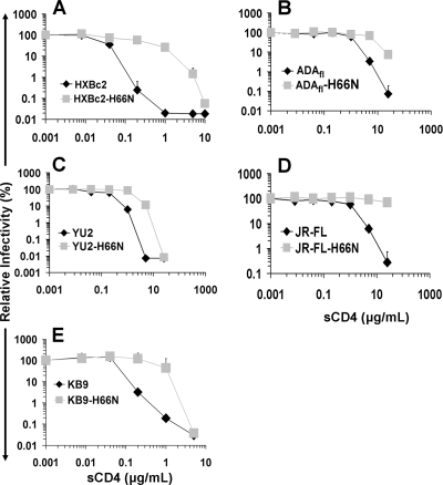
Recombinant HIV-1 encoding luciferase was pseudotyped with the wild-type and H66N variants of the above-described envelope glycoproteins and used to infect Cf2Th canine thymic epithelial cells expressing human CD4 and the appropriate human chemokine receptor. The infections were carried out after the viruses were incubated at 37°C for 1 h without treatment or with increasing concentrations of sCD4. The basal levels of infection in the absence of sCD4 treatment were similar for viruses with the wild-type and H66N envelope glycoproteins (see Fig. S2A in the supplemental material). As expected (55, 75, 78, 80, 93), viruses with the wild-type HXBc2 envelope glycoproteins exhibited the greatest sensitivity to sCD4, with a 90% inhibitory concentration (IC90) of approximately 1.2 nM (Fig. 1A). Recombinant HIV-1 viruses with the wild-type envelope glycoproteins from the R5 primary isolates were significantly more resistant to sCD4, with IC90 values ranging from 14 to 80 nM (Fig. 1B to D). Viruses with the dual-tropic KB9 envelope glycoproteins exhibited intermediate levels of sCD4 sensitivity (IC90 of 3 nM) (Fig. 1E). Thus, as previously reported (5, 16, 57, 82), viruses with envelope glycoproteins derived from TCLA and primary HIV-1 strains differ in sCD4 sensitivity.
FIG. 1.
Sensitivity to sCD4 neutralization. (A to E) Single-round recombinant viruses bearing either the wild-type envelope glycoproteins (HXBc2, ADAfl, YU2, JR-FL, or KB9) or the H66N-modified envelope glycoproteins (KB9-H66N, YU2-H66N, JR-FL-H66N, ADAfl-H66N, or HXBc2-H66N) were equalized based on RT activity and incubated with serial dilutions of sCD4 at 37°C for 1 h. The viruses were then added to Cf2Th cells expressing CD4 and the appropriate coreceptor. Infectivity was assessed by measuring the luciferase activity in the total cell lysate. Infectivity at each dilution of sCD4 tested is shown as the percentage of infection seen in the absence of sCD4. Data are representative of results from three independent experiments performed in triplicate, with means and standard deviations indicated.
Viruses with the H66N envelope glycoproteins were more resistant to sCD4 than viruses bearing the wild-type HIV-1 envelope glycoproteins. Depending on the particular envelope glycoproteins, the degree of sCD4 resistance varied from approximately 7-fold to 33-fold. Thus, the H66N change confers some degree of sCD4 resistance to viruses with a variety of HIV-1 envelope glycoproteins, in the context of infection of CD4-positive cells.
Effect of the H66N change on HIV-1 infection of CD4-negative cells.
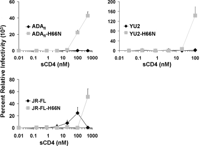
To examine the effect of the H66N change on HIV-1 infection of CD4-negative, CCR5-expressing cells, the recombinant viruses with wild-type or H66N R5 envelope glycoproteins were incubated with Cf2Th-CCR5 cells along with different concentrations of sCD4. In the absence of sCD4, the level of infection of the Cf2Th-CCR5 cells by any of the viruses was near the background for the assay (Fig. 2). The addition of sCD4 resulted in enhanced infection by viruses with the wild-type JR-FL envelope glycoproteins but not by viruses with the wild-type ADAfl or YU2 envelope glycoproteins. The lack of enhancement of the viruses with the wild-type ADAfl or YU2 envelope glycoproteins was not due to a lack of sCD4 binding to the envelope glycoproteins, because the infectivity of these viruses for CD4-positive target cells was markedly inhibited by incubation with sCD4 in this same concentration range (Fig. 1B and C).
FIG. 2.
Effect of H66N on sCD4-induced HIV-1 infection of CD4− CCR5+ cells. Single-round viruses bearing either the wild-type envelope glycoproteins (ADAfl, YU2, or JR-FL) or the H66N-modified envelope glycoproteins (ADAfl-H66N, YU2-H66N, or JR-FL-H66N) were equalized according to RT activity and incubated with serial dilutions of sCD4 at 37°C for 1 h. The viruses were then added to Cf2Th-CCR5 cells that do not express CD4, and infectivity was assessed by measuring the luciferase activity in the total cell lysate. Infectivity at each dilution of sCD4 tested is shown as the percentage of infection seen in the absence of sCD4. Infectivity of the viruses with Cf2Th-CCR5 cells in the absence of sCD4 was close to the background for the assay. Data are representative of results from three independent experiments performed in triplicate, with means and standard deviations indicated.
Recombinant viruses bearing the H66N envelope glycoproteins exhibited greater enhancement by sCD4 than viruses with the respective wild-type envelope glycoproteins. As the H66N and wild-type viruses were normalized by RT measurements and demonstrated nearly equivalent levels of infectivity with Cf2Th-CD4/CCR5 cells (see Fig. S2A in the supplemental material), we conclude that the H66N change results in an increase in sCD4 enhancement of infection of CD4-negative, CCR5-expressing target cells.
Effects of H66N on gp120 ligand binding and induction of conformational changes.
The above-described results suggest that the H66N change facilitates the enhancing effects of sCD4 binding on HIV-1 infection, diminishes the negative consequences of sCD4 binding on infection, or both. To investigate the first possibility, we used isothermal titration microcalorimetry to assess the effect of H66N on sCD4 binding to gp120 and on the ability of sCD4 to induce conformational changes in gp120. We studied the binding of sCD4 to the wild-type and H66N gp120 glycoproteins from HIV-1KB9 (Table 1). The H66N mutant gp120 exhibited a slight (∼2-fold) decrease in affinity for sCD4 compared with that of the wild-type gp120 glycoprotein. Very small decreases in the magnitudes of the entropic and enthalpic changes associated with sCD4 binding were seen for the H66N gp120 glycoproteins, compared with the values observed for the wild-type HIV-1KB9 gp120. The slight reduction in affinity for sCD4 associated with the H66N change was corroborated by SPR analysis (Table 2). Of note, the slight difference in sCD4 affinity between the wild-type and H66N HIV-1KB9 gp120 glycoproteins arises entirely from a difference in off-rate of binding.
TABLE 1.
Thermodynamic values associated with gp120-sCD4 interaction
| gp120 protein | Kd (nM) | ΔG (kcal/mol)a | ΔH (kcal/mol)b | −TΔS (kcal/mol)c |
|---|---|---|---|---|
| KB9 | 11 | −10.8 | −40.9 | +30.1 |
| KB9-H66N | 21 | −10.5 | −39.6 | +29.1 |
| ADA N197S | 25 | −10.4 | −28.0 | +17.6 |
| ADA N197S-H66N | 116 | −9.5 | −19.0 | +9.5 |
ΔG, free energy change.
ΔH, enthalpy change.
T, temperature; ΔS, entropy change.
TABLE 2.
SPR measurements of the effect of the H66N change on gp120-sCD4 binding
| gp120 protein | Kona (M−1 s−1) | Koffb (s−1) | Kd (nM) |
|---|---|---|---|
| KB9 | 1.1 × 10⁴ | 1.9 × 10⁻⁴ | 17 |
| KB9-H66N | 1.1 × 10⁴ | 2.8 × 10⁻⁴ | 24 |
| ADA N197S | 9.3 × 10³ | 6.4 × 10⁻⁴ | 69 |
| ADA N197S-H66N | 7.3 × 10³ | 1.1 × 10⁻³ | 140 |
Kon, on-rate constant.
Koff, off-rate constant.
We wished to examine the effect of H66N on CD4 binding by a gp120 envelope glycoprotein derived from a CD4-independent HIV-1 isolate, ADA N197S, which can enter CCR5-expressing cells lacking CD4 (43, 44). The loss of a glycosylation site at asparagine 197 has been shown to allow the ADA N197S envelope glycoproteins to bind CCR5 and to mediate infection of CCR5-expressing cells without prior interaction with CD4 (44). Similarly to our observations with the CD4-dependent KB9 envelope glycoprotein, the H66N change in the ADA N197S gp120 glycoprotein resulted in a reduction in affinity for sCD4 (Tables 1 and 2). Compared with the values observed for ADA N197S gp120, the magnitudes of the entropic and enthalpic changes associated with sCD4 binding to the ADA N197S-H66N gp120 were decreased. As was observed for KB9 gp120, the reduction in affinity for sCD4 resulting from the H66N change in ADA N197S gp120 was primarily due to an increased off-rate. Therefore, in both CD4-dependent and CD4-independent envelope glycoproteins, the H66N change slightly decreases affinity for CD4, apparently by destabilizing the gp120-CD4 complex and allowing more-rapid dissociation of gp120 and CD4.
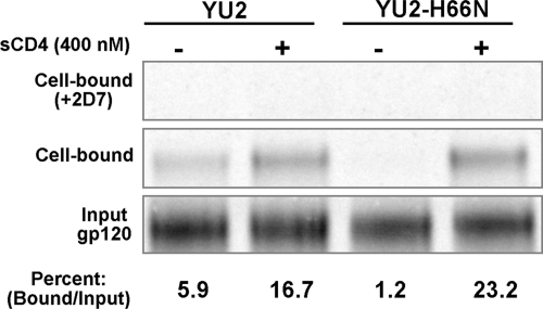
A major functional consequence of CD4 binding to the HIV-1 gp120 glycoprotein is the induction of a conformation capable of binding the chemokine receptor CCR5 or CXCR4 (33, 50, 86, 94). To examine whether H66N influences this induction, wild-type and H66N gp120 glycoproteins from HIV-1YU2 were radiolabeled and incubated with Cf2Th-CCR5 cells in the absence or presence of sCD4. After the cells were washed and lysed, bound gp120 was detected by immunoprecipitation. In the presence of a high concentration of sCD4, the wild-type and H66N gp120 glycoproteins bound to the CCR5-expressing cells comparably (Fig. 3, middle). In control experiments, the Cf2Th-CCR5 cells were incubated with an anti-CCR5 antibody, 2D7, to demonstrate the dependence of gp120 binding on the CCR5 protein (Fig. 3, top). In the absence of sCD4, the wild-type YU2 gp120 glycoprotein bound at low but detectable levels to the Cf2Th-CCR5 cells; this low level of binding was not seen for the H66N gp120 glycoprotein (Fig. 3, middle). These results suggest that the wild-type YU2 gp120 glycoprotein spontaneously assumes the CD4-bound conformation more efficiently than the H66N counterpart. The H66N change apparently destabilizes the CD4-bound conformation in the absence of CD4 but does allow efficient CCR5 binding following CD4 engagement.
FIG. 3.
Effect of H66N on CCR5 binding. Radiolabeled YU2 and YU2-H66N gp120 envelope glycoproteins were incubated with Cf2Th-CCR5 cells in the presence (+) or absence (−) of 400 nM sCD4. Bound and input gp120 glycoproteins were immunoprecipitated and analyzed by SDS-PAGE. The percentage of the input gp120 bound to the Cf2Th-CCR5 cells is indicated beneath each lane. In control experiments, the anti-CCR5 antibody 2D7 was included in the incubation to demonstrate the dependence of gp120 binding on CCR5 (top).
Effect of H66N on CD4-independent HIV-1 infection.
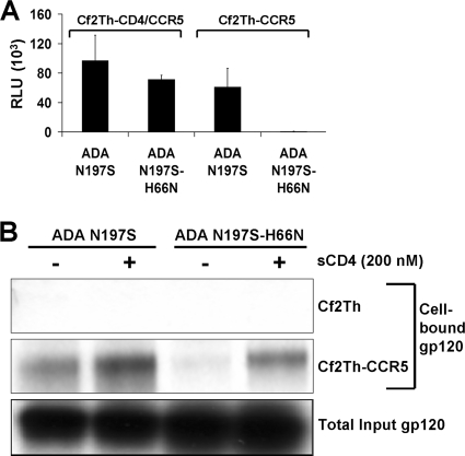
We wished to examine the effect of the H66N change on infection by CD4-independent HIV-1. Recombinant HIV-1 viruses bearing the ADA N197S or ADA N197S-H66N envelope glycoproteins were compared with respect to their abilities to infect Cf2Th-CD4/CCR5 and Cf2Th-CCR5 cells (Fig. 4A). Both viruses efficiently infected Cf2Th-CD4/CCR5 cells. Consistent with the results obtained with CD4-dependent HIV-1 envelope glycoproteins, the H66N change exerted only a small effect on the efficiency of infection of cells expressing CD4 and a chemokine receptor. In contrast, although viruses with the ADA N197S envelope glycoproteins efficiently infected Cf2Th-CCR5 cells, as expected (44), no infection of these CD4-negative cells by viruses with the ADA N197S-H66N envelope glycoproteins was detected. Thus, the H66N change eliminates the ability of otherwise CD4-independent envelope glycoproteins to mediate infection of target cells lacking CD4, even though the ability to infect CD4-expressing cells is retained.
FIG. 4.
Effect of the H66N change on CD4-independent CCR5 binding and infection. (A) The infectivity of recombinant, luciferase-expressing viruses with the ADA N197S CD4-independent envelope glycoproteins or the ADA N197S-H66N envelope glycoproteins was measured using Cf2Th-CD4/CCR5 or Cf2Th-CCR5 cells. The means and standard deviations of infectivity, reported as relative luciferase units (RLU), from a representative experiment performed in triplicate are shown. (B) Radiolabeled ADA N197S or ADA N197S-H66N gp120 envelope glycoproteins were incubated with control Cf2Th cells or Cf2Th-CCR5 cells in the presence (+) or absence (−) of 200 nM sCD4. Bound gp120 and input gp120 were immunoprecipitated and separated by SDS-PAGE.
The gp120 glycoproteins of CD4-independent HIV-1 variants can efficiently bind the chemokine receptor, without prior interaction with CD4 (44). To examine the effect of the H66N change on this process, radiolabeled ADA N197S and ADA N197S-H66N gp120 glycoproteins were incubated with Cf2Th-CCR5 cells in the absence or presence of sCD4. Studies using CCR5 ligands have demonstrated that gp120 binding to Cf2Th-CCR5 cells is dependent upon CCR5 (86, 94). Consistent with this, no gp120 binding to Cf2Th cells that do not express CCR5 was detected in either the presence or the absence of sCD4 (Fig. 4B). In the absence of sCD4, the ADA N197S gp120 glycoprotein, but not the ADA N197S-H66N gp120 glycoprotein, bound the Cf2Th-CCR5 cells. Both gp120 glycoproteins bound the Cf2Th-CCR5 cells efficiently following incubation with sCD4, with a mild decrease in binding for the ADA N197S-H66N gp120 compared with that for the ADA N197S glycoprotein. These results suggest that the H66N change decreases the ability of the ADA N197S gp120 glycoprotein to bind CCR5 spontaneously but does not prevent CCR5 binding once CD4 is engaged.
The change in asparagine 197 in the ADA N197S mutant removes a glycan from the V1/V2 stem, thereby increasing the flexibility of the gp120 V1/V2 variable loops and contributing to CD4 independence (44). A CD4-independent phenotype can also be achieved by deleting the V1 and V2 variable loops of the ADA gp120 glycoprotein (43). To examine whether the phenotype of H66N is dependent on the integrity of the gp120 V1/V2 loops, the H66N change was introduced into an HIV-1 envelope glycoprotein, ADA ΔV1/V2, in which the gp120 V1 and V2 loops were deleted. As shown in Fig. S3A in the supplemental material, viruses with the ADA ΔV1/V2 envelope glycoproteins efficiently infected Cf2Th-CCR5 cells, as expected (43). However, the infectivity of viruses with the ADA ΔV1/V2-H66N envelope glycoproteins was significantly lower using these cells. We conclude that the H66N change decreases the ability of CD4-independent HIV-1 envelope glycoproteins to infect CD4-negative cells expressing CCR5 and that this phenotype does not depend on the presence of the gp120 V1 and V2 variable loops.
Effect of the H66N change on antibody recognition and neutralization.
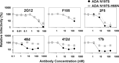
Distinct conformations of the HIV-1 gp120 glycoproteins are recognized by particular groups of monoclonal antibodies. For example, CD4i antibodies recognize the CD4-bound conformation of HIV-1 gp120 (13, 84, 96), whereas most CD4 binding site antibodies recognize conformations of gp120 other than that induced by CD4 binding (97). Because antibody binding to the HIV-1 envelope glycoprotein leads to neutralization (40, 65, 69, 88, 98), we assessed the conformational state of the ADA N197S and ADA N197S-H66N envelope glycoproteins by measuring the sensitivities of viruses with these envelope glycoproteins to neutralization by several monoclonal antibodies. Viruses with the ADA N197S and ADA N197S-H66N envelope glycoproteins exhibited similar degrees of sensitivity to the carbohydrate-directed antibody 2G12 (72, 87) and to F105, an antibody directed against the CD4 binding site of gp120 (67, 85) (Fig. 5). The viruses with the ADA N197S envelope glycoproteins were slightly more sensitive to neutralization by the 2F5 antibody than viruses with the ADA N197S-H66N envelope glycoproteins. The 2F5 antibody recognizes a membrane-proximal epitope in the gp41 ectodomain (9, 15, 60).
FIG. 5.
Effect of H66N on HIV-1 neutralization by monoclonal antibodies. Single-round recombinant viruses bearing the ADA N197S or ADA N197S-H66N envelope glycoproteins were incubated for 1 h at 37°C with monoclonal antibodies directed against a gp120 carbohydrate-dependent epitope (2G12), the gp120 CD4 binding site (F105), the gp41 membrane-proximal external region (2F5), and gp120 CD4i epitopes (48d, 412d, and 17b). Viruses were then added to Cf2Th-CD4/CCR5 cells, and infection was measured by assaying luciferase activity in the target cell lysate. The infectivity relative to that seen in the absence of antibody is reported. Data are representative of results from three independent experiments performed in triplicate, with means and standard deviations indicated.
In contrast to the results with the antibodies described above, the viruses with the ADA N197S-H66N envelope glycoproteins were significantly more resistant to neutralization by three CD4i antibodies, 48d, 412d, and 17b (Fig. 5). Because of steric constraints, HIV-1 neutralization by CD4i antibodies must occur prior to the engagement of CD4 on the target cell by virus (49, 83). Thus, our results suggest that, in the absence of bound ligand, the ADA N197S-H66N envelope glycoproteins on the virus are recognized less efficiently than the ADA N197S envelope glycoproteins by CD4i antibodies.
To obtain more quantitative information on the effect of the H66N change on recognition by CD4i antibodies, we used an SPR assay to measure the binding of gp120 glycoproteins to the 17b antibody captured on a Biacore chip. In the experiment with results shown in Fig. S4A in the supplemental material, the same concentrations of ADA N197S and ADA N197S-H66N gp120 glycoproteins were incubated in the absence or presence of sCD4 prior to exposure to the captured 17b antibody. In the absence of sCD4, the binding of the ADA N197S-H66N gp120 glycoprotein to the 17b antibody was much lower than that of the ADA N197S glycoprotein. The H66N change reduced the affinity of the ADA N197S gp120 glycoprotein for the 17b antibody by more than 60-fold (see Fig. S4B in the supplemental material). This decreased affinity resulted from a slower association between gp120 and 17b as well as a faster dissociation of the complex. Similar results were obtained for the wild-type and H66N variants of the KB9 gp120 glycoprotein, which is a CD4-dependent envelope glycoprotein (see Fig. S4C in the supplemental material). Thus, in the absence of sCD4, HIV-1 gp120 with the H66N change is recognized only weakly by the 17b antibody. In the presence of sCD4, however, the ADA N197S-H66N gp120 glycoprotein bound the 17b antibody nearly as efficiently as the ADA N197S glycoprotein (see Fig. S4B in the supplemental material).
In summary, the H66N change decreases the efficiency with which unliganded HIV-1 gp120 samples the CD4-bound conformation recognized by CCR5 and the CD4i antibodies.
Effects of H66N on sensitivity of CD4-independent envelope glycoproteins to sCD4.
The sensitivity of CD4-independent viruses to neutralization by sCD4 was examined. These experiments were conducted with both CD4-positive and CD4-negative target cells.
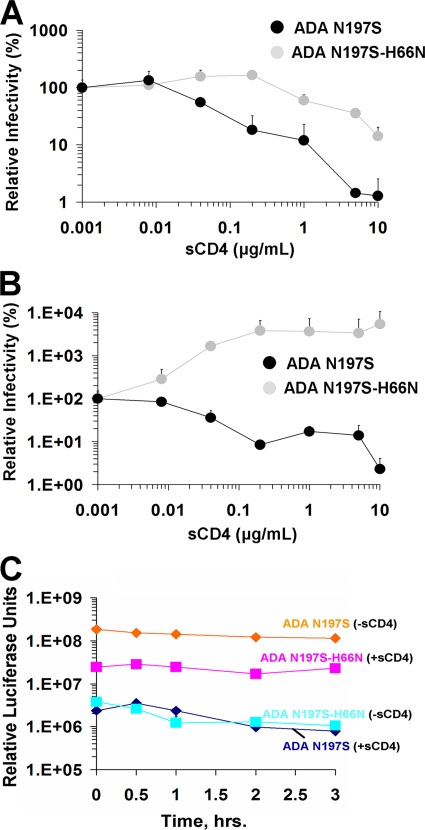
The basal levels of infectivity of viruses with the ADA N197S and ADA ΔV1/V2 envelope glycoproteins and their H66N counterparts were comparable with Cf2Th-CD4/CCR5 target cells (see Fig. S2B in the supplemental material). Both two-domain and four-domain sCD4 proteins inhibited the ADA N197S-H66N viruses less efficiently than the ADA N197S viruses (Fig. 6A and data not shown). In fact, mild enhancement of the virus with the ADA N197S-H66N envelope glycoproteins was evident at low concentrations of sCD4. Likewise, the viruses with the ADA ΔV1/V2-H66N envelope glycoproteins were significantly more resistant to sCD4 than the viruses with the ADA ΔV1/V2 envelope glycoproteins (see Fig. S3B in the supplemental material). Thus, H66N confers a degree of sCD4 resistance to both CD4-dependent and CD4-independent HIV-1 viruses in the course of infection of CD4-expressing target cells.
FIG. 6.
Effect of H66N on sCD4 sensitivity of CD4-independent viruses. (A) Single-round recombinant viruses carrying either the ADA N197S or the ADA N197S-H66N envelope glycoproteins were incubated with serial dilutions of four-domain sCD4 at 37°C for 1 h. Infectivity was measured with Cf2Th-CD4/CCR5 cells and is reported at each dilution of sCD4 as the percentage of infectivity observed without sCD4 treatment of the viruses. Data are representative of results from three independent experiments performed in triplicate, with means and standard deviations indicated. (B) Single-round recombinant viruses carrying the ADA N197S or ADA N197S-H66N envelope glycoproteins were incubated with serial dilutions of four-domain sCD4 at 37°C for 1 h. The infectivity of the viruses was measured with Cf2Th-CCR5 cells. Infectivity at each dilution of sCD4 is given as the percentage of infectivity observed without sCD4. Data are representative of results from three independent experiments performed in triplicate, with means and standard deviations indicated. (C) Single-round recombinant viruses bearing the ADA N197S or ADA N197S-H66N envelope glycoproteins were incubated with (+) or without (−) 200 nM sCD4 at 37°C for 1 h. Unbound sCD4 was removed by pelleting virus at 41,000 rpm in an SW-41 rotor for 30 min at 4°C. Pelleted virus was resuspended in 10 volumes of medium without sCD4 and repelleted twice to ensure removal of unbound sCD4. Virus was then resuspended in medium and incubated at 37°C for different amounts of time before an aliquot was removed and added to Cf2Th-CCR5 cells to measure infectivity. Data are representative of results from three independent experiments performed in triplicate, with means and standard deviations indicated.
In CD4-negative target cells expressing only CCR5, the basal levels of infection of the viruses with the ADA N197S and ADA ΔV1/V2 envelope glycoproteins were significantly higher than those of viruses with the H66N-modified envelope glycoproteins (Fig. 4A; also see Fig. S3C in the supplemental material). Incubation with sCD4 neutralized the viruses with the ADA N197S and ADA ΔV1/V2 envelope glycoproteins but enhanced the infection of viruses with the ADA N197S-H66N and ADA ΔV1/V2-H66N envelope glycoproteins (Fig. 6B; also see Fig. S3C in the supplemental material). Two-domain sCD4 and four-domain sCD4 exhibited similar effects in this assay (data not shown). In the setting of CD4-independent infection, the potential inhibitory effects of sCD4 resulting from competition with membrane-bound CD4 for gp120 binding are eliminated. Thus, the observed sCD4 inhibition of the CD4-independent viruses with the ADA N197S and ADA ΔV1/V2 envelope glycoproteins results solely from detrimental effects of sCD4 binding on these envelope glycoproteins. The H66N change apparently prevents or diminishes these detrimental effects and reveals the activating effects of sCD4 binding on virus entry. The sCD4 activation of infection of CD4-negative cells by CD4-independent viruses with the H66N change is reminiscent of that seen for viruses with natural HIV-1 envelope glycoproteins modified by H66N (Fig. 2).
Stability of the sCD4-induced state of the viral envelope glycoproteins.
The above-described studies support the existence of both positive and negative effects of sCD4 binding on HIV-1 entry; the phenotypes of the H66N variants provided an opportunity to examine the stability of the sCD4-induced envelope glycoprotein states. Recombinant viruses with the ADA N197S or ADA N197S-H66N envelope glycoproteins were incubated with sCD4 for 1 h at 37°C, and then the viruses were pelleted and washed repeatedly to ensure removal of sCD4 from the medium. The resuspended viruses were incubated at 37°C and, at different times, added to Cf2Th-CCR5 cells. The resultant levels of infection are shown in Fig. 6C. In agreement with the above-described observations, sCD4 enhanced the infection of viruses with the ADA N197S-H66N envelope glycoproteins by 10-fold but inhibited infection by viruses with the ADA N197S envelope glycoproteins by more than 40-fold. The activated state of the ADA N197S-H66N envelope glycoproteins persisted for at least 3 h, the duration of the experiment. The inhibitory effect of sCD4 on viruses with the ADA N197S envelope glycoproteins occurred by the time of the first measurement of infection and persisted thereafter. Thus, the sCD4-activated state of the ADA N197S-H66N envelope glycoproteins and the sCD4-induced, functionally inactivated state of the ADA N197S envelope glycoproteins are both long-lived.
Effect of the H66N change on sCD4 binding to the envelope glycoprotein trimer and induction of gp120 shedding.
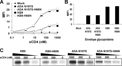
The above-described results suggest that the major effect of the H66N change on the resistance of HIV-1 to sCD4 neutralization is mediated by stabilization of a sCD4-induced activated form of the envelope glycoproteins, minimizing the probability of transitions to less functional states. To examine this possibility further, we studied the binding of sCD4 to HIV-1 envelope glycoprotein trimers expressed on the surface of 293T cells. The gp41 cytoplasmic tail was deleted from these envelope glycoproteins to increase cell surface expression (22, 26, 31, 56, 58, 59). The levels of binding of sCD4 to the ADA N197S and ADA N197S-H66N envelope glycoproteins at 37°C were similar (Fig. 7A). A control experiment using the 2G12 antibody indicated that the levels of cell surface expression of the two envelope glycoproteins were comparable (Fig. 7B). We conclude that minimal differences in binding of sCD4 exist between the ADA N197S and ADA N197S-H66N trimeric envelope glycoproteins, corroborating the results obtained with monomeric gp120 variants (see above). Additional experiments conducted at room temperature with the KB9 and ADA N197S envelope glycoproteins and their H66N-modified counterparts yielded similar results (see Fig. S5 in the supplemental material).
FIG. 7.
Effect of H66N on sCD4 binding and induction of gp120 shedding. (A) 293T cells transiently expressing cytoplasmic-tail-deleted ADA N197S, ADA N197S-H66N, KB9, or KB9-H66N envelope glycoproteins were incubated at 37°C for 1 h with sCD4 at the indicated concentrations. After the cells were washed, the bound sCD4 was detected by FACS analysis with OKT4, a fluorescein isothiocyanate-conjugated anti-CD4 antibody. The mean fluorescence intensity (MFI) values at different sCD4 concentrations are shown. Mock-transfected cells (Mock) were included as a negative control. (B) 293T cells expressing the HIV-1 envelope glycoproteins described for panel A were incubated with 50 μg/ml 2G12 antibody. After the cells were washed, the bound antibody was detected with phycoerythrin-conjugated rabbit anti-human IgG. (C) 293T cells transiently expressing cytoplasmic-tail-deleted KB9 or KB9-H66N envelope glycoproteins or ADA N197S or ADA N197S-H66N envelope glycoproteins were radiolabeled and incubated either in the absence of sCD4 or with 200 or 1,000 nM sCD4 at 37°C for 1 h. The gp120 shed into the supernatant was precipitated by a polyclonal mixture of sera from HIV-1-infected individuals and analyzed by SDS-PAGE.
One detrimental effect of sCD4 binding is the shedding of gp120 from the HIV-1 envelope glycoprotein trimer (22, 26, 31, 56, 58, 59). To test whether the H66N change affects the propensity of the HIV-1 envelope glycoproteins to shed gp120 in response to CD4 binding, we expressed radiolabeled functional envelope glycoproteins on the surface of 293T cells. The gp41 cytoplasmic tail was removed from these envelope glycoproteins to enhance cell surface expression (28, 101). Cells expressing KB9, KB9-H66N, ADA N197S, and ADA N197S-H66N envelope glycoproteins were incubated with different concentrations (200 and 1,000 nM) of sCD4 at 37°C for 1 h. Media were harvested and examined for shed gp120 by immunoprecipitation with a mixture of sera from HIV-1-infected individuals. The addition of sCD4 resulted in an increase in the shedding of gp120 into the medium. The H66N change in both the KB9 and ADA N197S envelope glycoproteins decreased the amount of shed gp120 (Fig. 7C). Expression levels and processing efficiencies of the various envelope glycoproteins were equivalent (see Fig. S6 in the supplemental material). These results suggest that the H66N change allows efficient sCD4 binding to the HIV-1 envelope glycoprotein trimer but decreases the degree of gp120 shedding consequent to sCD4 binding.
Effects of the H66N change on susceptibility to a small-molecule CD4 mimic.
NBD-556 is a low-molecular-weight compound that can induce conformational changes in the HIV-1 gp120 glycoprotein similar to those that occur upon CD4 binding (52, 74, 103). NBD-556 can enhance HIV-1 infection of CD4-negative, CCR5-positive cells and weakly inhibits infection of CD4-expressing cells (52, 74, 103).
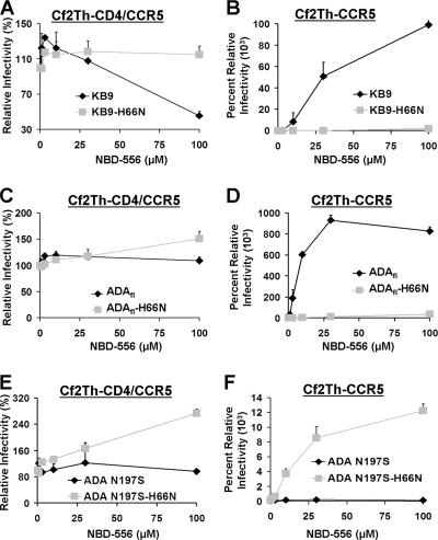
The effects of NBD-556 on the infectivity of viruses with the wild-type KB9 or KB9 H66N envelope glycoproteins were examined using Cf2Th-CD4/CCR5 and Cf2Th-CCR5 target cells. NBD-556 inhibited the infection of viruses with the KB9 envelope glycoproteins in Cf2Th-CD4/CCR5 cells but did not significantly affect the infectivity of viruses with the KB9-H66N envelope glycoproteins (Fig. 8A). With the CD4-negative Cf2Th-CCR5 cells, NBD-556 dramatically enhanced infection of viruses bearing the wild-type KB9 envelope glycoproteins but did not affect the infectivity of viruses with the KB9-H66N envelope glycoproteins (Fig. 8B). These results indicate that the H66N change renders the HIV-1 envelope glycoproteins resistant to both the negative and the positive effects of NBD-556 on viruses with the corresponding wild-type envelope glycoproteins.
FIG. 8.
Effect of a CD4-mimetic compound, NBD-556, on virus infectivity. Single-round recombinant viruses bearing the KB9 or KB9-H66N envelope glycoproteins (A and B), the ADAfl or ADAfl-H66N envelope glycoproteins (C and D), or the ADA N197S or ADA N197S-H66N envelope glycoproteins (E and F) were incubated with serial dilutions of NBD-556. Infectivity was measured using Cf2Th-CD4/CCR5 (A, C, and E) and Cf2Th-CCR5 (B, D, and F) cells. Infectivity is reported as the percentage of infection observed in the absence of the compound. Data are representative of results from three independent experiments performed in triplicate, with means and standard deviations indicated.
One explanation for the above-described observations is that the H66N change interferes with the binding of NBD-556 to the gp120 glycoprotein. To test this, isothermal titration microcalorimetry studies were conducted with NBD-556 and the wild-type and H66N gp120 glycoproteins from the KB9 HIV-1 strain (Table 3). The H66N change in gp120 reduced the NBD-556 binding affinity to below the detection limits of the assay, explaining the resistance of the virus with the KB9 H66N envelope glycoproteins to the effects of NBD-556. This result is also consistent with the observations made using another virus with the CD4-dependent ADA envelope glycoproteins, in which the H66N change rendered the virus resistant to the effects of NBD-556 (Fig. 8C and D). In contrast, for the CD4-independent ADA N197S envelope glycoproteins, where removal of a sugar in the V1/V2 stem allows CCR5 binding without prior binding to CD4 (43), NBD-556 binding and entry enhancement were not eliminated by the H66N change (Fig. 8E and F; Table 3). Thus, histidine 66 promotes the transition of free gp120 into the CD4-bound conformation, a process that contributes to the ability of NBD-556 to bind gp120 and induce this conformation. The CD4-independent gp120 glycoprotein has a greater propensity to make the transition into the CD4-bound state than the CD4-dependent envelope glycoprotein. As a likely consequence, the H66N change is less effective in preventing the sampling of the CD4-bound conformation by the CD4-independent envelope glycoprotein.
TABLE 3.
Thermodynamic values associated with the binding of gp120 and NBD-556
| gp120 protein | Kd (μM) | ΔG (kcal/mol) | ΔH (kcal/mol) | −TΔSa (kcal/mol) |
|---|---|---|---|---|
| KB9 | 1.7 | −7.8 | −22.6 | +14.8 |
| KB9-H66N | NBb | NB | NB | NB |
| ADA N197S | 0.40 | −8.7 | −12.0 | +3.3 |
| ADA N197S-H66N | 0.64 | −8.4 | −13.8 | +5.4 |
T, temperature; ΔS, entropy change.
NB, no detectable binding.
DISCUSSION
The H66N gp120 variant of HIV-1KB9 was derived by repeated selection of viruses by extremes of temperature; the effect of the change in histidine 66 on the temperature stability of HIV-1 has been described elsewhere (37). Here, we demonstrate that the H66N change in gp120 affects several of the biological properties of HIV-1 related to the induction of the CD4-bound state and the functional consequences of CD4 binding. The phenotypic effects of the H66N change in the envelope glycoproteins derived from several different HIV-1 strains were similar. This observation and the highly conserved nature of histidine 66 (see Fig. S1 in the supplemental material) support the generality of the results and suggest that HIV-1 strains have conserved mechanisms whereby changes in receptor binding trigger conformational transitions. Some of the CD4-induced conformational transitions, such as those that allow CCR5 binding or result in the exposure of the HR1 groove in the gp41 ectodomain, play positive roles in infection (12, 27, 45). Other CD4-induced transitions lead to inactivation of the HIV-1 envelope glycoprotein trimer and, in extreme cases, involve the shedding of gp120 (30, 31, 56).
For most wild-type, primary HIV-1 envelope glycoproteins, competence for CCR5 binding and priming of the gp120-gp41 interaction occur only rarely in a spontaneous fashion; thus, CD4 binding is required to allow these events, and virus entry, to proceed (17). CD4-independent HIV-1 variants generated in the laboratory negotiate these transitions spontaneously, thus bypassing the need for CD4 binding (43, 44). The H66N change reverts the CD4-independent envelope glycoproteins back to envelope glycoproteins with CD4-dependent phenotypes. This reversion is evident in a decrease in spontaneous gp120 binding to CCR5; in reduced infection of CD4-negative, CCR5-expressing cells; and in decreased sensitivity to CD4i antibodies, which recognize the CD4-bound conformation of gp120 (13, 84, 96). These properties suggest that the unliganded CD4-independent envelope glycoproteins with asparagine 66 spontaneously sample the CD4-bound conformation less than their counterparts with histidine 66. This implies that, in the absence of sCD4, the H66N change either destabilizes the CD4-bound conformation or stabilizes unliganded gp120 conformations other than the CD4-bound state. We favor the former model for two reasons: (i) it is more consistent with our observation that the H66N change selectively decreases sensitivity to neutralization by CD4i antibodies and does not affect sensitivity to the F105 CD4 binding site antibody, and (ii) it proposes a positive function for the highly conserved histidine 66, i.e., stabilization of the spontaneously sampled CD4-bound conformation. Such stabilization could assist the flexible, unliganded HIV-1 gp120 glycoprotein to make secondary contacts with CD4 after the initial, weak contact of the two molecules (27, 45, 77, 92). This explanation is consistent with the observation that alteration of histidine 66 primarily increases the dissociation of gp120-sCD4 complexes, with no apparent effect on the rate of association of the two proteins.
Both CD4-dependent and CD4-independent HIV-1 envelope glycoproteins with the H66N change exhibited decreased susceptibility to the detrimental effects of sCD4 on virus infection. Some part of this resistance to sCD4 inhibition may arise as a consequence of the small differences in ability to bind CD4 between wild-type and H66N gp120 glycoproteins. If the ∼2-fold difference in CD4-binding affinity between monomeric wild-type and H66N gp120 glycoproteins applies, the levels of occupancy of the envelope glycoprotein trimers should be similar at high CD4 concentrations. Indeed, the levels of binding of sCD4 to wild-type and H66N envelope glycoprotein complexes expressed on cell surfaces were indistinguishable. These and other results suggest that the H66N envelope glycoproteins differ from their unaltered counterparts in the consequences of CD4 binding. The CD4-bound HIV-1 envelope glycoproteins represent metastable intermediates in the virus entry process and make further transitions that either promote virus entry (activation pathway) or lead to nonfunctional states (inactivation pathway[s]) (30, 31, 56). The variables that determine which of these pathways predominates are still being elucidated but presumably include the availability and binding affinity of the chemokine receptor and the intrinsic strength of the interactions that stabilize the CD4-bound state of the envelope glycoprotein trimer. It is noteworthy that when CD4 and CCR5 were present at high concentrations on the target cells, there were minimal differences in infectivity of viruses with unmodified and H66N envelope glycoproteins. In contrast, with CD4− CCR5+ target cells, when sCD4 was first incubated with virus so that the time between CD4 binding and chemokine receptor binding was prolonged, dramatic differences in infectivity between the wild-type and H66N viruses were evident. Whereas the wild-type viruses were inactivated by sCD4 under these circumstances, infection by viruses with the H66N envelope glycoproteins was enhanced. This observation suggests that the H66N change may decrease the efficiency of envelope glycoprotein transitions to less functional states, thus favoring transitions to membrane-fusogenic states promoted by chemokine receptor binding. In other studies, wild-type HIV-1 envelope glycoproteins induced to assume the CD4-bound conformation at a distance from a target cell were shown to undergo a short-lived activation followed by irreversible inactivation (30). By limiting the ability of the HIV-1 envelope glycoproteins to assume the full CD4-bound state, the H66N change may decrease the likelihood of negotiating transitions into inactive states. This model is consistent with the H66N envelope glycoprotein being selected on the basis of improved temperature stability of the virus (37). Thus, resistance to generic destabilizing influences, such as temperature extremes, can be achieved by avoidance of the labile CD4-bound conformation.
The other interesting phenotype associated with the H66N change in CD4-dependent envelope glycoproteins is resistance to the CD4-mimetic compound NBD-556. This drug inhibits virus entry into cells by competitive inhibition of CD4 binding (103) and by prematurely triggering metastable activated envelope glycoprotein states that decay into inactive forms (30). Binding of NBD-556 to gp120 induces conformational changes similar to those associated with CD4 binding, albeit to a lesser extent (66). When introduced into the gp120 glycoprotein of a CD4-dependent virus, the H66N change completely eliminated binding to NBD-556, whereas this change only slightly reduced binding affinity for CD4 itself. Apparently, the contribution of histidine 66 to the achievement of the CD4-bound state is critical for the ability of a small molecule like NBD-556 to induce this state in a CD4-dependent envelope glycoprotein. Presumably, the greater number of gp120 contacts made by CD4 compensates more effectively for the loss of the histidine 66 contribution to the gp120 transition to the CD4-bound state.
Of interest, the H66N derivative of a gp120 glycoprotein from a CD4-independent HIV-1 bound NBD-556 efficiently. Apparently, the gp120 change (N197S or ΔV1/V2) that allows CD4 independence compensates for the lack of histidine 66 and allows NBD-556 to bind gp120 and drive it into the CD4-bound conformation. The magnitudes of the NBD-556-induced entropic and enthalpic changes in CD4-independent gp120 are lower than those observed for CD4-dependent gp120, consistent with a model in which the former gp120 glycoproteins have spontaneously negotiated a portion of the conformational transition to the CD4-bound state.
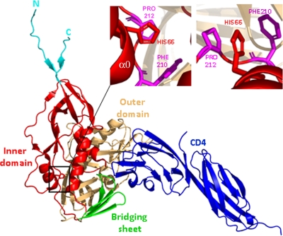
What is the structural explanation for the observed effects of the H66N change? As the H66N alteration can exert phenotypes independently of the gp120 V1/V2 variable loops, insights into histidine 66 function should be possible without consideration of the V1/V2 loops, which have been deleted from the “gp120 core” proteins for which structures are currently available (11, 47, 48). Although CD4-bound structures of the HIV-1 gp120 core and unliganded structures of the SIV gp120 core have been determined, the gp120 N-terminal segment containing histidine 66 was deleted from these constructs to allow crystallization (11, 48). Recently, however, the structure of the HIV-1 gp120 glycoprotein with intact termini has been solved in the CD4-bound state (Pancera et al., unpublished). The disulfide-bonded loop in which histidine 66 resides is part of the gp120 inner domain (48, 95) (Fig. 9). In the envelope glycoprotein complex, this disulfide-bonded loop is thought to be located near the trimer axis and thus may be proximal to the gp41 ectodomain. Although some elements of the gp120 inner domain are thought to contact gp41 (Pancera et al., unpublished), the histidine 66 side chain is not surface exposed in the CD4-bound conformation. Rather, histidine 66 projects from the α0 helix in the disulfide-bonded loop into the interior of the inner domain, contacting residues phenylalanine 210 and proline 212 (Fig. 9). Modeling of the wild-type and H66N gp120 glycoproteins suggests that histidine 66 makes stacking interactions with phenylalanine 210 and proline 212 that are predicted to stabilize the CD4-bound conformation (see Fig. S7 in the supplemental material). These interactions are lost upon substitution of an asparagine residue at position 66, resulting in destabilization of the CD4-bound state. The modeling predictions are consistent with multiple observations, discussed above, that suggest that the H66N gp120 spontaneously samples the CD4-bound conformation less efficiently than the wild-type gp120 glycoprotein. Examination of the gp120-CD4 complex structure shown in Fig. 9 rules out any direct contact between histidine 66 and CD4; based on recent mapping of the NBD-556 binding site (52), histidine 66 is not likely to contact this compound either. Thus, the effects of the H66N change on CD4/NBD-556 interactions with the HIV-1 envelope glycoproteins likely arise indirectly, by influencing associations within the inner domain that promote the attainment of the CD4-bound state. Of interest, the H66N change does not affect the on-rate of the CD4 interaction with gp120, which is thought to involve contacts primarily between CD4 and the gp120 outer domain (54). Rather, H66N increases the dissociation rate of the gp120-CD4 complex. This observation is consistent with a model in which the folding of the gp120 inner domain and bridging sheet stabilizes the complex with CD4. Thus, histidine 66, by contributing to interactions within the gp120 inner domain, helps gp120 to achieve the CD4-bound conformation from its previously unliganded state.
FIG. 9.
Structure of the HIV-1 gp120 glycoprotein with complete N and C termini in the CD4-bound conformation. The structure of the HIV-1 gp120 glycoprotein bound to two-domain CD4 (Pancera et al., unpublished) is shown, from the approximate perspective of the trimer axis in the assembled envelope glycoprotein trimer. The gp120 domains and the N and C termini are depicted. The inner domain and the N and C termini have been implicated in the noncovalent interaction with the gp41 ectodomain (32, 100). The α0 helix, in which histidine 66 resides, is distant from the CD4-binding region but near the gp41-interactive surface of gp120. In the inset panels, the stacking interactions of histidine 66 (in the α0 helix) with proline 212 and phenylalanine 210 are shown from two perspectives. The images were created with PyMOL (18).
How might the H66N change prolong the stability of the activated state induced on the viral envelope glycoproteins by sCD4? The longevity of this activated state of infectivity has recently been shown to be correlated with the duration of sCD4-induced exposure of gp41 ectodomain sequences (30). Stabilization of this activated intermediate would presumably require modification of the gp120-gp41 interactions involved in governing conformational transitions in the envelope glycoproteins. Histidine 66 is strategically positioned between the putative gp41-interactive surface of gp120 and the binding site for CD4 and is thus potentially available to influence the consequences of CD4 binding on gp41 conformation. However, in the CD4-bound state, the histidine 66 side chain is completely buried and therefore cannot contact gp41. It is possible that the interactions of histidine 66 with other inner domain residues could indirectly influence the spatial relationship of gp120 and gp41. Alternatively, in the unliganded gp120 glycoprotein in the trimeric envelope glycoprotein complex, histidine 66 and the surrounding disulfide-bonded loop may exist in a conformation that allows more direct contacts with the gp41 glycoprotein. Future studies will examine these possibilities.
Supplementary Material
Acknowledgments
We thank Yvette McLaughlin and Elizabeth Carpelan for manuscript preparation and William Schief and Darrel Hurt for assistance with H66N modeling.
This work was supported by NIH grants (AI24755, AI39420, and AI40895), by a Center for HIV/AIDS Vaccine Immunology grant (AI67854), by a Center for AIDS Research grant (AI42848), by an unrestricted research grant from the Bristol-Myers Squibb Foundation, by a gift from the late William F. McCarty-Cooper, and by funds from the International AIDS Vaccine Initiative.
Footnotes
Published ahead of print on 17 June 2009.
Supplemental material for this article may be found at http://jvi.asm.org/.
REFERENCES
- 1.Adachi, A., H. E. Gendelman, S. Koenig, T. Folks, R. Willey, A. Rabson, and M. A. Martin. 1986. Production of acquired immunodeficiency syndrome-associated retrovirus in human and nonhuman cells transfected with an infectious molecular clone. J. Virol. 59284-291. [DOI] [PMC free article] [PubMed] [Google Scholar]
- 2.Aguilar-Cordova, E., J. Chinen, L. Donehower, D. E. Lewis, and J. W. Belmont. 1994. A sensitive reporter cell line for HIV-1 tat activity, HIV-1 inhibitors, and T cell activation effects. AIDS Res. Hum. Retrovir. 10295-301. [DOI] [PubMed] [Google Scholar]
- 3.Alkhatib, G., C. Combadiere, C. C. Broder, Y. Feng, P. E. Kennedy, P. M. Murphy, and E. A. Berger. 1996. CC CKR5: a RANTES, MIP-1alpha, MIP-1beta receptor as a fusion cofactor for macrophage-tropic HIV-1. Science 2721955-1958. [DOI] [PubMed] [Google Scholar]
- 4.Allan, J. S., J. Strauss, and D. W. Buck. 1990. Enhancement of SIV infection with soluble receptor molecules. Science 2471084-1088. [DOI] [PubMed] [Google Scholar]
- 5.Ashkenazi, A., D. H. Smith, S. A. Marsters, L. Riddle, T. J. Gregory, D. D. Ho, and D. J. Capon. 1991. Resistance of primary isolates of human immunodeficiency virus type 1 to soluble CD4 is independent of CD4-rgp120 binding affinity. Proc. Natl. Acad. Sci. USA 887056-7060. [DOI] [PMC free article] [PubMed] [Google Scholar]
- 6.Barre-Sinoussi, F., J. C. Chermann, F. Rey, M. T. Nugeyre, S. Chamaret, J. Gruest, C. Dauguet, C. Axler-Blin, F. Vezinet-Brun, C. Rouzioux, W. Rozenbaum, and L. Montagnier. 1983. Isolation of a T-lymphotropic retrovirus from a patient at risk for acquired immune deficiency syndrome (AIDS). Science 220868-871. [DOI] [PubMed] [Google Scholar]
- 7.Brunger, A. T. 2007. Version 1.2 of the crystallography and NMR system. Nat. Protoc. 22728-2733. [DOI] [PubMed] [Google Scholar]
- 8.Brunger, A. T., P. D. Adams, G. M. Clore, W. L. DeLano, P. Gros, R. W. Grosse-Kunstleve, J. S. Jiang, J. Kuszewski, M. Nilges, N. S. Pannu, R. J. Read, L. M. Rice, T. Simonson, and G. L. Warren. 1998. Crystallography & NMR system: a new software suite for macromolecular structure determination. Acta Crystallogr. D 54905-921. [DOI] [PubMed] [Google Scholar]
- 9.Cao, J., L. Bergeron, E. Helseth, M. Thali, H. Repke, and J. Sodroski. 1993. Effects of amino acid changes in the extracellular domain of the human immunodeficiency virus type 1 gp41 envelope glycoprotein. J. Virol. 672747-2755. [DOI] [PMC free article] [PubMed] [Google Scholar]
- 10.Chan, D. C., D. Fass, J. M. Berger, and P. S. Kim. 1997. Core structure of gp41 from the HIV envelope glycoprotein. Cell 89263-273. [DOI] [PubMed] [Google Scholar]
- 11.Chen, B., E. M. Vogan, H. Gong, J. J. Skehel, D. C. Wiley, and S. C. Harrison. 2005. Structure of an unliganded simian immunodeficiency virus gp120 core. Nature 433834-841. [DOI] [PubMed] [Google Scholar]
- 12.Choe, H., M. Farzan, Y. Sun, N. Sullivan, B. Rollins, P. D. Ponath, L. Wu, C. R. Mackay, G. LaRosa, W. Newman, N. Gerard, C. Gerard, and J. Sodroski. 1996. The beta-chemokine receptors CCR3 and CCR5 facilitate infection by primary HIV-1 isolates. Cell 851135-1148. [DOI] [PubMed] [Google Scholar]
- 13.Choe, H., W. Li, P. L. Wright, N. Vasilieva, M. Venturi, C. C. Huang, C. Grundner, T. Dorfman, M. B. Zwick, L. Wang, E. S. Rosenberg, P. D. Kwong, D. R. Burton, J. E. Robinson, J. G. Sodroski, and M. Farzan. 2003. Tyrosine sulfation of human antibodies contributes to recognition of the CCR5 binding region of HIV-1 gp120. Cell 114161-170. [DOI] [PubMed] [Google Scholar]
- 14.Clapham, P. R., A. McKnight, and R. A. Weiss. 1992. Human immunodeficiency virus type 2 infection and fusion of CD4-negative human cell lines: induction and enhancement by soluble CD4. J. Virol. 663531-3537. [DOI] [PMC free article] [PubMed] [Google Scholar]
- 15.Conley, A. J., J. A. Kessler II, L. J. Boots, J. S. Tung, B. A. Arnold, P. M. Keller, A. R. Shaw, and E. A. Emini. 1994. Neutralization of divergent human immunodeficiency virus type 1 variants and primary isolates by IAM-41-2F5, an anti-gp41 human monoclonal antibody. Proc. Natl. Acad. Sci. USA 913348-3352. [DOI] [PMC free article] [PubMed] [Google Scholar]
- 16.Daar, E. S., X. L. Li, T. Moudgil, and D. D. Ho. 1990. High concentrations of recombinant soluble CD4 are required to neutralize primary human immunodeficiency virus type 1 isolates. Proc. Natl. Acad. Sci. USA 876574-6578. [DOI] [PMC free article] [PubMed] [Google Scholar]
- 17.Dalgleish, A. G., P. C. Beverley, P. R. Clapham, D. H. Crawford, M. F. Greaves, and R. A. Weiss. 1984. The CD4 (T4) antigen is an essential component of the receptor for the AIDS retrovirus. Nature 312763-767. [DOI] [PubMed] [Google Scholar]
- 18.DeLano, W. L. 2002. The PyMOL molecular graphics system on the World Wide Web. http://www.pymol.org.
- 19.Deng, H., R. Liu, W. Ellmeier, S. Choe, D. Unutmaz, M. Burkhart, P. Di Marzio, S. Marmon, R. E. Sutton, C. M. Hill, C. B. Davis, S. C. Peiper, T. J. Schall, D. R. Littman, and N. R. Landau. 1996. Identification of a major co-receptor for primary isolates of HIV-1. Nature 381661-666. [DOI] [PubMed] [Google Scholar]
- 20.Derdeyn, C. A., J. M. Decker, J. N. Sfakianos, X. Wu, W. A. O'Brien, L. Ratner, J. C. Kappes, G. M. Shaw, and E. Hunter. 2000. Sensitivity of human immunodeficiency virus type 1 to the fusion inhibitor T-20 is modulated by coreceptor specificity defined by the V3 loop of gp120. J. Virol. 748358-8367. [DOI] [PMC free article] [PubMed] [Google Scholar]
- 21.Derdeyn, C. A., J. M. Decker, J. N. Sfakianos, Z. Zhang, W. A. O'Brien, L. Ratner, G. M. Shaw, and E. Hunter. 2001. Sensitivity of human immunodeficiency virus type 1 to fusion inhibitors targeted to the gp41 first heptad repeat involves distinct regions of gp41 and is consistently modulated by gp120 interactions with the coreceptor. J. Virol. 758605-8614. [DOI] [PMC free article] [PubMed] [Google Scholar]
- 22.Dimitrov, D. S., K. Hillman, J. Manischewitz, R. Blumenthal, and H. Golding. 1992. Kinetics of soluble CD4 binding to cells expressing human immunodeficiency virus type 1 envelope glycoprotein. J. Virol. 66132-138. [DOI] [PMC free article] [PubMed] [Google Scholar]
- 23.Dragic, T., V. Litwin, G. P. Allaway, S. R. Martin, Y. Huang, K. A. Nagashima, C. Cayanan, P. J. Maddon, R. A. Koup, J. P. Moore, and W. A. Paxton. 1996. HIV-1 entry into CD4+ cells is mediated by the chemokine receptor CC-CKR-5. Nature 381667-673. [DOI] [PubMed] [Google Scholar]
- 24.Emsley, P., and K. Cowtan. 2004. Coot: model-building tools for molecular graphics. Acta Crystallogr. D 602126-2132. [DOI] [PubMed] [Google Scholar]
- 25.Feng, Y., C. C. Broder, P. E. Kennedy, and E. A. Berger. 1996. HIV-1 entry cofactor: functional cDNA cloning of a seven-transmembrane, G protein-coupled receptor. Science 272872-877. [DOI] [PubMed] [Google Scholar]
- 26.Fu, Y. K., T. K. Hart, Z. L. Jonak, and P. J. Bugelski. 1993. Physicochemical dissociation of CD4-mediated syncytium formation and shedding of human immunodeficiency virus type 1 gp120. J. Virol. 673818-3825. [DOI] [PMC free article] [PubMed] [Google Scholar]
- 27.Furuta, R. A., C. T. Wild, Y. Weng, and C. D. Weiss. 1998. Capture of an early fusion-active conformation of HIV-1 gp41. Nat. Struct. Biol. 5276-279. [DOI] [PubMed] [Google Scholar]
- 28.Gabuzda, D. H., A. Lever, E. Terwilliger, and J. Sodroski. 1992. Effects of deletions in the cytoplasmic domain on biological functions of human immunodeficiency virus type 1 envelope glycoproteins. J. Virol. 663306-3315. [DOI] [PMC free article] [PubMed] [Google Scholar]
- 29.Gallo, R. C., S. Z. Salahuddin, M. Popovic, G. M. Shearer, M. Kaplan, B. F. Haynes, T. J. Palker, R. Redfield, J. Oleske, B. Safai, et al. 1984. Frequent detection and isolation of cytopathic retroviruses (HTLV-III) from patients with AIDS and at risk for AIDS. Science 224500-503. [DOI] [PubMed] [Google Scholar]
- 30.Haim, H., Z. Si, N. Madani, L. Wang, J. R. Courter, A. Princiotto, A. Kassa, M. DeGrace, K. McGee-Estrada, M. Mefford, D. Gabuzda, A. B. Smith III, and J. Sodroski. 2009. Soluble CD4 and CD4-mimetic compounds inhibit HIV-1 infection by induction of a short-lived activation state. PLoS Pathog. 5e10000360. [DOI] [PMC free article] [PubMed] [Google Scholar]
- 31.Hart, T. K., R. Kirsh, H. Ellens, R. W. Sweet, D. M. Lambert, S. R. Petteway, Jr., J. Leary, and P. J. Bugelski. 1991. Binding of soluble CD4 proteins to human immunodeficiency virus type 1 and infected cells induces release of envelope glycoprotein gp120. Proc. Natl. Acad. Sci. USA 882189-2193. [DOI] [PMC free article] [PubMed] [Google Scholar]
- 32.Helseth, E., M. Kowalski, D. Gabuzda, U. Olshevsky, W. Haseltine, and J. Sodroski. 1990. Rapid complementation assays measuring replicative potential of human immunodeficiency virus type 1 envelope glycoprotein mutants. J. Virol. 642416-2420. [DOI] [PMC free article] [PubMed] [Google Scholar]
- 33.Hill, C. M., H. Deng, D. Unutmaz, V. N. Kewalramani, L. Bastiani, M. K. Gorny, S. Zolla-Pazner, and D. R. Littman. 1997. Envelope glycoproteins from human immunodeficiency virus types 1 and 2 and simian immunodeficiency virus can use human CCR5 as a coreceptor for viral entry and make direct CD4-dependent interactions with this chemokine receptor. J. Virol. 716296-6304. [DOI] [PMC free article] [PubMed] [Google Scholar]
- 34.Hwang, S. S., T. J. Boyle, H. K. Lyerly, and B. R. Cullen. 1992. Identification of envelope V3 loop as the major determinant of CD4 neutralization sensitivity of HIV-1. Science 257535-537. [DOI] [PubMed] [Google Scholar]
- 35.Karlsson, G. B., F. Gao, J. Robinson, B. Hahn, and J. Sodroski. 1996. Increased envelope spike density and stability are not required for the neutralization resistance of primary human immunodeficiency viruses. J. Virol. 706136-6142. [DOI] [PMC free article] [PubMed] [Google Scholar]
- 36.Karlsson, G. B., M. Halloran, J. Li, I. W. Park, R. Gomila, K. A. Reimann, M. K. Axthelm, S. A. Iliff, N. L. Letvin, and J. Sodroski. 1997. Characterization of molecularly cloned simian-human immunodeficiency viruses causing rapid CD4+ lymphocyte depletion in rhesus monkeys. J. Virol. 714218-4225. [DOI] [PMC free article] [PubMed] [Google Scholar]
- 37.Kassa, A., A. Finzi, M. Pancera, J. R. Courter, A. B. Smith III, and J. Sodroski. 2009. Identification of a human immunodeficiency virus type 1 envelope glycoprotein variant resistant to cold inactivation. J. Virol. 834476-4488. [DOI] [PMC free article] [PubMed] [Google Scholar]
- 38.Kattenbelt, J. A., J. Meers, and A. R. Gould. 2006. Genome sequence of the thermostable Newcastle disease virus (strain I-2) reveals a possible phenotypic locus. Vet. Microbiol. 114134-141. [DOI] [PubMed] [Google Scholar]
- 39.King, D. J. 2001. Selection of thermostable Newcastle disease virus progeny from reference and vaccine strains. Avian Dis. 45512-516. [PubMed] [Google Scholar]
- 40.Klasse, P. J., and Q. J. Sattentau. 2002. Occupancy and mechanism in antibody-mediated neutralization of animal viruses. J. Gen. Virol. 832091-2108. [DOI] [PubMed] [Google Scholar]
- 41.Klatzmann, D., E. Champagne, S. Chamaret, J. Gruest, D. Guetard, T. Hercend, J. C. Gluckman, and L. Montagnier. 1984. T-lymphocyte T4 molecule behaves as the receptor for human retrovirus LAV. Nature 312767-768. [DOI] [PubMed] [Google Scholar]
- 42.Koito, A., G. Harrowe, J. A. Levy, and C. Cheng-Mayer. 1994. Functional role of the V1/V2 region of human immunodeficiency virus type 1 envelope glycoprotein gp120 in infection of primary macrophages and soluble CD4 neutralization. J. Virol. 682253-2259. [DOI] [PMC free article] [PubMed] [Google Scholar]
- 43.Kolchinsky, P., E. Kiprilov, P. Bartley, R. Rubinstein, and J. Sodroski. 2001. Loss of a single N-linked glycan allows CD4-independent human immunodeficiency virus type 1 infection by altering the position of the gp120 V1/V2 variable loops. J. Virol. 753435-3443. [DOI] [PMC free article] [PubMed] [Google Scholar]
- 44.Kolchinsky, P., T. Mirzabekov, M. Farzan, E. Kiprilov, M. Cayabyab, L. J. Mooney, H. Choe, and J. Sodroski. 1999. Adaptation of a CCR5-using, primary human immunodeficiency virus type 1 isolate for CD4-independent replication. J. Virol. 738120-8126. [DOI] [PMC free article] [PubMed] [Google Scholar]
- 45.Koshiba, T., and D. C. Chan. 2003. The prefusogenic intermediate of HIV-1 gp41 contains exposed C-peptide regions. J. Biol. Chem. 2787573-7579. [DOI] [PubMed] [Google Scholar]
- 46.Kuiken, C., T. Leitner, B. Foley, B. Hahn, P. Marx, F. McCutchan, S. Wolinsky, and B. Korber. 2008. HIV sequence compendium 2008. Los Alamos National Laboratory, Los Alamos, NM.
- 47.Kwong, P. D., R. Wyatt, E. Desjardins, J. Robinson, J. S. Culp, B. D. Hellmig, R. W. Sweet, J. Sodroski, and W. A. Hendrickson. 1999. Probability analysis of variational crystallization and its application to gp120, the exterior envelope glycoprotein of type 1 human immunodeficiency virus (HIV-1). J. Biol. Chem. 2744115-4123. [DOI] [PubMed] [Google Scholar]
- 48.Kwong, P. D., R. Wyatt, J. Robinson, R. W. Sweet, J. Sodroski, and W. A. Hendrickson. 1998. Structure of an HIV gp120 envelope glycoprotein in complex with the CD4 receptor and a neutralizing human antibody. Nature 393648-659. [DOI] [PMC free article] [PubMed] [Google Scholar]
- 49.Labrijn, A. F., P. Poignard, A. Raja, M. B. Zwick, K. Delgado, M. Franti, J. Binley, V. Vivona, C. Grundner, C. C. Huang, M. Venturi, C. J. Petropoulos, T. Wrin, D. S. Dimitrov, J. Robinson, P. D. Kwong, R. T. Wyatt, J. Sodroski, and D. R. Burton. 2003. Access of antibody molecules to the conserved coreceptor binding site on glycoprotein gp120 is sterically restricted on primary human immunodeficiency virus type 1. J. Virol. 7710557-10565. [DOI] [PMC free article] [PubMed] [Google Scholar]
- 50.Lapham, C. K., J. Ouyang, B. Chandrasekhar, N. Y. Nguyen, D. S. Dimitrov, and H. Golding. 1996. Evidence for cell-surface association between fusin and the CD4-gp120 complex in human cell lines. Science 274602-605. [DOI] [PubMed] [Google Scholar]
- 51.Liu, J., A. Bartesaghi, M. J. Borgnia, G. Sapiro, and S. Subramaniam. 2008. Molecular architecture of native HIV-1 gp120 trimers. Nature 455109-113. [DOI] [PMC free article] [PubMed] [Google Scholar]
- 52.Madani, N., A. Schön, A. M. Princiotto, J. M. Lalonde, J. R. Courter, T. Soeta, D. Ng, L. Wang, E. T. Brower, S. H. Xiang, Y. Do Kwon, C. C. Huang, R. Wyatt, P. D. Kwong, E. Freire, A. B. Smith III, and J. Sodroski. 2008. Small-molecule CD4 mimics interact with a highly conserved pocket on HIV-1 gp120. Structure 161689-1701. [DOI] [PMC free article] [PubMed] [Google Scholar]
- 53.Maddon, P. J., A. G. Dalgleish, J. S. McDougal, P. R. Clapham, R. A. Weiss, and R. Axel. 1986. The T4 gene encodes the AIDS virus receptor and is expressed in the immune system and the brain. Cell 47333-348. [DOI] [PubMed] [Google Scholar]
- 54.Martin, K. A., R. Wyatt, M. Farzan, H. Choe, L. Marcon, E. Desjardins, J. Robinson, J. Sodroski, C. Gerard, and N. P. Gerard. 1997. CD4-independent binding of SIV gp120 to rhesus CCR5. Science 2781470-1473. [DOI] [PubMed] [Google Scholar]
- 55.Modrow, S., B. H. Hahn, G. M. Shaw, R. C. Gallo, F. Wong-Staal, and H. Wolf. 1987. Computer-assisted analysis of envelope protein sequences of seven human immunodeficiency virus isolates: prediction of antigenic epitopes in conserved and variable regions. J. Virol. 61570-578. [DOI] [PMC free article] [PubMed] [Google Scholar]
- 56.Moore, J. P., and P. J. Klasse. 1992. Thermodynamic and kinetic analysis of sCD4 binding to HIV-1 virions and of gp120 dissociation. AIDS Res. Hum. Retrovir. 8443-450. [DOI] [PubMed] [Google Scholar]
- 57.Moore, J. P., J. A. McKeating, Y. X. Huang, A. Ashkenazi, and D. D. Ho. 1992. Virions of primary human immunodeficiency virus type 1 isolates resistant to soluble CD4 (sCD4) neutralization differ in sCD4 binding and glycoprotein gp120 retention from sCD4-sensitive isolates. J. Virol. 66235-243. [DOI] [PMC free article] [PubMed] [Google Scholar]
- 58.Moore, J. P., J. A. McKeating, W. A. Norton, and Q. J. Sattentau. 1991. Direct measurement of soluble CD4 binding to human immunodeficiency virus type 1 virions: gp120 dissociation and its implications for virus-cell binding and fusion reactions and their neutralization by soluble CD4. J. Virol. 651133-1140. [DOI] [PMC free article] [PubMed] [Google Scholar]
- 59.Moore, J. P., J. A. McKeating, R. A. Weiss, and Q. J. Sattentau. 1990. Dissociation of gp120 from HIV-1 virions induced by soluble CD4. Science 2501139-1142. [DOI] [PubMed] [Google Scholar]
- 60.Muster, T., F. Steindl, M. Purtscher, A. Trkola, A. Klima, G. Himmler, F. Ruker, and H. Katinger. 1993. A conserved neutralizing epitope on gp41 of human immunodeficiency virus type 1. J. Virol. 676642-6647. [DOI] [PMC free article] [PubMed] [Google Scholar]
- 61.Myszka, D. G., R. W. Sweet, P. Hensley, M. Brigham-Burke, P. D. Kwong, W. A. Hendrickson, R. Wyatt, J. Sodroski, and M. L. Doyle. 2000. Energetics of the HIV gp120-CD4 binding reaction. Proc. Natl. Acad. Sci. USA 979026-9031. [DOI] [PMC free article] [PubMed] [Google Scholar]
- 62.O'Brien, W. A., I. S. Chen, D. D. Ho, and E. S. Daar. 1992. Mapping genetic determinants for human immunodeficiency virus type 1 resistance to soluble CD4. J. Virol. 663125-3130. [DOI] [PMC free article] [PubMed] [Google Scholar]
- 63.Orloff, S. L., M. S. Kennedy, A. A. Belperron, P. J. Maddon, and J. S. McDougal. 1993. Two mechanisms of soluble CD4 (sCD4)-mediated inhibition of human immunodeficiency virus type 1 (HIV-1) infectivity and their relation to primary HIV-1 isolates with reduced sensitivity to sCD4. J. Virol. 671461-1471. [DOI] [PMC free article] [PubMed] [Google Scholar]
- 64.Parolin, C., B. Taddeo, G. Palu, and J. Sodroski. 1996. Use of cis- and trans-acting viral regulatory sequences to improve expression of human immunodeficiency virus vectors in human lymphocytes. Virology 222415-422. [DOI] [PubMed] [Google Scholar]
- 65.Parren, P. W., I. Mondor, D. Naniche, H. J. Ditzel, P. J. Klasse, D. R. Burton, and Q. J. Sattentau. 1998. Neutralization of human immunodeficiency virus type 1 by antibody to gp120 is determined primarily by occupancy of sites on the virion irrespective of epitope specificity. J. Virol. 723512-3519. [DOI] [PMC free article] [PubMed] [Google Scholar]
- 66.Popovic, M., M. G. Sarngadharan, E. Read, and R. C. Gallo. 1984. Detection, isolation, and continuous production of cytopathic retroviruses (HTLV-III) from patients with AIDS and pre-AIDS. Science 224497-500. [DOI] [PubMed] [Google Scholar]
- 67.Posner, M. R., T. Hideshima, T. Cannon, M. Mukherjee, K. H. Mayer, and R. A. Byrn. 1991. An IgG human monoclonal antibody that reacts with HIV-1/GP120, inhibits virus binding to cells, and neutralizes infection. J. Immunol. 1464325-4332. [PubMed] [Google Scholar]
- 68.Reimann, K. A., J. T. Li, R. Veazey, M. Halloran, I. W. Park, G. B. Karlsson, J. Sodroski, and N. L. Letvin. 1996. A chimeric simian/human immunodeficiency virus expressing a primary patient human immunodeficiency virus type 1 isolate env causes an AIDS-like disease after in vivo passage in rhesus monkeys. J. Virol. 706922-6928. [DOI] [PMC free article] [PubMed] [Google Scholar]
- 69.Ren, X., J. Sodroski, and X. Yang. 2005. An unrelated monoclonal antibody neutralizes human immunodeficiency virus type 1 by binding to an artificial epitope engineered in a functionally neutral region of the viral envelope glycoproteins. J. Virol. 795616-5624. [DOI] [PMC free article] [PubMed] [Google Scholar]
- 70.Rho, H. M., B. Poiesz, F. W. Ruscetti, and R. C. Gallo. 1981. Characterization of the reverse transcriptase from a new retrovirus (HTLV) produced by a human cutaneous T-cell lymphoma cell line. Virology 112355-360. [DOI] [PubMed] [Google Scholar]
- 71.Rizzuto, C. D., R. Wyatt, N. Hernandez-Ramos, Y. Sun, P. D. Kwong, W. A. Hendrickson, and J. Sodroski. 1998. A conserved HIV gp120 glycoprotein structure involved in chemokine receptor binding. Science 2801949-1953. [DOI] [PubMed] [Google Scholar]
- 72.Sanders, R. W., M. Venturi, L. Schiffner, R. Kalyanaraman, H. Katinger, K. O. Lloyd, P. D. Kwong, and J. P. Moore. 2002. The mannose-dependent epitope for neutralizing antibody 2G12 on human immunodeficiency virus type 1 glycoprotein gp120. J. Virol. 767293-7305. [DOI] [PMC free article] [PubMed] [Google Scholar]
- 73.Schenten, D., L. Marcon, G. B. Karlsson, C. Parolin, T. Kodama, N. Gerard, and J. Sodroski. 1999. Effects of soluble CD4 on simian immunodeficiency virus infection of CD4-positive and CD4-negative cells. J. Virol. 735373-5380. [DOI] [PMC free article] [PubMed] [Google Scholar]
- 74.Schön, A., N. Madani, J. C. Klein, A. Hubicki, D. Ng, X. Yang, A. B. Smith III, J. Sodroski, and E. Freire. 2006. Thermodynamics of binding of a low-molecular-weight CD4 mimetic to HIV-1 gp120. Biochemistry 4510973-10980. [DOI] [PMC free article] [PubMed] [Google Scholar]
- 75.Seillier-Moiseiwitsch, F., B. H. Margolin, and R. Swanstrom. 1994. Genetic variability of the human immunodeficiency virus: statistical and biological issues. Annu. Rev. Genet. 28559-596. [DOI] [PubMed] [Google Scholar]
- 76.Shiomi, H., T. Urasawa, S. Urasawa, N. Kobayashi, S. Abe, and K. Taniguchi. 2004. Isolation and characterisation of poliovirus mutants resistant to heating at 50 degrees Celsius for 30 min. J. Med. Virol. 74484-491. [DOI] [PubMed] [Google Scholar]
- 77.Si, Z., N. Madani, J. M. Cox, J. J. Chruma, J. C. Klein, A. Schön, N. Phan, L. Wang, A. C. Biorn, S. Cocklin, I. Chaiken, E. Freire, A. B. Smith III, and J. G. Sodroski. 2004. Small-molecule inhibitors of HIV-1 entry block receptor-induced conformational changes in the viral envelope glycoproteins. Proc. Natl. Acad. Sci. USA 1015036-5041. [DOI] [PMC free article] [PubMed] [Google Scholar]
- 78.Simmonds, P., P. Balfe, C. A. Ludlam, J. O. Bishop, and A. J. Brown. 1990. Analysis of sequence diversity in hypervariable regions of the external glycoprotein of human immunodeficiency virus type 1. J. Virol. 645840-5850. [DOI] [PMC free article] [PubMed] [Google Scholar]
- 79.Skehel, J. J., and D. C. Wiley. 1998. Coiled coils in both intracellular vesicle and viral membrane fusion. Cell 95871-874. [DOI] [PubMed] [Google Scholar]
- 80.Starcich, B. R., B. H. Hahn, G. M. Shaw, P. D. McNeely, S. Modrow, H. Wolf, E. S. Parks, W. P. Parks, S. F. Josephs, R. C. Gallo, et al. 1986. Identification and characterization of conserved and variable regions in the envelope gene of HTLV-III/LAV, the retrovirus of AIDS. Cell 45637-648. [DOI] [PubMed] [Google Scholar]
- 81.Sullivan, N., Y. Sun, J. Binley, J. Lee, C. F. Barbas III, P. W. Parren, D. R. Burton, and J. Sodroski. 1998. Determinants of human immunodeficiency virus type 1 envelope glycoprotein activation by soluble CD4 and monoclonal antibodies. J. Virol. 726332-6338. [DOI] [PMC free article] [PubMed] [Google Scholar]
- 82.Sullivan, N., Y. Sun, J. Li, W. Hofmann, and J. Sodroski. 1995. Replicative function and neutralization sensitivity of envelope glycoproteins from primary and T-cell line-passaged human immunodeficiency virus type 1 isolates. J. Virol. 694413-4422. [DOI] [PMC free article] [PubMed] [Google Scholar]
- 83.Sullivan, N., Y. Sun, Q. Sattentau, M. Thali, D. Wu, G. Denisova, J. Gershoni, J. Robinson, J. Moore, and J. Sodroski. 1998. CD4-induced conformational changes in the human immunodeficiency virus type 1 gp120 glycoprotein: consequences for virus entry and neutralization. J. Virol. 724694-4703. [DOI] [PMC free article] [PubMed] [Google Scholar]
- 84.Thali, M., J. P. Moore, C. Furman, M. Charles, D. D. Ho, J. Robinson, and J. Sodroski. 1993. Characterization of conserved human immunodeficiency virus type 1 gp120 neutralization epitopes exposed upon gp120-CD4 binding. J. Virol. 673978-3988. [DOI] [PMC free article] [PubMed] [Google Scholar]
- 85.Thali, M., U. Olshevsky, C. Furman, D. Gabuzda, M. Posner, and J. Sodroski. 1991. Characterization of a discontinuous human immunodeficiency virus type 1 gp120 epitope recognized by a broadly reactive neutralizing human monoclonal antibody. J. Virol. 656188-6193. [DOI] [PMC free article] [PubMed] [Google Scholar]
- 86.Trkola, A., T. Dragic, J. Arthos, J. M. Binley, W. C. Olson, G. P. Allaway, C. Cheng-Mayer, J. Robinson, P. J. Maddon, and J. P. Moore. 1996. CD4-dependent, antibody-sensitive interactions between HIV-1 and its co-receptor CCR-5. Nature 384184-187. [DOI] [PubMed] [Google Scholar]
- 87.Trkola, A., M. Purtscher, T. Muster, C. Ballaun, A. Buchacher, N. Sullivan, K. Srinivasan, J. Sodroski, J. P. Moore, and H. Katinger. 1996. Human monoclonal antibody 2G12 defines a distinctive neutralization epitope on the gp120 glycoprotein of human immunodeficiency virus type 1. J. Virol. 701100-1108. [DOI] [PMC free article] [PubMed] [Google Scholar]
- 88.Wei, X., J. M. Decker, S. Wang, H. Hui, J. C. Kappes, X. Wu, J. F. Salazar-Gonzalez, M. G. Salazar, J. M. Kilby, M. S. Saag, N. L. Komarova, M. A. Nowak, B. H. Hahn, P. D. Kwong, and G. M. Shaw. 2003. Antibody neutralization and escape by HIV-1. Nature 422307-312. [DOI] [PubMed] [Google Scholar]
- 89.Weissenhorn, W., A. Dessen, S. C. Harrison, J. J. Skehel, and D. C. Wiley. 1997. Atomic structure of the ectodomain from HIV-1 gp41. Nature 387426-430. [DOI] [PubMed] [Google Scholar]
- 90.Weissenhorn, W., S. A. Wharton, L. J. Calder, P. L. Earl, B. Moss, E. Aliprandis, J. J. Skehel, and D. C. Wiley. 1996. The ectodomain of HIV-1 env subunit gp41 forms a soluble, alpha-helical, rod-like oligomer in the absence of gp120 and the N-terminal fusion peptide. EMBO J. 151507-1514. [PMC free article] [PubMed] [Google Scholar]
- 91.Wild, C., T. Oas, C. McDanal, D. Bolognesi, and T. Matthews. 1992. A synthetic peptide inhibitor of human immunodeficiency virus replication: correlation between solution structure and viral inhibition. Proc. Natl. Acad. Sci. USA 8910537-10541. [DOI] [PMC free article] [PubMed] [Google Scholar]
- 92.Wild, C. T., D. C. Shugars, T. K. Greenwell, C. B. McDanal, and T. J. Matthews. 1994. Peptides corresponding to a predictive alpha-helical domain of human immunodeficiency virus type 1 gp41 are potent inhibitors of virus infection. Proc. Natl. Acad. Sci. USA 919770-9774. [DOI] [PMC free article] [PubMed] [Google Scholar]
- 93.Willey, R. L., R. A. Rutledge, S. Dias, T. Folks, T. Theodore, C. E. Buckler, and M. A. Martin. 1986. Identification of conserved and divergent domains within the envelope gene of the acquired immunodeficiency syndrome retrovirus. Proc. Natl. Acad. Sci. USA 835038-5042. [DOI] [PMC free article] [PubMed] [Google Scholar]
- 94.Wu, L., N. P. Gerard, R. Wyatt, H. Choe, C. Parolin, N. Ruffing, A. Borsetti, A. A. Cardoso, E. Desjardin, W. Newman, C. Gerard, and J. Sodroski. 1996. CD4-induced interaction of primary HIV-1 gp120 glycoproteins with the chemokine receptor CCR-5. Nature 384179-183. [DOI] [PubMed] [Google Scholar]
- 95.Wyatt, R., P. D. Kwong, E. Desjardins, R. W. Sweet, J. Robinson, W. A. Hendrickson, and J. G. Sodroski. 1998. The antigenic structure of the HIV gp120 envelope glycoprotein. Nature 393705-711. [DOI] [PubMed] [Google Scholar]
- 96.Xiang, S. H., N. Doka, R. K. Choudhary, J. Sodroski, and J. E. Robinson. 2002. Characterization of CD4-induced epitopes on the HIV type 1 gp120 envelope glycoprotein recognized by neutralizing human monoclonal antibodies. AIDS Res. Hum. Retrovir. 181207-1217. [DOI] [PubMed] [Google Scholar]
- 97.Xiang, S. H., P. D. Kwong, R. Gupta, C. D. Rizzuto, D. J. Casper, R. Wyatt, L. Wang, W. A. Hendrickson, M. L. Doyle, and J. Sodroski. 2002. Mutagenic stabilization and/or disruption of a CD4-bound state reveals distinct conformations of the human immunodeficiency virus type 1 gp120 envelope glycoprotein. J. Virol. 769888-9899. [DOI] [PMC free article] [PubMed] [Google Scholar]
- 98.Yang, X., S. Kurteva, S. Lee, and J. Sodroski. 2005. Stoichiometry of antibody neutralization of human immunodeficiency virus type 1. J. Virol. 793500-3508. [DOI] [PMC free article] [PubMed] [Google Scholar]
- 99.Yang, X., S. Kurteva, X. Ren, S. Lee, and J. Sodroski. 2006. Subunit stoichiometry of human immunodeficiency virus type 1 envelope glycoprotein trimers during virus entry into host cells. J. Virol. 804388-4395. [DOI] [PMC free article] [PubMed] [Google Scholar]
- 100.Yang, X., E. Mahony, G. H. Holm, A. Kassa, and J. Sodroski. 2003. Role of the gp120 inner domain beta-sandwich in the interaction between the human immunodeficiency virus envelope glycoprotein subunits. Virology 313117-125. [DOI] [PubMed] [Google Scholar]
- 101.Ye, L., Z. Bu, A. Vzorov, D. Taylor, R. W. Compans, and C. Yang. 2004. Surface stability and immunogenicity of the human immunodeficiency virus envelope glycoprotein: role of the cytoplasmic domain. J. Virol. 7813409-13419. [DOI] [PMC free article] [PubMed] [Google Scholar]
- 102.Young, C. S. 1975. Heat-stable variant of human adenovirus type 5: characterization and use in three-factor crosses. J. Virol. 151168-1175. [DOI] [PMC free article] [PubMed] [Google Scholar]
- 103.Zhao, Q., L. Ma, S. Jiang, H. Lu, S. Liu, Y. He, N. Strick, N. Neamati, and A. K. Debnath. 2005. Identification of N-phenyl-N′-(2,2,6,6-tetramethyl-piperidin-4-yl)-oxalamides as a new class of HIV-1 entry inhibitors that prevent gp120 binding to CD4. Virology 339213-225. [DOI] [PubMed] [Google Scholar]
Associated Data
This section collects any data citations, data availability statements, or supplementary materials included in this article.


