Abstract
A minor core protein, VP6, of bluetongue virus (BTV) possesses nucleoside triphosphatase, RNA binding, and helicase activities. Although the enzymatic functions of VP6 have been documented in vitro using purified protein, its definitive role in BTV replication remains unclear. In this study, using a recently developed T7 transcript-based reverse genetics system for BTV, we examined the importance of VP6 in virus replication. We show that VP6 is active early in replication, consistent with a role as part of the transcriptase or packaging complex, and that its action can be provided in trans by a newly developed complementary cell line. Furthermore, the genomic segment encoding VP6 was mutated to reveal the cis-acting sequences required for replication or packaging, which subsequently enabled the construction of a chimeric BTV expressing enhanced green fluorescent protein. These data confirm that one of the 10 genome segments of BTV can be replaced with a chimeric RNA containing the essential packaging and replication signals of BTV and the coding sequence of a foreign gene.
Bluetongue virus (BTV), the etiological agent of bluetongue disease of livestock, is a member of the genus Orbivirus of the family Reoviridae. BTV particles have three consecutive layers of proteins organized into two capsids, an outer capsid of two proteins (VP2 and VP5) and an inner capsid, or “core,” composed of two major proteins, VP7 and VP3, which encloses the three minor proteins VP1, VP4, and VP6, in addition to the viral genome. The viral genome is segmented and consists of 10 linear double-stranded RNA (dsRNA) molecules, classified from segment 1 (S1) to S10 in decreasing order of size (44, 45).
The replication of members of the Reoviridae, including BTV, occurs in two stages (48). In the first stage, shortly after cell entry, the outer capsid is removed to release a transcriptionally active core particle, which provides a compartment within which the 10 genome segments can be repeatedly transcribed by core-associated enzymes (15, 43). mRNA is synthesized from each of the 10 genome segments and released from the core particle into the host cell cytoplasm to function as templates for translation, as well as acting as templates for negative-strand viral-RNA synthesis (26, 43). Newly synthesized transcripts, released from the cores, initiate the primary replication cycle generating the replicase complex. Our current model is that during this early stage of replication the three minor enzymatic proteins, VP1 (polymerase), VP4 (capping enzyme), and VP6 (helicase), together with the genomic RNAs, are assembled with VP3, the major protein that forms the inner layer of the core (34, 35). In the second stage, VP7 is added to form the stable core particle, which subsequently acquires the two outer capsid proteins, VP2 and VP5, to form virion particles prior to release (3, 36).
In addition to the structural proteins, BTV also encodes three nonstructural (NS) proteins in infected cells, each of which is involved in virus replication, assembly, and morphogenesis (2, 18, 27, 30, 47).
The functions of both VP1 and VP4 have been confirmed by extensive in vitro analysis (6, 31, 41, 46), and structural studies have revealed them to be in close association and located at the fivefold vertices of the VP3 subcore (12, 28). The role of VP6 as an RNA-dependent ATPase with helicase activities has also been confirmed by in vitro assay (19, 39), but it has not been possible to determine the precise role of the protein in the virus replication process or its unambiguous location within the core. As a result, although VP6 most likely unwinds dsRNA either ahead of or behind the transcribing polymerase, it is also plausible that the helicase plays an entirely different role in virus assembly, for example, by assisting in the packaging of viral RNA into the capsid. Neither rotavirus nor bacteriophages with dsRNA genomes have an equivalent helicase activity inside the viral core. Thus, there is a fundamental question regarding the role of BTV VP6 in the biology of virus. Is VP6 an integral component of the transcription machinery or a packaging motor that is involved in translocating RNA into the nascent core particle?
Recently, a reverse genetics system has been developed for BTV in which the transfection of mRNAs of all 10 segments transcribed in vitro leads to the recovery of infectious virus (4). Thus, the roles of individual viral components within the replication cycle can now be addressed through the rescue of virus carrying specific mutations or by the introduction of conditionally lethal mutations in the context of a helper cell line. Such approaches have been used for a number of viruses (10, 14).
In this study, we utilized the BTV reverse genetics systems to investigate the role of VP6 in the virus replication cycle. We show that VP6 is active early in replication, consistent with a role as part of the transcriptase and packaging complex, and that its action can be provided in trans by a newly developed complementary cell line, BSR-VP6. Further, a series of the mutagenized RNA S9 encoding VP6 was used within the helper cell line to reveal the cis-acting sequences required for replication or packaging, enabling the construction of a recombinant BTV expressing enhanced green fluorescent protein (EGFP) in vivo.
MATERIALS AND METHODS
Cell lines and virus.
BSR cells (a strain of BHK-21 cells) were maintained in Dulbecco's modified Eagle's medium (DMEM) (Sigma) supplemented with 4% (vol/vol) fetal bovine serum (FBS) (Invitrogen). The stable BSR-VP6 cell line was grown in DMEM-4% FBS supplemented with 7.5 μg/ml of puromycin (Sigma). BTV serotype 1 (BTV-1) stock was obtained by infecting BSR cells at a multiplicity of infection of 0.1 and was harvested 3 to 4 days postinfection.
Preparation of dsRNA, purification of BTV virion and core particles, and synthesis of BTV core transcripts were performed as described previously (4, 5, 25, 43).
The BTV genome.
BTV-1 (South African reference strain) genome segments 1 to 8 and BTV-10 (U.S. isolate) genome segments 9 and 10 were used in this study. BTV-10 S10 was used as a marker, since its migration pattern is different than that of BTV-1 S10. In this study, S9 was also derived from BTV-10, since we used purified VP6 of BTV-10 in our previous studies (19, 39).
Construction of the BSR-VP6 cell line.
BSR cells were transfected with the VP6 expression vector pCAG-PBTV10VP6, which was produced by inserting the coding region (CDR) of BTV-10 S9 into the mammalian expression vector pCAG-PM (24), kindly provided by Y. Matsuura (Osaka University, Japan). After transfection, cells with integrated copies of the expression vector were selected by the addition of 7.5 μg/ml of puromycin (Sigma). The surviving clones were tested for the expression level of VP6 by immunoblotting analysis.
T7 plasmids for BTV transcripts and modified S9 transcripts.
T7 plasmids for BTV transcripts used in the reverse-genetics system were as described previously (4). Briefly, cDNA amplified from each segment was inserted into pUC19 (Fermentas) at the SmaI site with the T7 promoter at the 5′ end and a unique restriction enzyme site at the 3′ end (4, 6). Modification of S9 was generated using the available restriction sites in the S9 sequence of the T7 plasmid of BTV-10 S9, and the sequence of each modified T7 plasmid was confirmed. T7 plasmids for chimeric S9 transcripts were constructed by the insertion of several EGFP cassettes amplified from pEGFP-1 (Clontech) by using PCR with a combination of the following primers: NdeI-EGFP-F (5′-tcgcatatgGTGAGCAAGGGCGAGGAGCTGT-3′), PstI-EGFP-F (5′-aaaactgcagtgATGGTGAGCAAGGGCGAGGAGCTGT-3′), Acc65I-S9-EGFP-R (5′-gaaggtaccctggaccctTTACTTGTACAGCTCGTCCATGCCG-3′), and NcoI-EGFP-R (5′-tttccatggTTACTTGTACAGCTCGTCCA-3′).
Synthesis of BTV transcription from cDNA plasmid clones.
The synthesis of BTV transcription was done as described previously (4). The truncated S9 transcript, d1020-1049, was synthesized from the wild-type T7 plasmid of BTV-10 S9 by digestion with Acc65I. All capped T7 transcripts were synthesized using an mMessage mMachine T7 Ultra Kit (Ambion) according to the manufacturer's procedures. For synthesis of uncapped T7 transcripts, the RiboMax Large Scale RNA Production System-T7 (Promega) was used according to the manufacturer's procedures. The synthesized RNA transcripts were dissolved in nuclease-free water and stored at −80°C.
Transfection of cells with BTV transcripts.
BSR or BSR-VP6 monolayers in 12-well plates were transfected twice with BTV mRNAs using Lipofectamine 2000 reagent (Invitrogen) as described previously (4, 5) with slight modifications. The BTV transcripts were mixed in Opti-Mem (Invitrogen) containing 0.5 U/μl of RNasin Plus (Promega) before being mixed with Lipofectamine 2000 reagent in Opti-Mem containing 0.5 U/μl of RNasin Plus. The transfection mixture was incubated at 20°C for 20 min and then added directly to the cells. The first transfection was performed with a standard of 50 ng of each T7 transcript (representing five to seven genome segments), followed by a second transfection, again with 50 ng of each of the 10 T7 transcripts, 18 h after the first transfection. Six hours after the second transfection, the culture medium was replaced with a 1.5-ml overlay consisting of DMEM, 2% FBS, and 1.5% (wt/vol) agarose type VII (Sigma), and the plates were incubated at 35°C in 5% CO2 for 3 days to allow plaques to appear.
Immunofluorescence.
BSR-VP6 or BSR cells were seeded on eight-well chamber slides (Nunc). After overnight incubation, the cells were washed once with phosphate-buffered saline (PBS) (Oxoid) and fixed in 4% paraformaldehyde in PBS. The cells were permeabilized in 0.5% (vol/vol) Triton X-100 (Sigma) in PBS. Staining was performed using a guinea pig polyclonal antiserum raised to BTV-10 VP6, followed by Alexa Fluor 488 goat anti-guinea pig immunoglobulin G (IgG) (Invitrogen). Fluorescence was observed using a fluorescence microscope.
Immunoblotting.
Standard immunoblotting techniques were used with a rabbit polyclonal antiserum raised against BTV-10 VP6. Proteins were visualized using alkaline phosphatase-conjugated goat anti-rabbit IgG (Sigma).
Infectivity assay of BTVS9EGFP.
Confluent BSR or BSR-VP6 monolayers (1 × 10⁶ cells) were incubated with BTVS9EGFP, a helper cell line-dependent chimeric virus, serially diluted with DMEM in 12-well plates. After incubation at 20°C for 1 h, the culture medium was replaced with a 1.5-ml overlay consisting of DMEM, 2% FBS, and 1.5% agarose type VII. The plates were incubated at 35°C in 5% CO2 for 2 days to allow plaques to appear in BSR-VP6 cells.
Nucleotide sequence accession numbers.
All genome sequences were deposited in GenBank (accession numbers FJ969719, FJ969720, FJ969721, FJ969722, FJ969723, FJ969724, FJ969725, FJ969726, NC006008, and NC006015), as well as BTV-1 S9 and S10 (accession numbers FJ969727 and FJ969728).
RESULTS
Use of a reverse-genetics system to determine the effect of VP6 on virus replication.
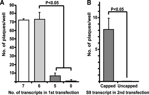
During ranging experiments performed as part of the optimization of the T7 transcript-based reverse-genetics system described for BTV (4), we observed that a second transfection of the complete set of 10 in vitro-synthesized BTV transcripts at 18 h after an initial transfection strongly stimulated the number of rescue events (M. Boyce and P. Roy, unpublished observation). To assess if all 10 transcripts were required in the primary transfection to enable such enhanced virus recovery, and to assess the role of VP6 in the enhancement, BSR cells were transfected with in vitro-synthesized T7 transcripts of only those BTV segments encoding products believed to be associated with primary BTV replication: VP1 (S1), VP4 (S4), VP6 (S9), VP3 (S3), NS1 (S6), and NS2 (S8). In parallel, a first transfection was performed with the same set of transcripts but lacking the VP6-encoding segment (S9). The transfected cells were subsequently transfected a second time 18 h posttransfection with all 10 BTV transcripts, and virus rescue was measured by plaque assay 3 days after the second transfection. Prior transfection with the six primary replication transcripts (S1, S4, S9, S3, S6, and S8) led to an ∼50-fold increase (1 to 3 plaques from no primary transcript versus 68 to 80 plaques from the six primary transcripts) in the number of rescue events compared to a single-transfection rescue (Fig. 1A). The stimulation of virus recovery was not observed when the first transfection mixture lacked the S9 transcript encoding VP6 (Fig. 1A). Moreover, a transfection of the complete six primary transcripts with the addition of a transcript encoding BTV core protein VP7 did not stimulate the number of plaques rescued over and above that observed with the six primary transcripts alone (Fig. 1A). We interpret these data to mean that the primary transfection allows the provision, within the transfected cells, of the proteins that form a competent replicase complex, which then efficiently concludes the replication cycle once the complete complement of BTV segments is added. VP6 is essential for the stimulation observed. To assess if the role of VP6 in stimulating virus recovery was due to the VP6 protein or the presence of the transcript, we carried out a further dual-transfection experiment in which the first transfection lacked S9, as before. However, in the second transfection, we included an uncapped S9 transcript in place of the normal capped variant. Uncapped transcripts should not be translated efficiently unless they are packaged into the assembling subcore, where they can be capped via the action of VP4 (23, 31, 32, 41). Plaque recovery was measured 3 days after the second transfection, as before. Inclusion of uncapped S9 transcript in the second transfection abolished virus rescue from transfected BSR cells, while transfection with the capped S9 transcript led to basal recovery, as expected (Fig. 1B). For these experiments, BTV-10 S9 was used to correspond to our prior studies (19, 39). However, no difference was observed when BTV-1 S9 was used in the reaction mixture instead of BTV-10 S9 (data not shown). These data strongly suggest that virus was not rescued due to lack of the VP6 protein and that VP6 is an essential component of the primary replication process.
FIG. 1.
Recovery of BTV from BSR cells doubly transfected with T7 BTV transcripts. (A) Total number of plaques recovered in each experiment when BSR cells were first transfected with seven transcripts, S1, S3, S4, S6, S7, S8, and S9; with six transcripts, S1, S3, S4, S6, S8 and S9; with five transcripts, S1, S3, S4, S6, and S8; or with no transcripts. All of the transcripts used were capped, and each was at 50 ng per transfection. In each case, the cells were then uniformly transfected a second time with a fixed amount (50 ng of each transcript/0.5 μg total RNA) of all 10 capped transcripts. The means plus standard deviations (SD) are shown. (B) Total number of plaques recovered in each experiment when BSR cells were transfected first with five capped transcripts (S1, S3, S4, S6, and S8) and subsequently with capped or uncapped S9, together with the nine remaining capped transcripts. The recovery of virus is shown as the number of plaques per well. The means and SD are shown.
A stable cell line expressing BTV VP6 that complements VP6 mutant viruses.
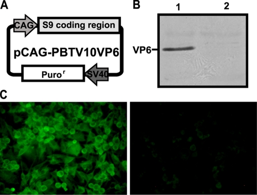
To further investigate the role of VP6 in BTV replication, we generated a stable cell line that expresses VP6 constitutively. To do this, a VP6 expression vector, pCAG-PBTV10VP6, was produced by inserting the CDR of S9 into the mammalian expression vector pCAG-PM (Fig. 2A). After transfection of BSR cells with pCAG-PBTV10VP6, cells with integrated copies of the expression vector were selected by the addition of puromycin. The surviving clones were tested for the expression level of VP6, and based on the intensity of immunoblot positivity, one clone (clone 22-2) was selected as a candidate for a complementary cell line expressing VP6 (Fig. 2B). Immunofluorescence analysis showed that the cells in this cell line (BSR-VP6) expressed VP6, which was detectable throughout the cytosol (Fig. 2C).
FIG. 2.
Generation of a stable cell line, BSR-VP6, expressing VP6 constitutively. (A) Schematic representation of the expression vector, pCAG-PBTV10VP6. VP6 protein was expressed under the control of the CAG promoter, whereas a puromycin resistance gene (Puror) was expressed under the control of the simian virus 40 early promoter. (B) Immunoblot of clone 22-2 with BSR-VP6 (lane 1) and BSR (lane 2) cells. The blot was probed using a rabbit polyclonal antiserum raised against BTV-10 VP6. (C) Immunofluorescence of BSR-VP6 cells (left) and BSR cells (right). Detection of VP6 was performed using a guinea pig polyclonal antiserum raised to BTV-10 VP6, followed by Alexa Fluor 488 goat anti-guinea pig IgG.
To determine if BSR-VP6 cells could provide active VP6 in trans, BSR-VP6 cells were transfected with five BTV T7 transcripts (encoding VP1, VP3, VP4, NS1, and NS2) lacking the transcript encoding VP6, followed 18 h later by a second transfection with all 10 T7 transcripts, as before. The provision of VP6 in trans from the cell line was sufficient to stimulate (at the same level as in Fig. 1A with six transcripts) rescued virus production despite the absence of an S9 transcript in the first transfection mixture (Fig. 3). When BSR-VP6 cells were transfected as described above but with an uncapped S9 transcript as the sole source of VP6 sequence in the second transfection mixture, virus rescue was as efficient as that observed with the use of capped transcripts (Fig. 3). In contrast, when S9 was omitted from both the first and second transfections of BSR-VP6 cells, no virus was rescued (data not shown). These data further confirm that VP6 is essential for early replication of BTV and demonstrate effective substitution of the virus-encoded protein by a complementing cell line. Additionally, unlike some other segmented RNA viruses, a complete set of 10 RNA segments is required for the recovery of infectious BTV (9, 11). This phenomenon has also been reported for reovirus (33).
FIG. 3.
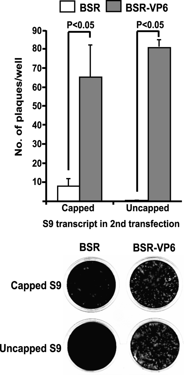
Recovery of BTV from T7 transcripts in BSR cells and BSR-VP6 cells. The recovery of BTV was compared in BSR cells and BSR-VP6 cells (top) transfected first with five capped transcripts (S1, S3, S4, S6, and S8) and subsequently with capped or uncapped S9, together with the nine remaining capped transcripts. Each transcript was at 50 ng per transfection. The recovery of virus is shown as the total number of plaques per well (mean plus SD). The plaque formation visualized in the plates is shown below.
Mapping the sequences of the S9 transcript is essential for virus rescue.
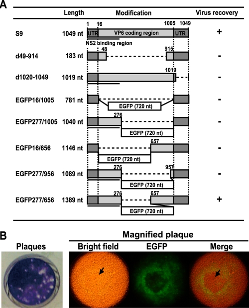
As VP6 protein expressed in the BSR-VP6 cell line functionally substituted for the normally S9-expressed protein in transfected cells, this cell line was used to investigate the cis-acting elements within the S9 RNA essential for packaging and replication. Reassortment between the genome segments of two different serotypes of BTV is commonly observed (8, 17, 37, 40), suggesting that some degree of variation in segments does not abrogate packaging or replication. To identify the sequence requirement for virus rescue, we constructed a series of truncated S9 transcripts, as well as insertion of a foreign gene into the S9 segment (Fig. 4A). In the first construct, the conserved terminal untranslated regions (UTRs) that are believed to be important for virus replication were kept intact while most of the CDR was deleted. This construct, d49-914, included 15 nucleotides (nt) from the 5′ end of the S9 CDR and 91 nt from the 3′ end, as well as both 5′ and 3′ UTRs (Fig. 4A). A second truncated S9 segment, d1020-1049, had 30 nt deleted from the 3′ terminus of S9 in order to confirm that the UTR is necessary for virus replication. This construct included the complete S9 CDR but lacked most of the 3′ UTR, including the conserved region (Fig. 4A). In further constructs, we inserted the EGFP gene at three positions with the S9 transcript to assess if virus consisting of a chimeric segment could be rescued as long as it retained specific regions of the segment. In the first chimeric S9 transcript, EGFP16/1005, the EGFP gene was flanked only by the 5′ and 3′ UTRs. In the second, EGFP277/1005, the EGFP gene was inserted between nt 276 and 1006 of S9. This transcript included both UTRs and the NS2 binding motif hypothesized to recruit single-stranded RNA (ssRNA) into the virus inclusion body (VIB) during core assembly (21, 22, 27). In the last construct, EGFP277/656, the EGFP gene was inserted between nt 276 and 657 of S9. This transcript included 276 nt from the 5′ end and 393 nt from the 3′ end of S9. In EGFP277/1005 and EGFP277/656, the EGFP gene was designed to express EGFP as a fusion protein with the N-terminal 87 amino acids of VP6. Note that a stop codon was inserted at the end of the EGFP sequence for each construct. To avoid the possibility that these proteins might interrupt early virus replication, all modified S9 transcripts were provided as uncapped ssRNAs in a mixture of other capped transcripts.
FIG. 4.
Mapping the sequences of the S9 transcripts essential for virus rescue. (A) Schematic representation of modified S9 transcripts. The NS2 binding region (nt 1 to 275) is shown as black bars. BSR-VP6 cells were transfected with five BTV T7 transcripts (S1, S3, S4, S6, and S8), followed by a second transfection 18 h later with uncapped modified S9 transcripts, together with nine other capped transcripts. Each transcript was at 50 ng per transfection. Virus recovery is shown as a plus in the right column. (B) Plaque formation and the expression of EGFP were observed in BSR-VP6 cells pretransfected with five capped transcripts (S1, S3, S4, S6, and S8), followed by a second transfection with uncapped chimeric S9 EGFP277/656, together with the nine remaining capped transcripts, using a fluorescence microscope. Upon observation of EGFP expression, the cells were stained with 0.2% (wt/vol) crystal violet (left). Cytopathic effect is clearly observed in a magnified plaque (indicated with arrows).
BSR-VP6 cells were transfected with five BTV T7 transcripts (encoding VP1, VP3, VP4, NS1, and NS2), followed by a second transfection 18 h later with each of the uncapped S9 variants, together with the nine remaining capped T7 transcripts. Of all the S9 variants assessed, virus recovery was observed only from BSR-VP6 cells transfected with EGFP277/656, where EGFP expression was clearly visible in each plaque (30 plaques per well) (Fig. 4B). No virus was recovered from BSR-VP6 cells transfected with the other two EGFP-containing chimeric S9 transcripts where the EGFP gene was either flanked only by the conserved 5′ and 3′ UTRs (EGFP16/1005) or by both UTRs and the NS2 binding motif (EGFP277/1005). Similarly, no virus was rescued when the modified S9 transcripts had both the 5′ and 3′ UTRs (d49/914) or only the 5′ UTR and the NS2 binding motif (d1020/1049). These results suggested that the UTR is necessary, but not sufficient, for the recovery of replicating BTV. Similarly, the minimum NS2 binding motif that was previously identified (21, 22) appears insufficient on its own to allow virus rescue, although this region is present in the successful S9 variant EGFP277/656. To validate this further, two other constructs were subsequently made. In one (EGFP16/656), we kept the intact 3′-terminal sequence, similar to EGFP277/656, but deleted the entire 5′-terminal CDR, including the NS2 recognition sequences. The second construct consisted of the same 5′ sequences as EGFP277/656; however, an additional deletion was made in the 3′-terminal region that had only 49 nt of the CDR sequences (EGFP277/956) instead of 349 nt of EGFP277/656. Thus, in EGFP16/656 and EGFP277/956, the EGFP gene was inserted between nt 15 and 657 and between nt 276 and 957, respectively. Both constructs failed to generate any virus in BSR-VP6 cells, in contrast to EGFP277/656. Altogether, these data strongly suggested that the packaging signal of the S9 segment is present within 276 nt at the 5′ end and within 93 nt to 393 nt at the 3′ end. In addition, our data clearly suggest there is no strict size limitation on the S9 transcript, as EGFP277/656 is considerably bigger than wild-type S9 (viz., 1,389 nt versus 1,049 nt). Foreign-gene incorporation into BTV appears viable as long as the essential cis-acting regions are present in the transgene. It was not possible to ascertain from these data whether the cis-acting sequence required for segment rescue acts at the stage of packaging or replication, although, as promoter elements are commonly thought to be restricted to the conserved ends of the segments, packaging may be the more plausible step. It is also unclear if a compatible segment could be rescued in addition to the full 10-segment complement for BTV as for lymphocytic choriomeningitis virus (9).
Characterization of BTVS9EGFP.
The results obtained in the complementing cell line BSR-VP6 showed that EGFP, expressed instead of VP6 in infected cells, could successfully generate BTVS9EGFP, a helper cell line-dependent chimeric virus, as recently reported for Ebola virus (14). As this is the first such chimeric virus reported for BTV, we characterized the virus further. BTVS9EGFP particles, produced in the BSR-VP6 cell line, were purified by sucrose gradient centrifugation, and both protein components and genomic dsRNA were analyzed. The results of the sodium dodecyl sulfate-polyacrylamide gel electrophoresis (Fig. 5A) confirmed that the virion particles contained all seven structural proteins, including VP6, provided by the helper cell line. The result of dsRNA extraction revealed that the EGFP-encoding modified S9 segment was packaged, in addition to the other nine segments (Fig. 5B). Furthermore, to examine the transcribing capability of the BTVS9EGFP core particle, core particles were purified and used for the synthesis of transcripts, which was compared with that of wild-type core. Approximately 10 μg of total ssRNA was produced from 50 μg of both wild-type BTV-1 core and BTVS9EGFP core (data not shown). To confirm the infectivity and helper dependence of the virus, BTVS9EGFP was used to infect both BSR and BSR-VP6 cells. Plaques were observed only in BSR-VP6 cells (Fig. 6A), and within these cells, the level of EGFP expressed was very high, indicative of a productive infection (Fig. 6B, left). Although no plaques were observed in unmodified BSR cells, expression of EGFP was observed, albeit at a low level compared to BSR-VP6 cells (Fig. 6B). This is consistent with a fully functional core able to synthesize the S9EGFP mRNA from virus but unable to amplify EGFP expression through further rounds of replication due to a lack of functional VP6 protein.
FIG. 5.
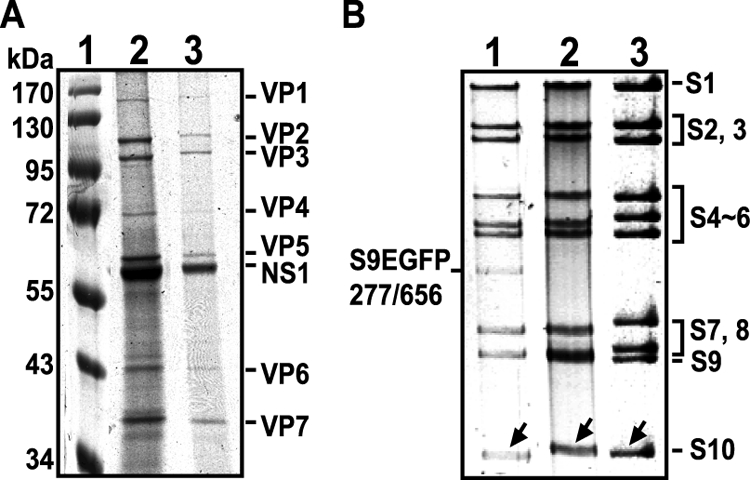
Characterization of the chimeric BTVS9EGFP. (A) Semipurified chimeric BTVS9EGFP and wild-type BTV-1 particles were resolved by sodium dodecyl sulfate-polyacrylamide gel electrophoresis. Lane 1, molecular mass marker; lane 2, semipurified BTVS9EGFP; lane 3, semipurified BTV-1. Note that the NS1 protein is copurified with virus in this one-step, discontinuous sucrose gradient. (B) Comparison of dsRNA segments purified from the chimeric BTVS9EGFP, wild-type BTV-1, and wild-type BTV-10. Lane 1, genomic dsRNA extracted from BSR-VP6 cells infected with BTVS9EGFP run on 9% nondenaturing polyacrylamide; lane 2, dsRNA purified from BSR cells infected with wild-type BTV-1; lane 3, dsRNA purified from BSR cells infected with wild-type BTV-10. Note that the migration patterns of S10 in BTV-1 and BTV-10 are different.
FIG. 6.
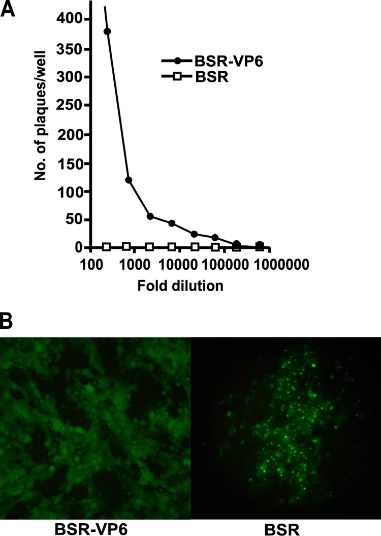
Infectivity assay of BTVS9EGFP. (A) Serial-dilution analysis of BTVS9EGFP. BSR-VP6 cells and BSR cells were infected with BTVS9EGFP stepwise diluted at one in three. The plaques were counted 2 days postinfection. (B) Expression of EGFP was observed in BSR-VP6 cells and BSR cells 2 days postinfection using a fluorescence microscope. (Left) BSR-VP6 cells infected with BTVS9EGFP. (Right) BSR cells infected with BTVS9EGFP. Note that compared to the BSR-VP6 cells, the BSR cells showed EGFP as distinct punctae.
DISCUSSION
The replication cycle of BTV and other members of the Reoviridae differs from those of other RNA viruses, as during virus disassembly, transcription complexes are not released from the capsid structure but remain within the inner capsid or core (36). In order to amplify the virus replication cycle, BTV likely assembles a similar early replicase complex, which subsequently acts to increase the levels of transcripts from all 10 genomic segments. Data in favor of an early replicase complex have been lacking, however, in part as a result of the early part of the cycle being occluded into VIBs primarily composed of NS2 (7, 18, 27, 42). We probed the assembly of an early replicase complex through the opportunistic finding that repeated transfection of BTV transcripts generated in vitro improved the recovery of infectious BTV by ∼50-fold compared to single transfection alone (4). We determined that an equivalent stimulation was possible using only six transcripts, encoding VP1, VP3, VP4, VP6, NS1, and NS2. These proteins represent the presumptive transcriptase complex (VP1, VP4, and VP6), the major subcore protein (VP3), and the two NS proteins associated with VIBs and virus replication. Interestingly, the addition of a transcript encoding VP7, which would allow completion of the core particle, did not further stimulate virus recovery, suggesting that a fully competent replicase complex requires only a substructure composed of VP3. Our data echo the findings in influenza A virus that the virus nucleocapsid protein is required for viral-mRNA synthesis by the polymerase complex (1, 16, 29), although an active BTV replicase complex appears to require more than the single VP3 structural component. The role played by the NS proteins was not investigated in our studies, but based on its RNA binding activity, NS2 may be responsible for the sequestering of early RNA complexes and may also protect the transcripts from turnover. Although the precise role of NS1 is not well defined yet, our recent data suggest that it may be responsible for enhancing BTV protein synthesis (Boyce and Roy, unpublished).
BTV is unique among the Reoviridae in encoding VP6, a protein with nucleoside triphosphatase, RNA binding, and helicase activities (19, 20, 39). We found that inclusion of an S9 transcript was essential for the observed stimulation of virus recovery by a primary transfection with six transcripts, supporting a role early in replication. Although these data do not directly address the role of VP6 as an RNA-packaging protein, we show positive data for the alternative explanation of its action: that it is an integral part of a transcription complex necessary for primary replication. This lessens the likelihood that VP6 plays another role in packaging, although it remains possible.
VP6 protein provided in trans by a constructed helper cell line, BSR-VP6, led to the recovery of virus at the same level as that observed with transfection of T7 transcript-based VP6. These data allowed the manipulation of the S9 segment for delineation of the cis-acting sequences required for successful incorporation into rescued virus. A similar system has recently been reported for Ebola virus (14). A helper cell line-dependent Ebola virus, requiring the VP30 product, has recently been described as a vaccine candidate using a reverse genetics system (13). Using a number of S9 deletions and insertions, we showed successful rescue of an EGFP-encoding segment, S9EGFP277/656, in the VP6 helper cell line. Alignment of the S9 variants revealed a requirement for the conserved ends of the segment and additional sequence from the 5′ and 3′ UTRs and CDR. These include the sequence mapped as binding NS2 protein and responsible for recruiting RNA to the VIB (18, 22). However, this sequence alone was not sufficient for rescue. We did not investigate the construction of a virus with fewer segments, as reported for influenza A virus, where a seven-segmented virus was recovered (11). However, a recent report on the reovirus packaging signal suggests that not only the packaging signals of 10 ssRNAs are important for genome assortment, but that a complete set of all 10 ssRNAs is also essential (33). The finite structure of the BTV core (12) also suggests a packaging limit for the virus, but as the S9 EGFP277/656 construct is marginally larger than S9 alone, some additional carriage capacity is clearly available. It will be interesting to attempt the construction of a virus carrying additional segments as, for example, a multiple-serotype vaccine candidate. We cannot distinguish the precise roles of the sequences required for rescue of S9 EGFP277/656. Packaging is the most plausible role, but a role in replication after packaging cannot be excluded. Our data suggest that capping of transcripts is not an essential packaging signal, as within BSR-VP6 cells, a second transfection with uncapped transcripts resulted in rescue as efficient as the use of capped transcripts. Further study is therefore necessary to define the precise components of the S9 packaging signal and to investigate the presence of equivalent structures on other segments. However, practical use of the S9 EGFP277/656 segment to produce an attenuated and marked single-round replication vaccine strain, a DISC vaccine (38), is now a reality.
Acknowledgments
We thank I. M. Jones (University of Reading, United Kingdom) and R. Noad (LSHTM) for critical reading of the manuscript.
This work is supported by the BBSRC (United Kingdom) and partly by NIH.
Footnotes
Published ahead of print on 24 June 2009.
REFERENCES
- 1.Baudin, F., C. Bach, S. Cusack, and R. W. Ruigrok. 1994. Structure of influenza virus RNP. I. Influenza virus nucleoprotein melts secondary structure in panhandle RNA and exposes the bases to the solvent. EMBO J. 133158-3165. [DOI] [PMC free article] [PubMed] [Google Scholar]
- 2.Beaton, A. R., J. Rodriguez, Y. K. Reddy, and P. Roy. 2002. The membrane trafficking protein calpactin forms a complex with bluetongue virus protein NS3 and mediates virus release. Proc. Natl. Acad. Sci. USA 9913154-13159. [DOI] [PMC free article] [PubMed] [Google Scholar]
- 3.Bhattacharya, B., R. J. Noad, and P. Roy. 2007. Interaction between Bluetongue virus outer capsid protein VP2 and vimentin is necessary for virus egress. Virol. J. 47. [DOI] [PMC free article] [PubMed] [Google Scholar]
- 4.Boyce, M., C. C. Celma, and P. Roy. 2008. Development of reverse genetics systems for bluetongue virus: recovery of infectious virus from synthetic RNA transcripts. J. Virol. 828339-8348. [DOI] [PMC free article] [PubMed] [Google Scholar]
- 5.Boyce, M., and P. Roy. 2007. Recovery of infectious bluetongue virus from RNA. J. Virol. 812179-2186. [DOI] [PMC free article] [PubMed] [Google Scholar]
- 6.Boyce, M., J. Wehrfritz, R. Noad, and P. Roy. 2004. Purified recombinant bluetongue virus VP1 exhibits RNA replicase activity. J. Virol. 783994-4002. [DOI] [PMC free article] [PubMed] [Google Scholar]
- 7.Brookes, S. M., A. D. Hyatt, and B. T. Eaton. 1993. Characterization of virus inclusion bodies in bluetongue virus-infected cells. J. Gen. Virol. 74525-530. [DOI] [PubMed] [Google Scholar]
- 8.de Mattos, C. C., C. A. de Mattos, B. I. Osburn, M. Ianconescu, and R. Kaufman. 1991. Evidence of genome segment 5 reassortment in bluetongue virus field isolates. Am. J. Vet. Res. 521794-1798. [PubMed] [Google Scholar]
- 9.Emonet, S. F., L. Garidou, D. B. McGavern, and J. C. de la Torre. 2009. Generation of recombinant lymphocytic choriomeningitis viruses with trisegmented genomes stably expressing two additional genes of interest. Proc. Natl. Acad. Sci. USA 1063473-3478. [DOI] [PMC free article] [PubMed] [Google Scholar]
- 10.Finke, S., K. Brzozka, and K. K. Conzelmann. 2004. Tracking fluorescence-labeled rabies virus: enhanced green fluorescent protein-tagged phosphoprotein P supports virus gene expression and formation of infectious particles. J. Virol. 7812333-12343. [DOI] [PMC free article] [PubMed] [Google Scholar]
- 11.Gao, Q., E. W. Brydon, and P. Palese. 2008. A seven-segmented influenza A virus expressing the influenza C virus glycoprotein HEF. J. Virol. 826419-6426. [DOI] [PMC free article] [PubMed] [Google Scholar]
- 12.Grimes, J. M., J. N. Burroughs, P. Gouet, J. M. Diprose, R. Malby, S. Zientara, P. P. Mertens, and D. I. Stuart. 1998. The atomic structure of the bluetongue virus core. Nature 395470-478. [DOI] [PubMed] [Google Scholar]
- 13.Halfmann, P., H. Ebihara, A. Marzi, Y. Hatta, S. Watanabe, M. Suresh, G. Neumann, H. Feldmann, and Y. Kawaoka. 2009. Replication-deficient ebola virus as a vaccine candidate. J. Virol. 833810-3815. [DOI] [PMC free article] [PubMed] [Google Scholar]
- 14.Halfmann, P., J. H. Kim, H. Ebihara, T. Noda, G. Neumann, H. Feldmann, and Y. Kawaoka. 2008. Generation of biologically contained Ebola viruses. Proc. Natl. Acad. Sci. USA 1051129-1133. [DOI] [PMC free article] [PubMed] [Google Scholar]
- 15.Huismans, H., A. A. van Dijk, and H. J. Els. 1987. Uncoating of parental bluetongue virus to core and subcore particles in infected L cells. Virology 157180-188. [DOI] [PubMed] [Google Scholar]
- 16.Inumaru, S., H. Ghiasi, and P. Roy. 1987. Expression of bluetongue virus group-specific antigen VP3 in insect cells by a baculovirus vector: its use for the detection of bluetongue virus antibodies. J. Gen. Virol. 681627-1635. [DOI] [PubMed] [Google Scholar]
- 17.Kahlon, J., K. Sugiyama, and P. Roy. 1983. Molecular basis of bluetongue virus neutralization. J. Virol. 48627-632. [DOI] [PMC free article] [PubMed] [Google Scholar]
- 18.Kar, A. K., B. Bhattacharya, and P. Roy. 2007. Bluetongue virus RNA binding protein NS2 is a modulator of viral replication and assembly. BMC Mol. Biol. 84. [DOI] [PMC free article] [PubMed] [Google Scholar]
- 19.Kar, A. K., and P. Roy. 2003. Defining the structure-function relationships of bluetongue virus helicase protein VP6. J. Virol. 7711347-11356. [DOI] [PMC free article] [PubMed] [Google Scholar]
- 20.Luking, A., U. Stahl, and U. Schmidt. 1998. The protein family of RNA helicases. Crit. Rev. Biochem. Mol. Biol. 33259-296. [DOI] [PubMed] [Google Scholar]
- 21.Lymperopoulos, K., R. Noad, S. Tosi, S. Nethisinghe, I. Brierley, and P. Roy. 2006. Specific binding of Bluetongue virus NS2 to different viral plus-strand RNAs. Virology 35317-26. [DOI] [PMC free article] [PubMed] [Google Scholar]
- 22.Lymperopoulos, K., C. Wirblich, I. Brierley, and P. Roy. 2003. Sequence specificity in the interaction of Bluetongue virus non-structural protein 2 (NS2) with viral RNA. J. Biol. Chem. 27831722-31730. [DOI] [PubMed] [Google Scholar]
- 23.Martinez-Costas, J., G. Sutton, N. Ramadevi, and P. Roy. 1998. Guanylyltransferase and RNA 5′-triphosphatase activities of the purified expressed VP4 protein of bluetongue virus. J. Mol.r Biol. 280859-866. [DOI] [PubMed] [Google Scholar]
- 24.Matsuo, E., H. Tani, C. Lim, Y. Komoda, T. Okamoto, H. Miyamoto, K. Moriishi, S. Yagi, A. H. Patel, T. Miyamura, and Y. Matsuura. 2006. Characterization of HCV-like particles produced in a human hepatoma cell line by a recombinant baculovirus. Biochem. Biophys. Res. Commun. 340200-208. [DOI] [PubMed] [Google Scholar]
- 25.Mertens, P. P., J. N. Burroughs, and J. Anderson. 1987. Purification and properties of virus particles, infectious subviral particles, and cores of bluetongue virus serotypes 1 and 4. Virology 157375-386. [DOI] [PubMed] [Google Scholar]
- 26.Mertens, P. P., and J. Diprose. 2004. The bluetongue virus core: a nano-scale transcription machine. Virus Res. 10129-43. [DOI] [PubMed] [Google Scholar]
- 27.Modrof, J., K. Lymperopoulos, and P. Roy. 2005. Phosphorylation of bluetongue virus nonstructural protein 2 is essential for formation of viral inclusion bodies. J. Virol. 7910023-10031. [DOI] [PMC free article] [PubMed] [Google Scholar]
- 28.Nason, E. L., R. Rothagel, S. K. Mukherjee, A. K. Kar, M. Forzan, B. V. V. Prasad, and P. Roy. 2004. Interactions between the inner and outer capsids of bluetongue virus. J. Virol. 788059-8067. [DOI] [PMC free article] [PubMed] [Google Scholar]
- 29.Newcomb, L. L., R. L. Kuo, Q. Ye, Y. Jiang, Y. J. Tao, and R. M. Krug. 2009. Interaction of the influenza a virus nucleocapsid protein with the viral RNA polymerase potentiates unprimed viral RNA replication. J. Virol. 8329-36. [DOI] [PMC free article] [PubMed] [Google Scholar]
- 30.Owens, R. J., C. Limn, and P. Roy. 2004. Role of an arbovirus nonstructural protein in cellular pathogenesis and virus release. J. Virol. 786649-6656. [DOI] [PMC free article] [PubMed] [Google Scholar]
- 31.Ramadevi, N., N. J. Burroughs, P. P. C. Mertens, I. M. Jones, and P. Roy. 1998. Capping and methylation of mRNA by purified recombinant VP4 protein of bluetongue virus. Proc. Natl. Acad. Sci. USA 9513537-13542. [DOI] [PMC free article] [PubMed] [Google Scholar]
- 32.Ramadevi, N., and P. Roy. 1998. Bluetongue virus core protein VP4 has nucleoside triphosphate phosphohydrolase activity. J. Gen. Virol. 792475-2480. [DOI] [PubMed] [Google Scholar]
- 33.Roner, M. R., and B. G. Steele. 2007. Localizing the reovirus packaging signals using an engineered m1 and s2 ssRNA. Virology 35889-97. [DOI] [PubMed] [Google Scholar]
- 34.Roy, P. 2008. Bluetongue virus: dissection of the polymerase complex. J. Gen. Virol. 891789-1804. [DOI] [PMC free article] [PubMed] [Google Scholar]
- 35.Roy, P. 2008. Functional mapping of bluetongue virus proteins and their interactions with host proteins during virus replication. Cell Biochem. Biophys. 50143-157. [DOI] [PubMed] [Google Scholar]
- 36.Roy, P. 1996. Orbivirus structure and assembly. Virology 2161-11. [DOI] [PubMed] [Google Scholar]
- 37.Roy, P. 1981. Structural characteristics of Spring Viremia of Carp Virus, p. 623-629. In D. H. L. Bishop and R. W. Compans (ed.), Replication of negative strand viruses. Elsevier, North Holland, Amsterdam, The Netherlands.
- 38.Roy, P., M. Boyce, and R. Noad. 2009. Prospects for improved bluetongue vaccines. Nat. Rev. Microbiol. 7120-128. [DOI] [PubMed] [Google Scholar]
- 39.Stauber, N., J. Martinez-Costas, G. Sutton, K. Monastyrskaya, and P. Roy. 1997. Bluetongue virus VP6 protein binds ATP and exhibits an RNA-dependent ATPase function and a helicase activity that catalyze the unwinding of double-stranded RNA substrates. J. Virol. 717220-7226. [DOI] [PMC free article] [PubMed] [Google Scholar]
- 40.Sugiyama, K., D. H. L. Bishop, and P. Roy. 1982. Analyses of the genomes of bluetongue viruses recovered from different states of the United States and at different times. Am. J. Epidemiol. 115332-347. [DOI] [PubMed] [Google Scholar]
- 41.Sutton, G., J. M. Grimes, D. I. Stuart, and P. Roy. 2007. Bluetongue virus VP4 is an RNA-capping assembly line. Nat. Struct. Mol. Biol. 14449-451. [DOI] [PubMed] [Google Scholar]
- 42.Thomas, C. P., T. F. Booth, and P. Roy. 1990. Synthesis of bluetongue virus-encoded phosphoprotein and formation of inclusion bodies by recombinant baculovirus in insect cells: it binds the single-stranded RNA species. J. Gen.l Virol. 712073-2083. [DOI] [PubMed] [Google Scholar]
- 43.Van Dijk, A. A., and H. Huismans. 1988. In vitro transcription and translation of bluetongue virus mRNA. J. Gen. Virol. 69573-581. [DOI] [PubMed] [Google Scholar]
- 44.Verwoerd, D. W. 1969. Purification and characterization of bluetongue virus. Virology 38203-212. [DOI] [PubMed] [Google Scholar]
- 45.Verwoerd, D. W., and H. Huismans. 1972. Studies on the in vitro and the in vivo transcription of the bluetongue virus genome. Onderstepoort J. Vet. Res. 39185-191. [PubMed] [Google Scholar]
- 46.Wehrfritz, J. M., M. Boyce, S. Mirza, and P. Roy. 2007. Reconstitution of Bluetongue virus polymerase activity from isolated domains based on a three-dimensional structural model. Biopolymers 8683-94. [DOI] [PMC free article] [PubMed] [Google Scholar]
- 47.Wirblich, C., B. Bhattacharya, and P. Roy. 2006. Nonstructural protein 3 of bluetongue virus assists virus release by recruiting ESCRT-I protein Tsg101. J. Virol. 80460-473. [DOI] [PMC free article] [PubMed] [Google Scholar]
- 48.Zarbl, H., and S. Millward. 1983. The reovirus multiplication cycle, p. 107-196. In W. Joklik (ed.), The Reoviridae. Springer, New York, NY.


