Abstract
Influenza virus has a set of ribonucleoproteins (RNPs) consisting of viral RNAs, influenza virus polymerase subunits, and nucleoprotein. Intracellular reconstitution of the whole set of RNPs via plasmid transfection results in the generation of influenza virus. By the use of reverse genetics and dual promoters, we constructed a 23.6-kb eight-unit plasmid that contains all the required constituents to generate influenza virus in chicken cells. Our “one-plasmid” system generated higher titers of influenza virus in chicken cells than the “eight-plasmid” system, enabling a simpler approach for generating vaccine seeds. Our study identified plasmid size as a potential limiting factor affecting transfection efficiency and hence the influenza viral yield from chicken cells.
The three recorded pandemics and most yearly global outbreaks of influenza are caused by influenza A virus. The virus belongs to the family Orthomyxoviridae and contains a segmented negative-strand RNA genome. Unlike with other RNA viruses, replication and transcription of influenza virus takes place in the host nucleus (12). The viral RNAs (vRNAs) are associated with influenza RNA polymerase B1, B2, and A proteins (PBl, PB2, PA) and nucleoprotein (NP) to make up a set of ribonucleoproteins (11, 15, 18).
Effort to construct recombinant influenza virus using modern genetic tools for potential application in vaccines has escalated since the early 1990s. The primary objective is to generate influenza virus from the least number of plasmid constructs that can be transfected into cultured cells to provide high viral yields. By combining intracellular synthesis of vRNAs and proteins, two reverse-genetics systems were established to generate influenza virus from cultured cells by cotransfection of 12 to 17 plasmids that together express influenza vRNA and mRNA. Although these techniques provided viral yield, the use of a large number of plasmid constructs still remained a disadvantage (6, 17).
The “ambisense” approach, which utilizes two promoters on a bidirectional transcription vector, is the first major breakthrough to reduce the number of plasmids required for virus generation (9). In this approach, the RNA polymerase I (PolI) promoter drives the synthesis of vRNA from a cDNA template, whereas the RNA PolII promoter drives the synthesis of mRNA from the same template in the opposite direction. A unidirectional PolI-PolII transcription system was also reported; however, it suffers from lower viral yield. A much improved method is the generation of influenza virus using a three-plasmid reverse-genetics system (16). Here, one plasmid carries eight PolI promoter-driven vRNA-transcribing cassettes, another plasmid encodes the three viral polymerase subunits, and the third plasmid provides NP. This three-plasmid system, although arduous to construct, yields higher titers of influenza virus than the earlier approaches (16).
Generation of influenza virus from any of the above-mentioned plasmid constructs requires transfection of DNA into cultured cells. In addition to various DNA transfection methods, bacterium-mediated gene transfer, known as bactofection, is a well-established method (8, 23). Salmonella enterica, Shigella spp., and other invasive bacteria are employed as vectors to deliver DNA vaccines into host cells (1, 4, 13, 21, 22). Inspired by both techniques, we wanted to develop a plasmid system that can be delivered in vivo by a bacterial carrier, resulting in the synthesis of viral proteins and vRNAs for recovering an attenuated influenza virus. The virus would serve as an attenuated form of influenza vaccine. The three-plasmid system is by far the most streamlined system for generating attenuated influenza virus (16). However, it may not be possible to ensure maintenance and delivery of all three plasmids by a single bacterium or their translocation into the host nucleus. Hence, developing a much more comprehensive plasmid system to generate influenza virus is highly desired.
As a first step toward achieving our long-term goal, we developed a single plasmid that carries all the genetic elements required to produce influenza virus in chicken cells. Recovery of virus from cultured chicken cells using this novel one-plasmid system was comparable to or better than that of the eight-plasmid system.
MATERIALS AND METHODS
Bacterial strain, enzymes, and plasmids.
EPI300 chemically competent Escherichia coli (Epicentre) was used for all DNA cloning experiments. Restriction enzyme SrfI was bought from Stratagene (La Jolla, CA). All other restriction enzymes were from New England Biolabs (Ipswich, MA). Plasmids pTM-PolI-WSN-All and pCAWS-NP were kindly provided by Yoshihiro Kawaoka (University of Wisconsin—Madison). Plasmid pYS1190 was a gift from Yixin Shi (Arizona State University). Plasmid constructs used in this study are listed in Table 1.
TABLE 1.
Plasmids used in this study
| Plasmid | Characteristic(s) | Reference |
|---|---|---|
| pcDNA3.1(−) | Eukaryotic expression vector carrying a CMV promoter and BGH polyadenylation signal | Invitrogen |
| pIRES-EGFP | Source of the EGFP gene | Clontech |
| pYS1190 | Source of the mCherry gene | Unpublished |
| pTM-PolI-WSN-All | Eight-unit plasmid for transcribing PB2, PB1, PA, NP, HA, NA, M, and NS vRNAs by means of HPI | 16 |
| pCAWS-NP | Eukaryotic expression of NP used as the helper plasmid | 16 |
| pYA3994 | Source of prokaryotic GFP cassette | Lab collection |
| pYA4464 | Vector with p15A ori sequence and Cmr cassette | Lab collection |
| pYA4749 | GFP expression vector with p15A ori constructed by fusing DNA segments from pYA3994 and pYA4464 | This study |
| pYA4337 | PB2 gene inserted into pcDNA3.1(−) | This study |
| pYA4338 | PB1 gene inserted into pcDNA3.1(−) | This study |
| pYA4339 | PA gene inserted into pcDNA3.1(−) | This study |
| pYA4379 | CPI and MTI cloned into pcDNA3.1(−) to create a bidirectional vector to synthesize vRNA by means of CPI and mRNA by means of the CMV promoter | This study |
| pYA4383 | PB2 cDNA cloned into pYA4379 to synthesize mRNA by means of the CMV promoter and vRNA by means of CPI | This study |
| pYA4384 | PB1 cDNA cloned into pYA4379 to synthesize mRNA by means of the CMV promoter and vRNA by means of CPI | This study |
| pYA4385 | PA cDNA cloned into pYA4379 to synthesize mRNA by means of the CMV promoter and vRNA by means of CPI | This study |
| pYA4386 | NP cDNA cloned into pYA4379 to synthesize mRNA by means of the CMV promoter and vRNA by means of CPI | This study |
| pYA4387 | EGFP gene cloned into pYA4379 to synthesize mRNA by means of the CMV promoter and antisense RNA (vRNA-like) by means of CPI | This study |
| pYA4380 | CPI and MTI cloned into modified pcDNA3.1(−) to synthesize vRNA | This study |
| pYA4388 | HA cDNA inserted into the AarI sites in pYA4380 to synthesize vRNA by means of CPI | This study |
| pYA4389 | NA cDNA cloned into pYA4380 to synthesize vRNA by means of CPI | This study |
| pYA4390 | M cDNA cloned into pYA4380 to synthesize vRNA by means of CPI | This study |
| pYA4391 | NS cDNA cloned into pYA4380 to synthesize vRNA by means of CPI | This study |
| pYA4392 | EGFP gene cloned into pYA4380 to transcribe antisense RNA (vRNA-like) by means of CPI | This study |
| pYA4688 | CPI replaced with HPI in pYA4392 to transcribe the EGFP gene into antisense RNA (vRNA-like) | This study |
| pYA4519 | Eight influenza cDNA cassettes cloned into one plasmid to synthesize vRNAs by means of CPI and PB2, PB1, PA, and NP mRNA by means of the CMV promoter | This study |
| pYA4731 | mCherry gene cloned between the CMV promoters and BGH terminator in pcDNA3.1(−) | This study |
| pYA4732 | CMV-mCherry-BGH cassette from pYA4731 inserted into pYA4519 at the SrfI site | This study |
Cell culture.
Chicken embryonic fibroblasts (CEFs) were prepared by standard trypsinization of decapitated 8-day embryos. CEFs, human embryonic kidney (HEK293) cells, and Madin-Darby canine kidney (MDCK) cells were maintained in Dulbecco's modified Eagle's medium supplemented with 10% fetal bovine serum, 100 U/ml penicillin, and 100 μg/ml streptomycin. To coculture CEFs and MDCK cells, each type of cell, grown in 75-cm2 flasks, was detached with trypsin, and one-third volume of each was mixed with growth medium to a total volume of 40 ml. The mixed cells were seeded into six-well plates at 3 ml per well. All cells were maintained at 37°C in 5% CO2.
Construction of CPI-based reporter plasmids.
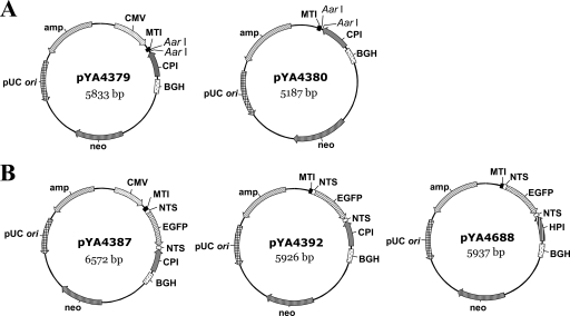
Plasmid pcDNA3.1(−) (Invitrogen, Carlsbad, CA) carrying the cytomegalovirus (CMV) promoter and the bovine growth hormone (BGH) polyadenylation signal, which together form the PolII promoter-terminator system, was used to construct the vector pYA4379. Briefly, the chicken PolI promoter (CPI) was amplified from chicken genomic DNA (14). The truncated (41-bp) murine PolI terminator (MTI) was amplified from plasmid pTM-PolI-WSN-All. By using unique enzyme sites introduced by PCR, the CPI region (nucleotides −415 to −1) and MTI (41 bp) were connected at the KpnI site and placed in between NheI and HindIII on pcDNA3.1(−) downstream of the CMV promoter to construct the bidirectional transcription vector pYA4379 (see Fig. 1A). The two AarI sites introduced in between CPI and MTI allow cloning of the insert without introduction of any additional nucleotides at either end. Plasmid pYA4380 was constructed by excising the CMV promoter fragment from pcDNA3.1(−) using enzymes SpeI and HindIII, followed by insertion of the CPI-MTI fusion product (see Fig. 1A).
FIG. 1.
Construction of dual- and single-promoter plasmids and their derivatives. (A) CPI and MTI, which together comprise the PolI promoter-terminator system, were cloned into the HindIII and NheI sites on the eukaryotic expression vector pcDNA3.1(−), which carries CMV promoter and BGH terminator sequences, to construct a bidirectional dual-promoter plasmid, pYA4379, or a variant of it, pYA4380, which lacks the CMV promoter element. (B) A 720-bp-long EGFP gene fragment flanked on the 5′ and 3′ ends with NTS from the M segment of WSN virus was cloned into the AarI sites in pYA4379 and pYA4380 to construct reporter plasmids pYA4387 and pYA4392, respectively. Plasmid pYA4688 was constructed by replacing CPI with HPI in pYA4392. Plasmids are not drawn to scale.
Plasmid pIRES-EGFP (Clontech, Mountain View, CA) was the source of the enhanced green fluorescent protein (EGFP) gene, used to measure promoter activities in plasmids pYA4379 and pYA4380. The EGFP gene was amplified by PCR from pIRES-EGFP using primers that introduce 5′ and 3′ nontranslating sequences (NTS) from the M segment of the influenza WSN virus. The 5′-NTS-EGFP-NTS-3′ fragment was cloned into the AarI sites in between CPI and MTI in plasmid pYA4379 and in pYA4380 to obtain plasmids pYA4387 and pYA4392, respectively (see Fig. 1B). Plasmid pYA4688 was derived from pYA4392 by replacing CPI with the human PolI promoter (HPI) obtained from pTM-PolI-WSN-All (see Fig. 1B). Genes encoding PB2, PB1, and PA were individually cloned into plasmid pcDNA3.1(−) to obtain plasmids pYA4337, pYA4338, and pYA4339, respectively. In transfection experiments, those three plasmids were used in combination with plasmid pCAWS-NP to provide viral polymerase and NP.
Construction of the eight-unit plasmid pYA4519.
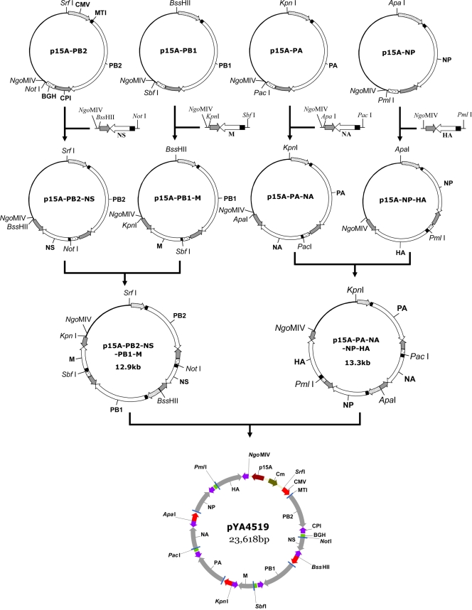
The eight-unit plasmid pYA4519 was constructed in four stages. (i) We first constructed eight one-unit plasmids. Plasmids pTM-PolI-WSN-All provides the whole set of genomic cDNAs of influenza A/WSN/33 virus. The cDNA fragments encoding PB2, PB1, PA, and NP were individually transferred into the AarI sites on pYA4379 to obtain plasmids pYA4383, pYA4384, pYA4385, and pYA4386, respectively (Table 1). cDNAs of hemagglutinin (HA), neuraminidase (NA), the matrix protein (M), and the nonstructural protein (NS) were similarly cloned into pYA4380 to obtain plasmids pYA4388, pYA4389, pYA4390, and pYA4391, respectively (Table 1). (ii) To construct the cloning vector p15A-T, a DNA fragment containing p15A ori and the Cmr gene from plasmid pYA4464 was fused with the GFP bacterial expression cassette that had been amplified from pYA3994 using primers that introduced an AhdI site on either end of the DNA fragment. The resulting plasmid was named pYA4749. The GFP cassette from plasmid pYA4749 was excised with AhdI, leaving behind a linear 2,530-bp vector with single 3′-T overhangs. This linear vector was called p15A-T and was used for convenient insertion of any DNA with single 3′-A overhangs. (iii) To clone viral mRNA expression cassettes into p15A-T, cDNA cassettes for PB2, PB1, PA, and NP, along with their promoter-terminator bidirectional elements, were individually amplified from pYA4383, pYA4384, pYA4385, and pYA4386, respectively. Purified fragments were each ligated with the p15A-T vector to obtain four one-unit plasmids: p15A-PB2, p15A-PB1, p15A-PA, and p15A-NP (see Fig. 3, upper panel). To construct two-unit plasmids, vRNA expression cassettes of the remaining four viral genes (HA, NA, M, and NS) were amplified from plasmids pYA4388, pYA4389, pYA4390, and pYA4391, respectively, and cloned into engineered unique restriction sites on each of the one-unit plasmids (see Fig. 3). As a stepwise incremental process, cDNA fragments from two two-unit plasmids were fused to obtain a four-unit plasmid. The structures of the two four-unit plasmids, p15A-PB2-NS-PB1-M (12.9 kb) and p15A-PA-NA-NP-HA (13.3 kb), are shown (see Fig. 3). (iv) To fuse eight cDNA cassettes into a single plasmid, the fragment containing the PA, NA, NP, and HA cassettes was excised from p15-PA-NA-NP-HA and cloned in between the KpnI and NgoMIV sites in the other four-unit plasmid to obtain the eight-unit plasmid pYA4519.
FIG. 3.
Stepwise construction of the eight-unit plasmid pYA4519. (A) PB2, PB1, PA, and NP bidirectional cassettes were amplified from pYA4379-derived plasmids, and each cassette was individually cloned into a single p15A-T vector to obtain four one-unit plasmids: p15A-PB2, p15A-PB1, p15A-PA, and p15A-NP. (B) The NS, M, NA, and HA vRNA-transcribing cassettes were amplified from pYA4380-derived plasmids by introducing compatible restriction sites and were each cloned into the one-unit plasmid to obtain four two-unit vectors: p15A-PB2-NS, p15A-PB1-M, p15A-PA-NA, and p15A-NP-HA. (C) Each of the two four-unit plasmids, p15A-PB2-NS-PB1-M and p15A-PA-NA-NP-HA, was constructed by fusing influenza virus cassettes from the two two-unit plasmids shown in panel B. (D) The fragment containing PA, NA, NP, and HA cassettes was excised using KpnI and NgoMIV and ligated into the compatible sites in the four-unit plasmid p15A-PB2-NS-PB1-M to obtain a 23.6-kb-long eight-unit plasmid, pYA4519, to transcribe the whole set of influenza vRNAs via CPI and to synthesize influenza virus RNA polymerase (PB1, PB2, PA) and NP by the CMV promoter. All constructs carry p15A ori. Plasmids are not drawn to scale. p15A-PB1, polymerase B1 cDNA cassette cloned into the p15A-T vector; p15A-PB2, polymerase B2 cDNA cassette cloned into the p15A-T vector; p15A-PA, polymerase A cDNA cassette cloned into the p15A-T vector; p15A-NP, NP cDNA cassette cloned into the p15A-T vector.
Plasmid pYA4519 is a 23.6-kb-long plasmid containing a unique restriction site between every two cassettes and a plasmid backbone to facilitate either any addition or the replacement of genes in this plasmid. A seven-unit plasmid was also constructed by deleting the HA expression cassette from pYA4519 to serve as a control. During the construction of four-unit, seven-unit, and eight-unit plasmids, large DNA fragments were stained with crystal violet to avoid the DNA-damaging effect of UV light (20).
The 711-bp mCherry gene was amplified from pYS1190 (Table 1) and cloned in between the CMV promoter and BGH terminator sequences on plasmid pcDNA3.1(−) to generate the plasmid pYA4731 (pcDNA-mCherry). The CMV-mCherry-BGH cassette was transferred to the SrfI site on plasmid pYA4519 to obtain pYA4732 (pYA4519-mCherry) (Table 1).
Transfection.
CEFs and HEK293 cells grown in six-well plates were transfected according to the manufacturer's instructions. Briefly, 2 μl of Lipofectamine 2000 (Invitrogen) per μg plasmid DNA was diluted in 100 μl of Opti-MEM. After a 5-min incubation at room temperature, the diluted transfection reagent was mixed with the DNA. After a 40-min incubation at room temperature, the transfection mix was added to prewashed cells. Three hours later, the transfection medium was replaced with Dulbecco's modified Eagle's medium supplemented with 10% fetal bovine serum. At 24 h posttransfection, images were acquired using a Zeiss AxioCam Mrc 5 camera mounted onto the Zeiss Axioskop 40 fluorescence microscope.
Virus generation.
For generation of influenza virus, CEFs or cocultured CEF and MDCK cells were transfected with plasmid DNA as described above. After 3 h of incubation, the transfection medium was replaced with 2 ml of Opti-MEM containing 0.3% bovine serum albumin, penicillin, and streptomycin. At 24 h posttransfection, each well was supplemented with 1 ml of Opti-MEM containing 2 μg/ml tosylsulfonyl phenylalanyl chloromethyl ketone-trypsin, 0.3% bovine serum albumin, penicillin, and streptomycin. At 3 to 6 days posttransfection, cell supernatants were titrated onto MDCK cell monolayers to estimate influenza virus titers. All experiments were done in triplicate.
RESULTS
EGFP expression in vectors with a dual- or single-promoter unit.
The bidirectional dual-promoter transcription vector pYA4379 was constructed by inserting PolI promoter-terminator elements in plasmid pcDNA3.1(−). Here, the CMV promoter and BGH polyadenylation signal together constitute the PolII promoter-terminator unit that synthesizes mRNA from cDNA, whereas CPI and MTI together constitute the PolI promoter-terminator unit that transcribes the antisense RNA of the target gene (Fig. 1A). Alternatively, the vector pYA4380, containing the PolI unit but lacking the CMV promoter, was created for the synthesis of antisense RNA alone (Fig. 1A). Plasmids pYA4387 and pYA4392 were derived from pYA4379 and pYA4380, respectively, by inserting the EGFP gene flanked with 5′ and 3′ NTS of the M segment from influenza A/WSN/33 virus (Fig. 1B). Another plasmid, pYA4688, was derived from pYA4392 (pYA4380-EGFP) by replacing CPI with HPI (Fig. 1B).
To test the promoter activity in each plasmid, CEFs were independently transfected with plasmids pYA4387 (CMV-MTI-EGFP-CPI-BGH) and pYA4392 (MTI-EGFP-CPI) and HEK293 cells were transfected with plasmid pYA4688 (MTI-EGFP-HPI) to monitor EGFP expression as a measure of promoter activity. CEFs transfected with pYA4387 were visibly green, confirming the synthesis of EGFP (Fig. 2A). As expected, the synthesis of EGFP was not observed in CEFs or in HEK293 cells when they were transfected with either pYA4392 or pYA4688 (data not shown), as only the vRNA-like antisense RNA was synthesized by the PolI unit in both cases. Synthesis of EGFP was restored only upon cotransfection with pYA4337 (PB2), pYA4338 (PB1), pYA4339 (PA), and pCAWS-NP, which together provide influenza RNA polymerase and the NP required for vRNA replication and transcription into mRNA (Fig. 2B and C). These data suggested that pYA4387 and pYA4392 (and thus the parent plasmids pYA4379 and pYA4380) carry functional promoter-terminator units and can transcribe the cloned cDNA into mRNA and/or vRNA-like molecules in CEFs. However, the percentage of cells expressing EGFP was higher in HEK293 than in CEFs (Fig. 2B and C).
FIG. 2.
EGFP synthesis as a measure of protein and vRNA synthesis. (A) CEFs transfected with pYA4387; (B) CEFs transfected with pYA4392 and four helper plasmids, pYA4337 (PB2), pYA4338 (PB1), pYA4339 (PA), and pCAWS-NP; (C) HEK293 cells transfected with pYA4688 and four helper plasmids, pYA4337 (PB2), pYA4338 (PB1), pYA4339 (PA), and pCAWS-NP. Images were taken 24 h posttransfection at a ×100 magnification.
The one-plasmid system pYA4519.
We chose influenza A/WSN/33 as our model virus, and the required cDNA cassettes for all eight segments were taken from plasmid pTM-PolI-WSN-All. Figure 3 outlines the sequential steps to construct the eight-unit plasmid pYA4519. Plasmid pYA4519 carries a p15A origin of replication adjacent to a chloramphenicol resistance marker and is designed to synthesize both vRNA and mRNA from cDNAs encoding PB1, PB2, PA, and NP and vRNA from cDNAs encoding HA, NA, M, and NS. The origin of replication, the resistance marker, or any of the viral cassettes on this plasmid can be conveniently replaced with any other phenotypic determinant to obtain reassortant influenza virus in cultured cells. Unique restriction sites also enable insertion of a reporter gene cassette to monitor the transfection efficiency of the plasmid. We tested this by replacing the WSN-HA expression cassette in pYA4519 with the one derived from the HA cDNA of a low-pathogenicity strain of avian influenza virus A/chicken/Texas/167280-4/02(H5N3), and the new plasmid was named pYA4691 (data not shown). The entire procedure was completed with moderate ease in less than 2 weeks. The eight-unit plasmid can be stably maintained at 37°C in E. coli strains containing a recA mutation.
Transfection efficiency of pYA4519.
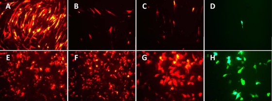
To determine the transfection efficiency of pYA4519, we introduced an mCherry eukaryotic expression cassette into pYA4519 to generate pYA4732 (pYA4519-mCherry) and compared the levels of expression of the reporter gene independently in CEFs and HEK293 cells. Levels of synthesis of the mCherry protein from plasmid pYA4731 (pcDNA-mCherry) were similar in both CEFs and HEK293 cells (Fig. 4A and E), suggesting comparable levels of transfection efficiency of the small plasmid in both cell lines. However, the level of mCherry synthesis from pYA4732 was much higher in HEK293 cells than in CEFs (Fig. 4B and F). Expression of mCherry from pYA4732 was comparable to that from the plasmid pYA4731 in the case of HEK293 cells (Fig. 4E and F), whereas in the case of CEFs, the efficiency decreased dramatically with the increase in plasmid size (Fig. 4A and B). Poor expression of the reporter mCherry gene from pYA4732 indirectly indicated lower expression of genes encoding polymerase subunits and the NP. To confirm this, we also cotransfected CEFs with pYA4732 and pYA4392 (CPI-EGFP-MTI) and cotransfected HEK293 with pYA4732 and pYA4688 (HPI-EGFP-MTI) and measured the synthesis of both mCherry (Fig. 4C and G) and EGFP (Fig. 4D and H) proteins from the same field. Since EGFP is expressed under the control of CPI on pYA4392 and of HPI on pYA4688 (resulting only in the generation of vRNA-like molecules), synthesis of a functional EGFP protein in either case is possible only in the presence of viral polymerase and the NP provided from plasmid pYA4732. We observed EGFP synthesis both in HEK293 and CEFs, but the percentage of HEK293 cells synthesizing both mCherry and EGFP was much greater than the percentage of CEFs (compare Fig. 4C and D with 4G and H). From these observations, we concluded that the expression of viral genes was indeed much lower in CEFs than in HEK cells, which may be a consequence of the poor import of the eight-unit plasmid into CEFs.
FIG. 4.
Transfection efficiency of the eight-unit plasmid. CEFs (A and B) and HEK293 cells (E and F) were transfected with plasmid pYA4731 (pcDNA-mCherry) (A and E) or plasmid pYA4732 (pYA4519-mCherry) (B and F). For CEFs cotransfected with pYA4732 and pYA4392 (pYA4380-EGFP), expression of mCherry (C) and EGFP (D) in CEFs was recorded from the same field. For HEK293 cells cotransfected with pYA4732 and pYA4688, expression of mCherry (G) and EGFP (H) was recorded from the same field. Images were taken 24 h posttransfection. Magnifications, ×100 (A to F) and ×200 (G and H).
Generation of influenza virus from plasmids.
Efficiencies of influenza virus recovery were compared between our one-unit eight-plasmid system and our novel one-plasmid (pYA4519) system. Cultured CEFs were either transfected with pYA4519 or cotransfected with eight one-unit plasmids (pYA4383 [PB2 mRNA and vRNA], pYA4384 [PB1 mRNA and vRNA], pYA4385 [PA mRNA and vRNA], pYA4386 [NP mRNA and vRNA], pYA4388 [HA vRNA], pYA4389 [NA vRNA], pYA4390 [M vRNA], and pYA4391 [NS vRNA]) to provide all the necessary viral components as described in Materials and Methods. The mean titers at 3 days and 6 days posttransfection were approximately 300 and 1,000 PFU/ml influenza viruses, respectively, when cells were transfected with pYA4519, whereas the virus yield using the eight-plasmid method estimated at the same time points was approximately 50 and 700 PFU/ml, respectively (Table 2). Virus yield was much higher in cocultured CEFs and MDCK cells transfected by plasmid pYA4519, with approximately 1 × 10⁴ PFU/ml and 1 × 10⁸ PFU/ml estimated on days 3 and 6 posttransfection, respectively (data not shown). This was expected, as MDCK cells are known to support the growth of influenza virus better than CEFs. We did not obtain any infectious virus particles from the supernatant collected from CEFs transfected by the seven-unit plasmid used as a control. In conclusion, recovery of influenza virus from pYA4519 was more efficient than from the eight-plasmid system when they were tested in CEFs.
TABLE 2.
Influenza A virus generation in CEFs in triplicate wells
| Plasmid(s) | Virus titer (PFU/ml) |
|||||
|---|---|---|---|---|---|---|
| 3rd day posttransfection |
6th day posttransfection |
|||||
| Well 1 | Well 2 | Well 3 | Well 1 | Well 2 | Well 3 | |
| Eight 1-unit plasmidsa | 40 | 60 | 60 | 1,280 | 440 | 480 |
| pYA4519 | 400 | 260 | 280 | 1,800 | 1,000 | 1,000 |
Plasmids pYA4383, pYA4384, pYA4385, pYA4386, pYA4388, pYA4389, pYA4390, and pYA4391.
DISCUSSION
The goal of this study was to construct the influenza virus genome on a single plasmid and rescue the virus from cultured chicken cells. We chose the influenza virus WSN strain as the model virus and, with the combination of reverse genetics and the dual-promoter system, successfully constructed an eight-unit plasmid, pYA4519 (Fig. 3). This plasmid was designed to produce influenza virus polymerase complex (PB1, PB2, and PA), NP, and eight vRNAs (PB1, PB2, PA, NP, HA, NA, M, and NS) in avian cells. By transfection, the “one-plasmid” system showed more-efficient virus generation in CEFs than our eight-plasmid system (Table 2). Generation of influenza virus from a minimal number of plasmid constructs has been a long-term challenge. In 2005, Neumann et al. (16) reported the generation of live influenza virus from a plasmid that carries only eight vRNA-transcribing cassettes by an intriguing mechanism. However, their plasmid system generates high viral titers only in certain cell lines. Through this study, we demonstrated recovery of influenza virus from a single plasmid encoding all necessary proteins and vRNAs. Since RNA PolI is species specific, the choice of host cell line used to generate influenza virus using our one-plasmid system depends on the PolI promoter used.
Intraplasmid recombination is a critical factor to be considered while constructing the entire genome of influenza virus on a single plasmid DNA. In an earlier study by Neumann et al., the authors reported the construction of plasmid pC-PolII-WSN-PB2-PB1-PA, which contains a pUC origin of replication and three PolII cassettes that encode influenza viral polymerase subunits (16). They were unsuccessful in including the NP-expressing cassette in their plasmid construct. We hypothesize that the use of four copies of the PolII promoter and terminator in their plasmid may have led to intraplasmid recombination (the adjacent PolII promoter and terminator result in ∼2-kb repetitive sequences). Use of a high-copy-number vector backbone (pUC ori, 500 to 700 copies per bacterial cell) may also have contributed to increased plasmid recombination and metabolic burden on the bacterium. To overcome these limitations, we used a low-copy-number vector (p15A ori, approximately 15 copies per bacterial cell) to construct our eight-unit plasmid. Care was also taken to minimize the number of CMV promoters used and to reduce the length of homologous sequences by alternate placement of a single-promoter and a dual-promoter cassette in our plasmid. These factors may have reduced intraplasmid recombination events in our eight-unit plasmid and ensured plasmid stability.
Factors such as the plasmid constructs and host cell line used affect the efficiency of virus recovery (14, 16, 17, 19), and our study provides additional evidence in their support. We compared transfection efficiencies between CEFs and HEK293 cells. Both cell types could be transfected with similar efficiencies when small-size plasmids were used (Fig. 4A and E). However, cotransfection of five plasmids resulted in increased EGFP expression in HEK293 cells but not in CEFs (Fig. 2B and C). We could not determine the underlying reason for this difference, but it may contribute to the higher viral yield in HEK293 cells. Transfection experiments involving the large-size plasmid pYA4732 (pYA4519-mCherry) (25.3 kb) indicated that HEK293 cells are better recipients than the CEFs (Fig. 4). This indicates that HEK293 cells not only are highly transfectable cells but also can be transfected efficiently with a large-size plasmid. This was one important criterion for the higher virus yield from the three-plasmid system (16). Nuclear translocation rather than cell entry is the rate-limiting step in the gene transfer process (24). Hence, certain factors in HEK293 cells seem to promote the nuclear translocation of a large plasmid more effectively than with CEFs. We are currently working toward improving the transport of pYA4519 into the nuclei of CEFs by including a nucleus-targeting sequence, such as the promoter/enhancer region of simian virus 40 (5).
Our one-plasmid system offers advantages in two different aspects of an avian influenza vaccine. First, this plasmid facilitates the design of a much simpler approach to develop influenza vaccine seeds. Currently, production of influenza vaccine seeds rely on the eight-plasmid system, in which the HA and NA segments are obtained from the epidemic strain and the viral backbone segments are taken from either the highly productive strain PR8 (A/Puerto Rico/8/34) or a cold-adapted strain (e.g., A/Ann Arbor/6/60) (2, 7, 10). Since the donor virus (either PR8 or a cold-adapted strain) cannot be altered, providing all the necessary backbone segments and proteins from a donor virus on a single plasmid would allow the establishment of a comprehensive three-plasmid (one-plus-two) approach. Alternatively, one could generate influenza vaccine seeds from a similar one-plasmid system and obtain a comparable viral yield. Second, our one-plasmid system provides a new platform for delivery of the influenza virus genome using a bacterial carrier. It is feasible to stably maintain a single plasmid in a bacterial cell and deliver it to the host cells, which is preferable to a system comprising multiple plasmids. We have demonstrated that the WSN HA cassette currently present in pYA4519 can be substituted for an HA cassette with moderate ease, and this flexibility enables us to meet any immediate need. Importantly, it offers an additional advantage of bacterium-mediated delivery of this plasmid, which is not easily possible with any of the existing plasmid systems.
The currently used influenza vaccines for human use are the inactivated and attenuated forms of the virus and are administered via either the intramuscular or the intranasal route. Manufacturing these vaccines using cell culture or embryonated chicken eggs is both expensive and a time-consuming process. We conceived an attenuated Salmonella-based influenza vaccine which is both inexpensive and easy to manufacture. The Salmonella carrier will harbor one plasmid encoding proteins and vRNAs for recovery of an attenuated influenza virus. We hope that oral, intranasal, or intramuscular administration of recombinant Salmonella will lead to intracellular delivery of the plasmid DNA, which subsequently would be expressed to produce an attenuated influenza vaccine in vivo. Construction of the one-plasmid system is the first significant step toward our goal, and it allows us to test this hypothesis in chickens using Salmonella as the delivery vector. Our laboratory has been successful in constructing recombinant attenuated strains of Salmonella enterica serovar Typhimurium that are designed for enhanced antigen delivery in the host and ensure delayed lysis of the pathogen to inhibit long-term host colonization (3). Studies are under way to make further improvements to the Salmonella strain to ensure effective delivery of plasmid DNA into the host cells.
Acknowledgments
We thank Yoshihiro Kawaoka for providing plasmids pTM-PolI-WSN-All and pCAWS-NP and Yixin Shi for providing plasmids pYS1190. We also thank Wei Xin, Shifeng Wang, and Melha Mellata for their suggestions on plasmid construction. We are grateful to Praveen Alamuri for critically editing the manuscript.
This work was supported by grant AI065779 from the National Institutes of Health.
Footnotes
Published ahead of print on 8 July 2009.
REFERENCES
- 1.Ascon, M. A., D. M. Hone, N. Walters, and D. W. Pascual. 1998. Oral immunization with a Salmonella Typhimurium vaccine vector expressing recombinant enterotoxigenic Escherichia coli K99 fimbriae elicits elevated antibody titers for protective immunity. Infect. Immun. 66:5470-5476. [DOI] [PMC free article] [PubMed] [Google Scholar]
- 2.Chen, Z., A. Aspelund, G. Kemble, and H. Jin. 2006. Genetic mapping of the cold-adapted phenotype of B/Ann Arbor/1/66, the master donor virus for live attenuated influenza vaccines (FluMist). Virology 345:416-423. [DOI] [PubMed] [Google Scholar]
- 3.Curtiss, R., III, S. Y. Wanda, B. M. Gunn, X. Zhang, S. A. Tinge, V. Ananthnarayan, H. Mo, S. Wang, and W. Kong. 2009. Salmonella enterica serovar Typhimurium strains with regulated delayed attenuation in vivo. Infect. Immun. 77:1071-1082. [DOI] [PMC free article] [PubMed] [Google Scholar]
- 4.Darji, A., C. A. Guzman, B. Gerstel, P. Wachholz, K. N. Timmis, J. Wehland, T. Chakraborty, and S. Weiss. 1997. Oral somatic transgene vaccination using attenuated S. Typhimurium. Cell 91:765-775. [DOI] [PubMed] [Google Scholar]
- 5.Dean, D. A. 1997. Import of plasmid DNA into the nucleus is sequence specific. Exp. Cell Res. 230:293-302. [DOI] [PubMed] [Google Scholar]
- 6.Fodor, E., L. Devenish, O. G. Engelhardt, P. Palese, G. G. Brownlee, and A. Garcia-Sastre. 1999. Rescue of influenza A virus from recombinant DNA. J. Virol. 73:9679-9682. [DOI] [PMC free article] [PubMed] [Google Scholar]
- 7.Gerdil, C. 2003. The annual production cycle for influenza vaccine. Vaccine 21:1776-1779. [DOI] [PubMed] [Google Scholar]
- 8.Grillot-Courvalin, C., S. Goussard, and P. Courvalin. 1999. Bacteria as gene delivery vectors for mammalian cells. Curr. Opin. Biotechnol. 10:477-481. [DOI] [PubMed] [Google Scholar]
- 9.Hoffmann, E., G. Neumann, G. Hobom, R. G. Webster, and Y. Kawaoka. 2000. “Ambisense” approach for the generation of influenza A virus: vRNA and mRNA synthesis from one template. Virology 267:310-317. [DOI] [PubMed] [Google Scholar]
- 10.Jin, H., B. Lu, H. Zhou, C. Ma, J. Zhao, C. F. Yang, G. Kemble, and H. Greenberg. 2003. Multiple amino acid residues confer temperature sensitivity to human influenza virus vaccine strains (FluMist) derived from cold-adapted A/Ann Arbor/6/60. Virology 306:18-24. [DOI] [PubMed] [Google Scholar]
- 11.Klumpp, K., R. W. Ruigrok, and F. Baudin. 1997. Roles of the influenza virus polymerase and nucleoprotein in forming a functional RNP structure. EMBO J. 16:1248-1257. [DOI] [PMC free article] [PubMed] [Google Scholar]
- 12.Lamb, R. A., and P. W. Choppin. 1983. The gene structure and replication of influenza virus. Annu. Rev. Biochem. 52:467-506. [DOI] [PubMed] [Google Scholar]
- 13.Lewis, G. K. 2007. Live-attenuated Salmonella as a prototype vaccine vector for passenger immunogens in humans: are we there yet? Expert Rev. Vaccines 6:431-440. [DOI] [PubMed] [Google Scholar]
- 14.Massin, P., P. Rodrigues, M. Marasescu, S. van der Werf, and N. Naffakh. 2005. Cloning of the chicken RNA polymerase I promoter and use for reverse genetics of influenza A viruses in avian cells. J. Virol. 79:13811-13816. [DOI] [PMC free article] [PubMed] [Google Scholar]
- 15.Murti, K. G., R. G. Webster, and I. M. Jones. 1988. Localization of RNA polymerases on influenza viral ribonucleoproteins by immunogold labeling. Virology 164:562-566. [DOI] [PubMed] [Google Scholar]
- 16.Neumann, G., K. Fujii, Y. Kino, and Y. Kawaoka. 2005. An improved reverse genetics system for influenza A virus generation and its implications for vaccine production. Proc. Natl. Acad. Sci. USA 102:16825-16829. [DOI] [PMC free article] [PubMed] [Google Scholar]
- 17.Neumann, G., T. Watanabe, H. Ito, S. Watanabe, H. Goto, P. Gao, M. Hughes, D. R. Perez, R. Donis, E. Hoffmann, G. Hobom, and Y. Kawaoka. 1999. Generation of influenza A viruses entirely from cloned cDNAs. Proc. Natl. Acad. Sci. USA 96:9345-9350. [DOI] [PMC free article] [PubMed] [Google Scholar]
- 18.Noda, T., H. Sagara, A. Yen, A. Takada, H. Kida, R. H. Cheng, and Y. Kawaoka. 2006. Architecture of ribonucleoprotein complexes in influenza A virus particles. Nature 439:490-492. [DOI] [PubMed] [Google Scholar]
- 19.Ozaki, H., E. A. Govorkova, C. Li, X. Xiong, R. G. Webster, and R. J. Webby. 2004. Generation of high-yielding influenza A viruses in African green monkey kidney (Vero) cells by reverse genetics. J. Virol. 78:1851-1857. [DOI] [PMC free article] [PubMed] [Google Scholar]
- 20.Rand, K. N. 1996. Crystal violet can be used to visualize DNA bands during gel electrophoresis and to improve cloning efficiency. Tech. Tips Online 1(1):23-24. http://www.science-direct.com/science/journal/13662120. [Google Scholar]
- 21.Schoen, C., J. Stritzker, W. Goebel, and S. Pilgrim. 2004. Bacteria as DNA vaccine carriers for genetic immunization. Int. J. Med. Microbiol. 294:319-335. [DOI] [PubMed] [Google Scholar]
- 22.Sizemore, D. R., A. A. Branstrom, and J. C. Sadoff. 1995. Attenuated Shigella as a DNA delivery vehicle for DNA-mediated immunization. Science 270:299-302. [DOI] [PubMed] [Google Scholar]
- 23.Weiss, S., and S. Krusch. 2001. Bacteria-mediated transfer of eukaryotic expression plasmids into mammalian host cells. Biol. Chem. 382:533-541. [DOI] [PubMed] [Google Scholar]
- 24.Zhou, R., R. C. Geiger, and D. A. Dean. 2004. Intracellular trafficking of nucleic acids. Expert Opin. Drug Deliv. 1:127-140. [DOI] [PMC free article] [PubMed] [Google Scholar]


