

Abstract
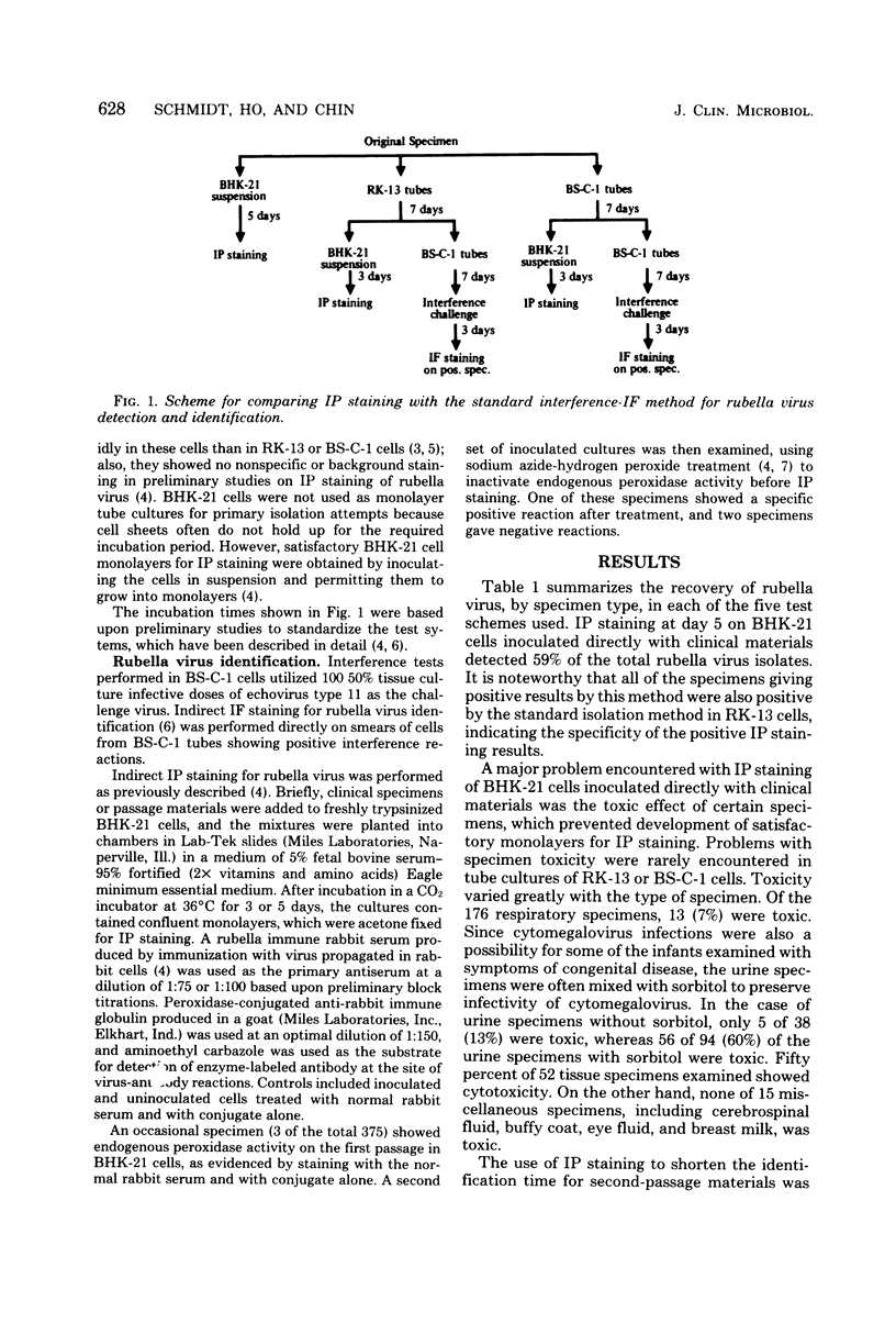
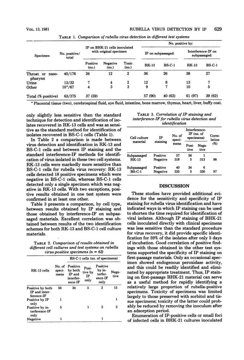
Efforts were made to shorten the time required for detection of rubella virus in clinical materials through the use of immunoperoxidase (IP) staining. Comparative studies were performed in which specimens were inoculated in parallel into BHK-21 hamster kidney cells, which were examined by IP staining at 5 days, and into BK-13 and BS-C-1 cells, which were examined in two ways, viz., by subpassage at 7 days into BHK-21 cells and IP staining 3 days later and by subpassage at 7 days into BS-C-1 cells followed by interference testing and immunofluorescence (IF) staining on positive materials (standard method). Direct inoculation into BHK-21 cells with IP staining at 5 days permitted detection and identification of 59% of the 63 positive specimens. Toxicity of some specimens preserved with sorbitol and of certain tissue specimens reduced the number of satisfactory examinations which could be performed in this system. Virus detection and identification by IP staining on subpassaged RK-13 and BS-C-1 materials, requiring a total of 18 days, was comparable to the longer interference-IF method, requiring 17 days. Results obtained by IP staining and interference-IF showed 98% correlation on RK-13 materials and 97% correlation on BS-C-1 materials. IP staining on inoculated BHK-21 cells can be a useful method for rapid identification of a relatively high proportion of rubella-positive specimens, particularly if sorbitol-preserved specimens are avoided, and IP staining on subpassaged RK-13 and BS-C-1 materials is a highly satisfactory alternative to the longer interference-IF method.
Full text
PDF
Selected References
These references are in PubMed. This may not be the complete list of references from this article.
- Schmidt N. J., Dennis J., Lennette E. H. Comparison of immunofluorescence and immunoperoxidase staining for identification of rubella virus isolates. J Clin Microbiol. 1978 Jun;7(6):576–583. doi: 10.1128/jcm.7.6.576-583.1978. [DOI] [PMC free article] [PubMed] [Google Scholar]
- Schmidt N. J., Dennis J., Lennette E. H. Hemadsorption and hemadsorption inhibition tests for rubella virus. Arch Gesamte Virusforsch. 1968;25(3):308–320. doi: 10.1007/BF01556559. [DOI] [PubMed] [Google Scholar]
- Schmidt N. J., Lennette E. H. Rubella complement-fixing antigens derived from the fluid and cellular phases of infected BHK-21 cells: extraction of cell-associated antigen with alkaline buffers. J Immunol. 1966 Dec;97(6):815–821. [PubMed] [Google Scholar]
- Schmidt N. J., Lennette E. H., Woodie J. D., Ho H. H. Identification of rubella virus isolates by immunofluorescent staining, and a comparison of the sensitivity of three cell culture systems for recovery of virus. J Lab Clin Med. 1966 Sep;68(3):502–509. [PubMed] [Google Scholar]