Abstract
Herpes simplex virus type-1 (HSV-1) causes significant health problems from periodic skin and corneal lesions to encephalitis. It is also considered a cofactor in the development of age-related secondary glaucoma. Inhibition of HSV-1 at the stage of viral entry generates a unique opportunity for preventative and/or therapeutic intervention. Here we provide evidence that a sugar-binding antiviral protein, cyanovirin-N (CV-N), can act as a potent inhibitor of HSV-1 entry into natural target cells. Inhibition of entry was independent of HSV-1 gD receptor usage and it was observed in transformed as well as primary cell cultures. Evidence presented herein suggests that CV-N can not only block virus entry to cells but also, it is capable of significantly inhibiting membrane fusion mediated by HSV glycoproteins. While CV-N treated virions were significantly deficient in entering into cells, HSV-1 glycoproteins-expressing cells pretreated with CV-N demonstrated reduced cell-to-cell fusion and polykaryocytes formation. The observation that CV-N can block both entry as well as membrane fusion suggests a stronger potential for this compound in anti-viral therapy against HSV-1.
Keywords: Cyanovirin-N, Microbicide, HSV-1, viral entry
1. Introduction
Herpes simplex virus type-1 (HSV-1) infection is the most common cause of infectious blindness in developed countries (Liesegang et al., 1989; Liesegang, 2001). Following an initial infection in epithelial cells, HSV establishes latency in the host sensory nerve ganglia (Spear, 1993; Spear and Longnecker, 2003). It has recently been reported that over 90 % of the trigeminal ganglia examined post-mortem in a sampling of the American population contained HSV-1 (Hill et al., 2008). The virus emerges sporadically from latency and causes lesions on mucosal epithelium, skin, and the cornea, among other locations. Prolonged or multiple recurrent episodes of corneal infections can result in vision impairment or blindness, due to the development of herpetic stromal keratitis (HSK) (Kaye et al., 2000). This is typically characterized by inflammation leading to scarring, thinning, and vascularization of the corneal stroma (Eisenberg et al., 1985; Ellison et al., 2003). Patients with corneal HSV infection risk lifelong recurrent corneal disease. HSK accounts for 20–48% of all recurrent ocular HSV infection leading to significant vision loss in many patients (Liesegang et al., 1989; Kaye et al., 2000). HSV infection may also lead to several other diseases including retinitis, meningitis, and encephalitis.
Primary infection begins with the entry of HSV into host cells. It is a complex process that is initiated by specific interaction of viral envelope glycoproteins and host cell surface receptors (Spear, 1993; Spear et al., 2000). Both HSV-1 and HSV-2 use glycoproteins B and C (gB and gC, respectively) to mediate their initial attachment to cell surface heparan sulfate proteoglycans (HSPG) (WuDunn and Spear, 1989; Shieh et al., 1992; Herold et al., 1991). Binding of herpesviruses to HSPG likely precedes a conformational change that brings viral glycoprotein D (gD) to the binding domain of host cell surface gD receptors (Whitbeck et al., 1999; Krummenacher et al., 1998, 1999, 2000). Thereafter, a concerted action involving gD, its receptor, three additional HSV glycoproteins; gB, gH, and gL, and possibly an additional gH co-receptor trigger fusion of the viral envelope with the plasma membrane of host cells (Scnalan et al., 2003; Spear and Longnecker, 2003; Perez-Romero et al., 2005). Subsequently viral capsids and tegument proteins are released into the cytoplasm of the host cell.
The gD receptors include cell-surface molecules derived from three structurally unrelated families. These include a member of the tumor necrosis factor (TNF) receptor family, two members of the nectin family of receptors, and the product of certain 3-OSTs, 3-O-sulfated heparan sulfate (3-OS HS) (Spear 1993; Spear and Longnecker, 2003). Herpesvirus entry mediator (HVEM or TNFRSF14) principally mediates entry of HSV-1 and HSV-2 (Montgomery et al., 1996; Marsters et al., 1997; Kwon et al., 1997) into human T lymphocytes and is expressed in many fetal and adult human tissues including the lung, liver, kidney, and lymphoid tissues (Montgomery et al., 1996) and human trabecular meshwork (Tiwari et al., 2007). Nectin-1 and nectin-2, also known as herpesvirus entry proteins C and B (HveC and HveB), respectively, belong to the immunoglobulin superfamily (Cocchi et al., 1998; Milne et al., 2001; Shukla et al., 2000). Both nectin-1 and nectin-2 mediate entry of HSV-1 and HSV-2, but only nectin-1 mediates bovineherpesvirus-1 (BHV-1) entry (Martinez and Spear, 2002; Warner et al., 1998). HSV-1entry mediating activity of nectin-2 is limited to some mutant strains only (Warner at al., 1998; Lopez et al., 2000). Nectin-1 is extensively expressed in human cells of epithelial and neuronal and ocular origin (Richart et al., 2003; Tiwari et al., 2008), while nectin-2 is widely expressed in many human tissues, but with only limited expression in neuronal cells and keratinocytes. The non-protein receptor, 3-OS HS, is expressed in multiple human cell lines (e.g. neuronal and endothelial cells) and mediates entry of HSV-1, but not HSV-2 (Shukla at al., 1999; Shukla and Spear, 2001; Tiwari et al., 2004; Tiwari et al., 2006; Tiwari et al., 2007).
Recently a novel 11-kb antiviral protein cyanovirin-N (CV-N) originally isolated from the cyanobacterium Nostoc ellipsosporum was shown to have potent anti-human immunodeficiency virus (HIV) activity. Its mechanism of action is based on the specific targeting of high mannose oligosaccharides oligomannose-8 (Man-8) and oligomannose-9 (Man-9) on the HIV envelope glycoproteins gp120 and gp41 (O‘Keefe et al., 2000; Bolmstedt et al., 2001; Shenoy et al., 2001). Similar oligosaccharides are known to be present on other viruses, including Ebola, Influenza, and Hepatitis C viruses (O’Keefe et al., 2003; Barrientos et al., 2003). Previous efforts to determine the efficacy of CV-N’s inhibition of HSV-1 entry into target cells have yielded conflicting results (Boyd et al., 1997; O’Keefe et al., 2003). Here, we demonstrate that CV-N significantly inhibits HSV-1 entry into natural target cells of human ocular origin at non-cytotoxic nanomolar concentrations. In addition, we show that CV-N also impairs the viral glycoprotein induced cell-to-cell fusion. These data demonstrate that targeting the HSV-1 envelope glycoproteins is a new and promising approach in the development of antiviral therapies to herpes simplex virus infection.
2. Materials and Methods
2.1. Cells, viruses and Cyanovirin-N
Wild-type CHO-K1 cells were grown in Ham’s F12 (Invitrogen Corp, Carlsbad, CA) supplemented with 10% fetal bovine serum (FBS), while African green monkey kidney (Vero) cells were grown in Dulbecco’s Modified Eagles Medium (DMEM) (Invitrogen Corp.) supplemented with 5% FBS. Cultures of HeLa and RPE cells were grown in L-glutamine containing DMEM (Invitrogen Corp.) supplemented with 10% FBS. As previously described, cultures of human corneal fibroblasts (CF) were derived from the stroma of corneal tissues obtained from the Illinois Eye Bank, Chicago, IL, using institution approved protocol and culture conditions in accordance with the Declaration of Helsinki). CF from the 4th passage was used for the study was kindly provided by Dr. Yue (University of Illinois at Chicago). Recombinant β-galactosidase-expressing HSV-1(KOS) gL86 were used (Montgomery et al., 1996). P.G. Spear (Northwestern University) provided wild-type CHO-K1 cells. GFP expressing HSV-1 (K26GFP) was provided by P. Desai (Johns Hopkins University, Baltimore). The viral stocks were propagated at low multiplicity of infection (MOI) in complementing cell lines, titered on Vero cells and stored at −80°C. Cyanovirin-N (CV-N) used in this study was generous gift of Dr. T. Mori (National Cancer Institute, Bethesda, Maryland).
2.2. Viral Entry Assay
Viral entry assays were based on quantitation of β-galactosidase expressed from the viral genome in which β-galactosidase expression is inducible by HSV infection (Montgomery et al., 1996). Cells were transiently transfected in 6-well tissue culture dishes, using Lipofectamine 2000 with plasmids expressing HSV-1 entry receptors (necitn-1, HVEM and 3-OST-3 expression plasmids) at 1.5 µg per well in 1 ml. At 24 hr post-transfection, cells were re-plated in 96-well tissue culture dishes (2 × 10 4 cells per well) at least 16 hr prior to infection. Cells were washed and exposed to serially diluted pre-incubated virus with CV-N or 1 × PBS at two fold dilutions in 50 µl of phosphate-buffered saline (PBS) containing 0.1% glucose (G) and 1% heat inactivated calf serum (CS) for 6 hr at 37°C before solubilization in 100µl of PBS containing 0.5% NP-40 and the β-galactosidase substrate, o-nitro-phenyl-β-D-galactopyranoside (ONPG; ImmunoPure, PIERCE, Rockford, IL, 3 mg/ml). The enzymatic activity was monitored at 410 nm by spectrophotometry (Molecular Devices spectra MAX 190, Sunnyvale, CA) at several time points after the addition of ONPG in order to define the interval over which the generation of the product was linear with time. In parallel experiment the cells were preincubated with CV-N for 90 min before the HSV-1 infection. Inhibitory effect of CV-N on HSV-1 entry in cells were also confirmed by 5-bromo-4-chloro-3-indolyl-β-D-galactopyranoside (X-gal) staining. The cells were grown in Lab-Tek chamber slides (Nunc, Inc., Naperville, IL). After 6 hr of infection with reporter virus treated with CV-N or left untreated with1 × PBS, cells were washed with PBS and fixed with 2% formaldehyde and 0.2% glutaradehyde at room temperature for 15 min. The cells were then washed with PBS and permeabilized with 2 mM MgCl2, 0.01% deoxycholate and 0.02% nonidet NP-40 for 15 min. After rinsing with PBS, 1.5 ml of 1.0 mg/ml X-gal in ferricyanide buffer was added to each well and the blue color developed in the cells was examined. Microscopy was performed using 20 × objective of the inverted microscope (Zeiss, Axiovert 100M). The slide book version 3.0 (imaging software) was used for images. All experiments were repeated a minimum of three times unless otherwise noted.
2.3. Viral binding assay
Purified GFP-expressing HSV-1 (K26 GFP) were pre-incubated with CV-N or with 1 × PBS for 90 min at room temperature were used to the gD receptor expressing CHO-K1 cells or naturally susceptible cells (HeLa, Vero and human CF) grown in Microtest 96-well assay plates (BD Falcon). All cells were incubated at 4°C for 1 hr, washed five times to remove unbound virus, and finally replace with warm medium for further incubation. Viral binding measured as relative fluorescence units (RFU) per treatment were determined by using GENios Pro plate reader (TECAN) at 480-nm excitation and 520-nm emission spectrum. Measurements of 4 replicates of CV-N treated and untreated samples were performed. Data were expressed as mean ± standard deviation (SD).
2.4. Virus-free Cell-to-Cell Fusion Assay
In this experiment, the CHO-K1 cells (grown in F-12 Ham, Invitrogen) designated “effector” cells were co-transfected with plasmids expressing four HSV-1(KOS) glycoproteins, pPEP98 (gB), pPEP99 (gD), pPEP100 (gH) and pPEP101 (gL), along with the plasmid pT7EMCLuc that expresses firefly luciferase gene under the T7 promoter (Pertel et al., 2001). Wild-type CHO-K1 cells express cell surface HS but lack functional gD receptors, therefore transiently transfected with plasmids expressing entry receptors nectin-1 (pBG38) , HVEM (pBec10) and/or 3-OST-3 (pDS43) (Shukla et al., 1999; Pertel et al., 2001). Wild type CHO-K1 cultured cells expressing HSV-1 entry receptors or naturally susceptible cells (HeLa, Vero and human CF) considered as “target cells” were co-transfected with pCAGT7 plasmid that expresses T7 RNA polymerase using chicken actin promoter and CMV enhancer (Tiwari et al., 2007). CV-N untreated effector cells expressing pT7EMCLuc and HSV-1 essential glycoproteins and the target cells expressing gD receptors transfected with T7 RNA polymerase were used as the positive control. CV-N treated effectors cells were used for the test. For fusion, at 18 hr post transfection, the target and the effector cells were mixed together (1:1 ratio) and co-cultivated in a 24 well- tissue culture plates (Nunc, Inc.). The activation of the reporter luciferase gene as a measure of cell fusion was determined using reporter lysis assay (Promega) at 24 hr post mixing.
2.5. Fluorescent-labeled cell fusion assay and quantification of polykaryocytes
In this experiment CHO-K1 effector cells were co-transfected with plasmids expressing four HSV-1(KOS) glycoproteins (gB, gD, gH-gL) along with the plasmid pT7EMCLuc that expresses firefly luciferase gene under the T7 promoter plus pDSRed-N1 plasmid (BD Falcon) constructs. The target CHO-K1 cells expressing gD receptor (3-OST-3 modified 3OS HS) were co-transfected with pCAGT7 plasmid that expresses T7 RNA polymerase using chicken actin promoter and CMV enhancer plus green fluorescent expression plasmid. During co-transfection effector and target cells were balanced with empty vector plasmid pCDNA3.1 to keep equal amount of DNA in both cell-types. Before co-culture, effector cells were pre-incubated with 10 nM CV-N or 1× PBS for 90 min. Then both populations of effector and target cells were cultured in 1:1 ratio for 24 hrs. The cells were then fixed and mounted in Vectorshield mounting medium (Vector Laboratories, Inc. Burlingame, CA). Leica confocal microscope SP2 was used at 40 × magnification. A group of multinucleate cells (8–10 joint cells) were scored positive for polykaryocytes formation.
3. Results
3.1. CV-N significantly blocks HSV-1 entry into gD receptor expressing CHO-K1 cells
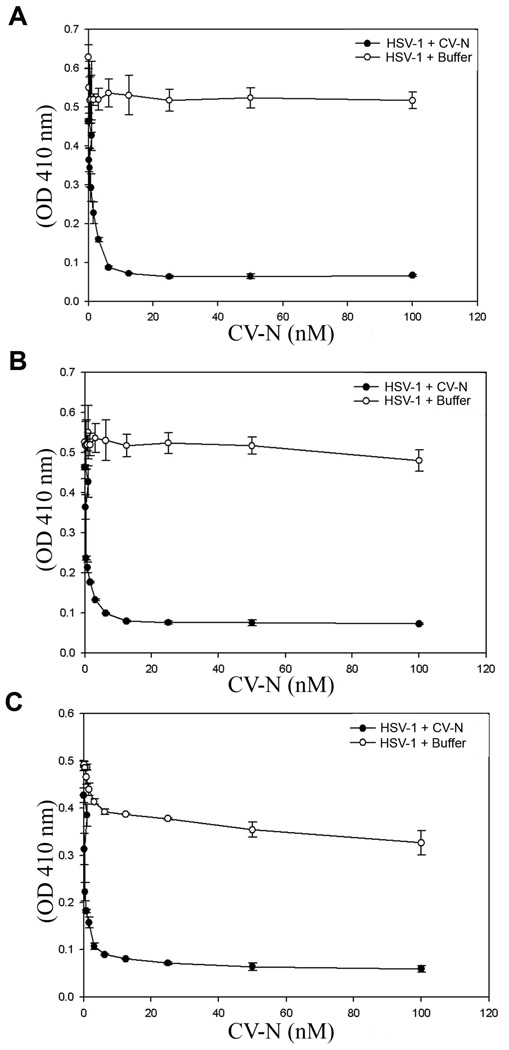
To determine the effect of CV-N on HSV-1 entry, we first tested the ability of HSV-1, in presence and absence of CV-N, to infect CHO-K1 cells expressing gD receptors. HSV-1 entry into cell was determined by using β-galactosidase expressing HSV-1 reporter virus (gL86). As shown in Fig. 1 HSV-1 pre-incubated with CV-N significantly blocked viral entry in a dose dependent manner in CHO-K1 cells expressing gD receptors (nectin-1, HVEM and 3-OST-3 modified 3OS HS). The blocking activity of CV-N was seen at low nano-molar concentrations and clearly, entry was inhibited irrespective of the gD receptors used. The inhibition seen was not due to the ability of CV-N to block reporter assay since strong enzyme activity was observed in CV-N treated cells transfected with a β-galactosidase expression plasmid (data not shown) (Montgomery et al., 1996).Taken together, the results indicated that the role of CV-N in HSV-1 entry blocking is not receptor specific phenomenon.
Figure 1. Cyanovirin-N (CV-N) blocks herpes simplex virus type-1 (HSV-1) entry into CHO-K1 cells expressing gD receptors.
In this experiment, β-galactosidase-expressing recombinant virus HSV-1 (KOS) HSV-1 gL86 (25 pfu/cell) was pre-incubated with CV-N at indicated concentration or mock treated with 1 × phosphate saline buffer (PBS) for 90 min at room temperature. After 90 min the virus was incubated with CHO-K1 cells expressing gD receptors: nectin-1 (A), HVEM (B) and 3-OST-3 expressing cells (C). After 6 hr, the cells were washed, permeabilized and incubated with ONPG substrate (3.0 mg/ml) for quantitation of β -galactosidase activity expressed from the encoded viral genome. The enzymatic activity was measured at an optical density of 410 nm (OD 410). In this and other figures each value shown is the mean of three or more determinations (± SD; standard deviation) from four independent experiments. Mock treated HSV-1 with PBS was used as a control.
3.2. CV-N significantly blocks HSV-1 entry into natural target cells
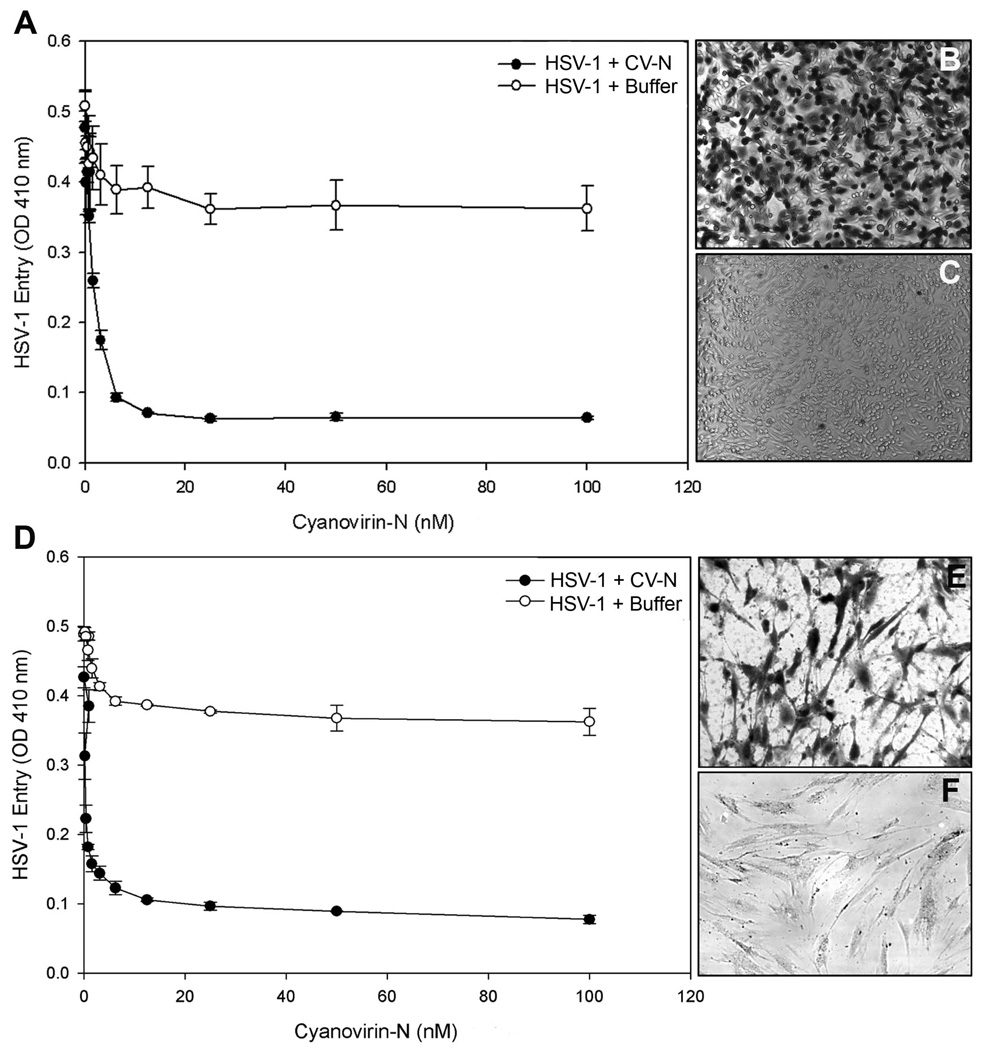
Next, to confirm blocking activity of CV-N on HSV-1 entry, we used natural target cells. We used HeLa and primary cultures of human corneal fibroblasts (CF). Human CF is a natural target cell line that has been shown previously to express 3-O sulfated heparan sulfate as a receptor (Tiwari et al., 2007; Tiwari et al., 2008). As shown in Fig. 2 (panel A and panel D) HSV-1 virions pre-treated with CV-N (50 nM) showed significant reduction of entry in both HeLa and CF. These results were further confirmed by X-gal assay. As demonstrated in panel C and F (Fig 2.) the HSV-1 treatment with CV-N significantly reduced the number of blue cells in both HeLa and CF cells (panels C and F). While corresponding untreated virus were able to infect all the cells as 100% cells turned blue (panels B and E). Taken together, the results indicated the role of CV-N in HSV-1 entry blocking is also observed in natural target cells including primary cells cultured from the human cornea.
Figure 2. Cyanovirin-N blocks herpes simplex virus type-1 (HSV-1) entry into natural target cells.
In this experiment HeLa (A–C) and primary cultures of human corneal fibroblasts (CF) (D–F) were used. The β-galactosidase-expressing recombinant virus HSV-1 (KOS) HSV-1 gL86 (5 pfu/cell) was pre-incubated with CV-N at indicated concentration or mock treated with 1 × PBS for 90 min at room temperature. After 90 min of CV-N treatment the virus was incubated with HeLa (A) and human CF (D). After 6 hr, the cells were washed, permeabilized and incubated with ONPG substrate (3.0 mg/ml) for quantitation of β-galactosidase activity expressed from the input viral genome signals that virus has entered the cell. The recombinant virus HSV-1 KOS gL86 is produced by inserting the E.coli lacZ gene driven by the HSV-1 ICP4 promoter in place of the thymidine kinase gene of KOS (Montgomery et al., 2009). The enzymatic activity was measured at an optical density of 410 nm (OD 410). In this figure each value shown is the mean of three or more determinations (±SD; standard deviation). Mock treated HSV-1 with PBS was used as a control. Confirmation of HSV-1 blocking activity of CV-N into natural target cells was further confirmed by X-gal (1.0 mg/ml) staining. Virus incubated with CV-N blocks viral entry (panels C and F), while virus incubated with 1 × PBS showed 100% cells infected B and E). Blue cells (representing viral entry) were seen as shown. Microscopy was performed using a 20 × objective of Zeiss Axiovert 100. The slide book version 3.0 software was used for images.
3.3. CV-N interacts with HSV-1 envelope glycoproteins
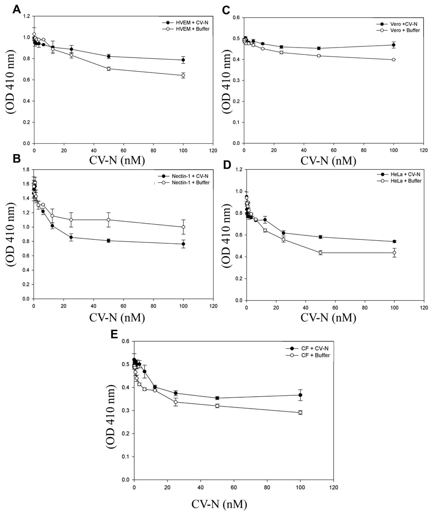
Subsequently, we asked whether the inhibitory activity of CV-N on HSV-1 entry occurs at the cellular receptor level or it could be attributed to viral glycoproteins. To answer this question, instead of preincubating HSV-1 with CV-N, we first preincubated cells with CN-V for 90 min and then infected target cells (HeLa, Vero, RPE and CF) with 25 pfu/cell of HSV-1(KOS). As shown in Fig. 3 pre-incubation of the cells with CV-N had relatively minor effects on HSV-1 entry, suggesting that anti-HSV-1 activity of CV-N is likely exerted on glycoproteins expressed on viral envelopes.
Figure 3. Cyanovirin-N interacts with HSV-1 envelope glycoproteins to block viral entry.
In this experiment CHO-K1 cells expressing entry receptor (A–C) and naturally infectible cells HeLa (D) and human CF (E) were pre-incubated with indicated concentration of CV-N or mock treated for 90 min at room temperature followed by addition of β-galactosidase-expressing recombinant virus HSV-1 (KOS) HSV-1 gL86 (5 pfu/cell) in the 96 well plates. After 6 hr, the cells were washed, permeabilized and incubated with ONPG substrate (3.0 mg/ml) for quantitation of β-galactosidase activity expressed from the input viral genome. The enzymatic activity was measured at an optical density of 410 nm (OD 410).
3.4. CV-N significantly affects HSV-1 binding to cells
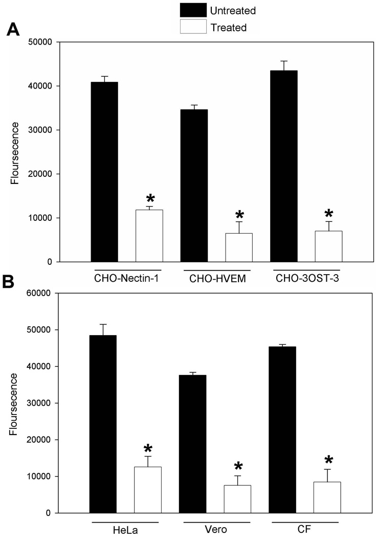
Because CV-N blocked HSV-1 entry, we next tested its ability to affect viral binding to the cells. To determine the difference between CV-N treated versus untreated virus on attachment or binding we used GFP-expressing HSV-1 (K26GFP). GFP was fused in frame with the UL35 ORF to generate a VP26-GFP fusion protein in HSV-1 KOS (Desai and Person, 1998). As shown in Fig. 4 the GFP signal on cell surface was significantly weaker when virus was treated with CV-N in both gD receptors-expressing CHO-K1 cells (panel A) and natural target cells (panel B). This data clearly indicated that CV-N significantly affects viral entry at attachment step. In a parallel experiment the post-treatment of CV-N after 45 min of HSV-1 infection had no effect on viral binding or attachment on the cells (data not shown).
Figure 4. Cyanovirin-N blocks HSV-1 binding to the cells.
Green fluorescent protein (GFP) expressing HSV-1 (K26 GFP) was pre-incubated with 10 nM CV-N or with 1 × phosphate buffer saline (PBS) at for 90 min at room temperature. The mixture was allowed to incubate with CHO-K1 cells expressing gD receptors (panel A) or natural target cells (panel B) for 1 hr at 37°C followed by a 20 mM citrate buffer (sodium citrate and citric acid, pH 3.0), treatment to remove unbound viruses. The fluorescent output as a result of viral binding to the cells was recorded using Tecan spectrophotometer is presented. In this figures each value shown is the mean of three or more determinations (±SD; standard deviation) from three independent experiments. GFP-expressing HSV-1 pre-incubated with 1 × PBS was used as a control.
3.5. CV-N treatment inhibits HSV-1 glycoprotein mediated cell-to-cell fusion and polykaryocytyes formation
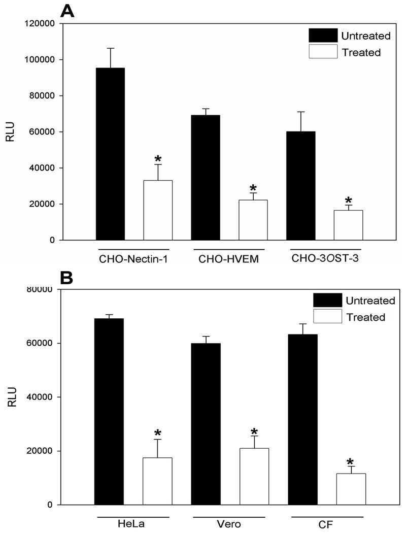
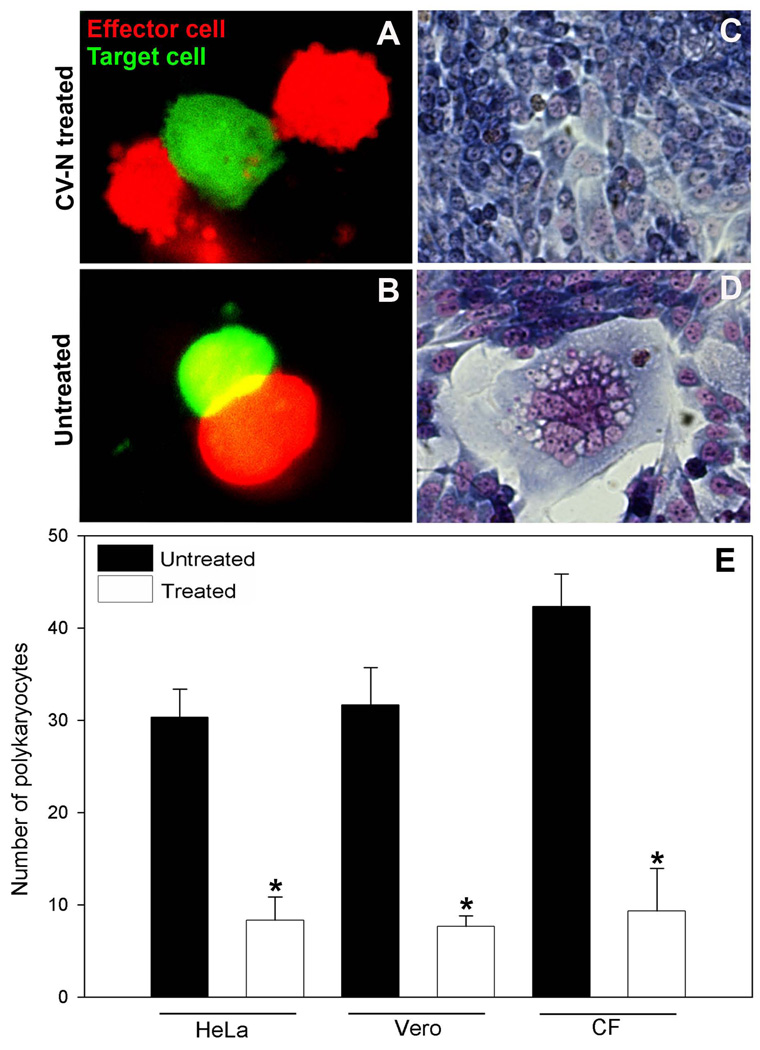
Finally, we tested the role of CV-N during HSV-1 glycoproteins mediated cell-to-cell fusion. Cell-to-cell fusion has been used to demonstrate the viral and cellular requirements during virus-cell interactions and also as means of viral spread (Pertel et al., 2001). We sought to determine whether CV-N interaction with HSV-1 envelope glycoproteins essential for viral entry may also affect cell-to-cell fusion. Surprisingly, effector cells expressing HSV-1 glycoproteins treated with CV-N impaired the cell-to-cell fusion in both CHO-K1 cells expressing specific gD receptors or naturally susceptible cells (Fig 5). This result was further confirmed during fluorescent -labeled cell fusion assay, where HSV-1 glycoprotein expressing effector cells co-transfected with pDSRed N1 fluorescent plasmid incubated with CV-N for 90 min failed to fuse with GFP-expressing CHO-3OST-3 target cells. In contrast, the control CV-N untreated effector red-cells fused (yellow color) with green target cells (Fig. 6). This response was further observed when polykaryocytes formation was estimated. Again CV-N treated effector cells failed to form polykaryons when co-cultured with target cells, while in control untreated effector cells efficiently showed larger number of polykaryons. Inhibitory fusion activity CV-N has been previously reported for human Herpesvirus 6 and measles virus (Dey et al., 2000).
Figure 5. HSV-1 glycoproteins induced cell to cell fusion is blocked by CV-N.
The “effector CHO-K1 cells” were transfected with expression plasmids for HSV-1 glycoproteins (gB, gD, gH-gL) along with T7 plasmid and mixed with either CHO-K1 cells expressing luciferase gene along with specific gD receptors (panel A) or with the natural target cells (HeLa, Vero and human CF; panel B). Membrane fusion as a means of viral spread was detected by monitoring luciferase activity. Relative luciferase units (RLUs) determined using a Sirius luminometer (Berthold detection systems). Error bars represent standard deviations. * P< 0.05, one way ANOVA.
Figure 6. Microscopic visualization of CV-N blocking activity of HSV-1 glycoprotein cell to cell fusion and polykaryocytes formation.
In this experiment effector CHO-K1 cells expressing four essential HSV-1 glycoproteins (gB, gD, gH-gL) were co-transfected (0.5 µg DNA per glycoprotein) with pDSRed N1 plasmid construct, while target CHO-K1 cells expressing 3-OST-3 (1.5 µg DNA) were co-transfected with GFP-expressing plasmid construct. Effector cells were pre-incubated with 10 nM CV-N or with 1 × PBS for 90 min at room temperature before the two pools of cells were co-cultured in 1:1 ratio for 24 hrs. Panels show the content mixing and cell fusion in presence (A) and absence of CV-N (B). In parallel polykaryocytes formation was also visualized in CV-N treated (panel C) and untreated (panel D) cell populations. Both effector and target cells were fixed with 2% formaldehyde and 0.2% glutaradehyde at room temperature for 30 min and then stained with Giemsa stain (Fluka) for 20 min. Shown are photographs of representative cells (Zeiss Axiovert 200) after 24 h in presence and absence of CV-N. (E). Quantification on number of polykaryocytes formation as a result of cell to cell fusion in presence and absence of CV-N in different cell types is presented. The numbers of multinucleated polykaryocytes were measured by counting 24 h after co-cultivating the effector HSV-1 glycoproteins expressing CHO-K1 cells with natural targets HeLa, Vero and primary cultures of human CF in presence (in white bar) and absence (in black bar) of CV-N. The values shown were from three independent experiments performed in triplicate. Error bars represent standard deviations. * P< 0.05, one way ANOVA.
4. Discussion
Viral entry into cell is the first critical step required for the onset of disease (Sieczkari and Whittaker, 2005). Hence viral entry into cell provides a unique opportunity to study virus cell interactions in detail and find novel ways to block viral cell interactions for therapeutic interventions (Dimitrov, 2004). Here we demonstrated that cyanobacterial protein cyanovirin-N (CV-N) affects both HSV-1 entry and viral glycoprotein mediated cell-to-cell fusion using in vitro cell culture models. The anti-HSV-1 activity of CV-N was not limited to any particular gD receptors. Our results showed that HSV-1 entry was significantly blocked in CHO-K1 cells expressing either protein receptors (nectin-1 and or HVEM) or a sugar receptor (3-OST-3 modified 3OS HS). Similar blocking was also observed in a natural target CF cells isolated from the human cornea which expresses 3OS HS as the prime gD receptor (Tiwari et al., 2006). The blocking of CV-N was more pronounced with pre-treatment of HSV-1 virions compared to pre-treatment of host cells. This result suggested that CV-N inhibition resulted predominantly from CV-N-virions interactions. Similar conclusions have been made in previous reports with HIV, Hepatitis C and Ebola virus entry (Boyd et al., 1997; Helle et al., 2006; Dey et al., 2000; Barrientos et al., 2003). It has been proposed that potent antiviral property of CV-N stems from the fact that it is sugar binding protein. In case of HIV, CV-N binds to envelope glycoprotein gp120 and gp41 that are rich in high-mannose oligosaccharides structures Man-8 and Man-9 (Boyd et al., 1997). Similar oligosaccharides are known to present on other viruses including Influenza virus, flaviviruses and herpesviruses (Cohen et al., 1983). Importantly, CV-N activity against HSV-1 was at low nanomolar concentrations. It remains to be investigated whether CV-N blocks viral attachment. Similarly, CV-N treatment may affect cell-to-cell fusion at the level of cell binding.
The observation that CV-N affected cell-to-cell fusion and polykaryocytes formation, it is likely that CV-N may have more pronounced effects in blocking HSV-1 infection in vivo as well, especially since it seems to affect the membrane fusion phenomena. The latter is required for both virus entry and cell-to-cell spread (Pertel et al., 2001). Therefore, CV-N becomes very relevant in the development of new preventative therapeutics. Recently, in vivo efficacy of CV-N gel in vaginal model of female macaques (Macaca fascicularis) was demonstrated without any cytotoxic or clinical adverse effect (Tsai et al., 2004).
The potent inhibitory activity of CV-N against HSV-1 and multiple other enveloped (Balzarini, 2007; Buffa et al., 2009) suggests an important possibility that seemingly unrelated viruses may share something in common that can be used for the development of “broad-spectrum” anti-viral. Our work with HSV-1 provide a new template for future investigation if CV-N has similar ability to block on genital herpes (HSV-2) virus entry in vaginal cell culture model.
Acknowledgments
This investigation was supported by NIH RO1 grants Al057860 (DS) and a grant from the Glaucoma Foundation (DS). The authors wish to thank confocal imaging facility in the Department of Ophthalmology at University of Illinois.
Footnotes
Publisher's Disclaimer: This is a PDF file of an unedited manuscript that has been accepted for publication. As a service to our customers we are providing this early version of the manuscript. The manuscript will undergo copyediting, typesetting, and review of the resulting proof before it is published in its final citable form. Please note that during the production process errors may be discovered which could affect the content, and all legal disclaimers that apply to the journal pertain.
References
- Balzarini J. Carbohydrate-binding agents: a potential future cornerstone for the chemotherapy of enveloped viruses? Antivir Chem Chemother. 2007;18:1–11. doi: 10.1177/095632020701800101. [DOI] [PubMed] [Google Scholar]
- Barrientos LG, O'Keefe BR, Bray M, Sanchez A, Gronenborn AM, Boyd MR. Cyanovirin-N binds to the viral surface glycoprotein, GP1,2 and inhibits infectivity of Ebola virus. Antiviral Res. 2003;58:47–56. doi: 10.1016/s0166-3542(02)00183-3. [DOI] [PubMed] [Google Scholar]
- Bolmstedt AJ, O'Keefe BR, Shenoy SR, McMahon JB, Boyd MR. Cyanovirin-N defines a new class of antiviral agent targeting N-linked, high-mannose glycans in an oligosaccharide-specific manner. Mol Pharmacol. 2001;59:949–954. doi: 10.1124/mol.59.5.949. [DOI] [PubMed] [Google Scholar]
- Boyd MR, Gustafson KR, McMahon JB, Shoemaker RH, O’Keefe BR, Mori T, Gulakowski RJ, Wu L, Rivera MI, Laurencot CM, Currens MJ, Cardellina JH, II, Buckheit RW, Jr, Nara PL, Pannell LK, Sowder RC, II, Henderson LE. Discovery of cyanovirin-N, a novel human immunodeficiency virus-inactivating protein that binds viral surface envelope glycoprotein gp120: potential applications to microbicide development. Antimicrob. Agents Chemother. 1997;41:1521–1530. doi: 10.1128/aac.41.7.1521. [DOI] [PMC free article] [PubMed] [Google Scholar]
- Buffa V, Stieh D, Mamhood N, Hu Q, Fletcher P, Shattock RJ. Cyanovirin-N potently inhibits human immunodeficiency virus type 1 infection in cellular and cervical explant models. J. Gen Virol. 2009;90:234–243. doi: 10.1099/vir.0.004358-0. [DOI] [PubMed] [Google Scholar]
- Cocchi F, Menotti L, Mirandola P, Lopez M, Campadelli-Fiume G. The ectodomain of a novel member of the immunoglobulin subfamily related to the poliovirus receptor has the attributes of a bona fide receptor for herpes simplex virus types 1 and 2 in human cells. J. Virol. 1998;72:9992–10002. doi: 10.1128/jvi.72.12.9992-10002.1998. [DOI] [PMC free article] [PubMed] [Google Scholar]
- Campadelli-Fiume G, Cocchi F, Menotti L, Lopez M. The novel receptors that mediate the entry of herpes simplex viruses and animal alphaherpesviruses into cells. Rev. Med. Virol. 2000;10:305–319. doi: 10.1002/1099-1654(200009/10)10:5<305::aid-rmv286>3.0.co;2-t. [DOI] [PubMed] [Google Scholar]
- Clement C, Tiwari V, Scanlan PM, Valyi-Nagy T, Yue BYJT, Shukla D. A novel role for phagocytosis-like uptake in herpes simplex virus entry. J. Cell Biol. 2006;174:1009–1021. doi: 10.1083/jcb.200509155. [DOI] [PMC free article] [PubMed] [Google Scholar]
- Cohen GH, Long D, Matthews JT, May M, Eisenberg R. Glycopeptides of the Type-Common Glycoprotein gD of Herpes Simplex Virus Types 1 and 2. J. Virol. 1983;46:679–689. doi: 10.1128/jvi.46.3.679-689.1983. [DOI] [PMC free article] [PubMed] [Google Scholar]
- Connolly SA, Whitbeck JC, Rux AH, Krummenacher C, van Drunen Little-van den Hurk S, Cohen GH, Eisenberg RJ. Glycoprotein D homologs in herpes simplex virus type 1, pseudorabies virus, and bovine herpes virus type 1 bind directly to human HveC (Nectin-1) with different affinities. Virology. 2001;280:7–18. doi: 10.1006/viro.2000.0747. [DOI] [PubMed] [Google Scholar]
- Desai P, Person S. Incorporation of the green fluorescent protein into the herpes simplex virus type 1 capsid. J. Virol. 1998;72:7563–7568. doi: 10.1128/jvi.72.9.7563-7568.1998. [DOI] [PMC free article] [PubMed] [Google Scholar]
- Dey B, Lerner DL, Lusso P, Boyd MR, Elder JH, Berger EA. Multiple antiviral activities of cyanovirin-N: blocking of human immunodeficiency virus type 1 gp120 interaction with CD4 and coreceptor and inhibition of diverse enveloped viruses. J. Viol. 2001;74:4562–4569. doi: 10.1128/jvi.74.10.4562-4569.2000. [DOI] [PMC free article] [PubMed] [Google Scholar]
- Dimitrov DS. Virus entry: Molecular mechanism and biomedical applications. Nature Rev. 2004;2:109–122. doi: 10.1038/nrmicro817. [DOI] [PMC free article] [PubMed] [Google Scholar]
- Eisenberg RJ, Long D, Ponce de Leon M, Matthews JT, Spear PG, Gibson MG, Lasky LA, Berman P, Golub E, Cohen GH. Localization of epitopes of gD of herpes simplex virus type 1 glycprotein D. J. Virol. 1985;53:634–644. doi: 10.1128/jvi.53.2.634-644.1985. [DOI] [PMC free article] [PubMed] [Google Scholar]
- Ellison AR, Yang L, Cevallos AV, Margolis TP. Analysis of the herpes simplex virus type 1 UL6 gene in patients with stromal keratitis. Virology. 2003;310:24–28. doi: 10.1016/s0042-6822(03)00075-8. [DOI] [PubMed] [Google Scholar]
- Helle F, Wychowski C, Vu-Dac N, Gustafson KR, Voisset C, Dubuisson J. Cyanovirin-N inhibits hepatitis C virus entry by binding to envelope protein glycans. J. Biol. Chem. 2006;281:25177–25183. doi: 10.1074/jbc.M602431200. [DOI] [PubMed] [Google Scholar]
- Herold BC, WuDunn D, Soltys N, Spear PG. Glycoprotein C of herpes simplex virus type 1 plays a principle role in the adsorption of virus to cells and in infectivity. J. Virol. 1991;65:1090–1098. doi: 10.1128/jvi.65.3.1090-1098.1991. [DOI] [PMC free article] [PubMed] [Google Scholar]
- Hill JM, Ball MJ, Neumann DM, Azcuy AM, Bhattacharjee PS, Bouhanik S, Clement C, Lukiw WJ, Foster TP, Kumar M, Kaufman HE, Thompson HW. The high prevalence of herpes simplex virus type 1 DNA in human trigeminal ganglia is not a function of age or gender. J. Virol. 2008;82:8230–8234. doi: 10.1128/JVI.00686-08. [DOI] [PMC free article] [PubMed] [Google Scholar]
- Kaye SB, Baker K, Bonshek R, Maseruka H, Grinfeld E, Tullo A, Easty DL, Hart CA. Human herpesviruses in the cornea. Br. J. Opthalmol. 2000;84:563–571. doi: 10.1136/bjo.84.6.563. [DOI] [PMC free article] [PubMed] [Google Scholar]
- Krummenacher C, Nicola AV, Whitbeck JC, Lou H, Hou W, Lambris JD, Geraghty RJ, Spear PG, Cohen GH, Eisenberg RJ. Herpes simplex virus glycoprotein D can bind to poliovirus receptor related protein 1 or herpesvirus entry mediator, two structurally unrelated mediators of virus entry. J. Virol. 1998;72:7064–7074. doi: 10.1128/jvi.72.9.7064-7074.1998. [DOI] [PMC free article] [PubMed] [Google Scholar]
- Krummenacher C, Rux AH, Whitbeck JC, Ponce-de-Leon M, Lou H, Baribaud I, Hou W, Zou C, Geraghty RJ, Spear PG, Eisenberg RJ, Cohen GH. The first immunoglobulin-like domain of HveC is sufficient to bind herpes simplex virus gD with full affinity while the third domain is involved in oligomerization of HveC. J. Virol. 1999;73:8127–8137. doi: 10.1128/jvi.73.10.8127-8137.1999. [DOI] [PMC free article] [PubMed] [Google Scholar]
- Krummenacher C, Baribaud I, Ponce de Leon M, Whitbeck JC, Lou H, Cohen GH, Eisenberg RJ. Localization of a binding site for herpes simplex virus glycoprotein D on herpesvirus entry mediator C by using antireceptor monoclonal antibodies. J. Virol. 2000;74:10863–10872. doi: 10.1128/jvi.74.23.10863-10872.2000. [DOI] [PMC free article] [PubMed] [Google Scholar]
- Kwon BS, Tan KB, Ni J, Oh KO, Lee ZH, Kim YJ, Wang S, Gentz R, Yu GL, Harrop J, Lyn SD, Silverman C, Porter TG, Truneh A, Young PR. A newly identified member of the tumor necrosis factor receptor superfamily with a wide tissue distribution and involvement in lymphocyte activation. J. Biol. Chem. 1997;272:14272–14276. doi: 10.1074/jbc.272.22.14272. [DOI] [PubMed] [Google Scholar]
- Liesegang TJ, Melton LJ, Daly PJ, Ilstrup DM. Epidemiology of ocular herpes simplex. Incidence in Rochester, Minn, 1950 through 1982. Arch. Ophthalmol. 1989;107:1155–1159. doi: 10.1001/archopht.1989.01070020221029. [DOI] [PubMed] [Google Scholar]
- Liesegang TJ. Herpes simplex virus epidemiology and ocular importance. Cornea. 2001;20:1–13. doi: 10.1097/00003226-200101000-00001. [DOI] [PubMed] [Google Scholar]
- Lopez M, Cocchi F, Menotti L, Avitabile E, Dubreuil P, Campadelli-Fiume G. Nectin2α (PRR2α or HveB) and nectin2δ are low-efficiency mediators for entry of herpes simplex virus mutants carrying the Leu25Pro substitution in glycoprotein D. J. Virol. 2000;74:1267–1274. doi: 10.1128/jvi.74.3.1267-1274.2000. [DOI] [PMC free article] [PubMed] [Google Scholar]
- Marsters SA, Ayres TM, Skubatch M, Gary CL, Rothe M, Ashkenazi A. Herpesvirus entry mediator, a member of the tumor necrosis factor receptor (TNFR) family, interacts with members of the TNFR-associated factor family and activates the transcription factors NF-kB and AP-1. J. Biol. Chem. 1997;272:14029–14032. doi: 10.1074/jbc.272.22.14029. [DOI] [PubMed] [Google Scholar]
- Martinez WM, Spear PG. Amino acid substitution in the V domain of nectin-1 (HveC) that impair entry activity for herpes simplex virus type 1 and 2 but not for pseudorabies virus or bovine herpesvirus 1. J Virol. 2002;76:7255–7262. doi: 10.1128/JVI.76.14.7255-7262.2002. [DOI] [PMC free article] [PubMed] [Google Scholar]
- Milne RS, Connolly SA, Krummenacher C, Eisenberg RJ, Cohen GH. Porcine, HveC, a member of the highly conserved HveC/nectin 1 family, is a functional alphaherpesvirus receptor. Virology. 2001;281:315–328. doi: 10.1006/viro.2000.0798. [DOI] [PubMed] [Google Scholar]
- Montgomery RI, Warner MS, Lum BJ, Spear PG. Herpes simplex virus-1 entry into cells mediated by a novel member of the TNF/NGF receptor family. Cell. 1996;87:427–436. doi: 10.1016/s0092-8674(00)81363-x. [DOI] [PubMed] [Google Scholar]
- Spear PG. Entry of alphaherpesviruses into cells. Semin Virol. 1993;4:167–180. [Google Scholar]
- O'Keefe BR, Shenoy SR, Xie D, Zhang W, Muschik JM, Currens MJ, Chaiken I, Boyd MR. Analysis of the interaction between the HIV-inactivating protein cyanovirin-N and soluble forms of the envelope glycoproteins gp120 and gp41. Mol Pharmacol. 2000;58:982–992. doi: 10.1124/mol.58.5.982. [DOI] [PubMed] [Google Scholar]
- O'Keefe BR, Smee DF, Turpin JA, Saucedo CJ, Gustafson KR, Mori T, Blakeslee D, Buckheit R, Boyd MR. Potent anti-influenza activity of cyanovirin-N and interactions with viral hemagglutinin. Antimicrob Agents Chemother. 2003;47:2518–2525. doi: 10.1128/AAC.47.8.2518-2525.2003. [DOI] [PMC free article] [PubMed] [Google Scholar]
- Perez-Romero P, Perez A, Capul A, Montgomery R, Fuller AO. Herpes simplex virus entry mediator associates in infected cells in a complex with viral proteins gD and at least gH. J. Virol. 2005;79:4540–4544. doi: 10.1128/JVI.79.7.4540-4544.2005. [DOI] [PMC free article] [PubMed] [Google Scholar]
- Pertel P, Fridberg A, Parish M, Spear PG. Cell fusion induced by herpes simplex virus glycoproteins gB, gD, and gH–gL requires a gD receptor but not necessarily heparan sulfate. Virology. 2001;279:313–324. doi: 10.1006/viro.2000.0713. [DOI] [PubMed] [Google Scholar]
- Richart SM, Simpson SA, Krummenacher C, Whitbeck JC, Pizer LI, Cohen GH, Eisenberg RJ, Wilcox CL. Entry of herpes simplex virus type 1 into primary sensory neurons in vitro is mediated by nectin-1/HveC. J Virol. 2003;77:3307–3311. doi: 10.1128/JVI.77.5.3307-3311.2003. [DOI] [PMC free article] [PubMed] [Google Scholar]
- Scanlan PM, Tiwari V, Bommireddy S, Shukla D. Cellular expression of gH confers resistance to herpes simplex virus type-1 entry. Virology. 2003;312:14–24. doi: 10.1016/s0042-6822(03)00176-4. [DOI] [PubMed] [Google Scholar]
- Shenoy SR, O'Keefe BR, Bolmstedt AJ, Cartner LK, Boyd MR. Selective interactions of the human immunodeficiency virus-inactivating protein cyanovirin-N with high-mannose oligosaccharides on gp120 and other glycoproteins. J Pharmacol Exp Ther. 2001;297:704–710. [PubMed] [Google Scholar]
- Shieh MT, WuDunn D, Montgomery RI, Esko JD, Spear PG. Cell surface receptors for herpes simplex virus are heparan sulfate proteoglycans. J. Cell Biol. 1992;116:1273–1281. doi: 10.1083/jcb.116.5.1273. [DOI] [PMC free article] [PubMed] [Google Scholar]
- Sieczkarski SB, Whittaker GR. Viral entry. Curr Top Microbiol Immunol. 2005;285:1–23. doi: 10.1007/3-540-26764-6_1. [DOI] [PubMed] [Google Scholar]
- Spear PG, Longnecker R. Herpesvirus entry: an update. J. Virol. 2003;77:10179–10185. doi: 10.1128/JVI.77.19.10179-10185.2003. [DOI] [PMC free article] [PubMed] [Google Scholar]
- Spear PG, Eisenberg RJ, Cohen GH. Three classes of cell surface receptors for alphaherpesvirus entry. Virology. 2000;275:1–8. doi: 10.1006/viro.2000.0529. [DOI] [PubMed] [Google Scholar]
- Shukla D, Dal Canto MC, Rowe CL, Spear PG. Striking similarity of murine nectin-1α to human nectin-1α (Hve C) in sequence and activity as a glycoprotein D receptor for alphaherpesvirus entry. J. Virol. 2000;74:11773–11781. doi: 10.1128/jvi.74.24.11773-11781.2000. [DOI] [PMC free article] [PubMed] [Google Scholar]
- Shukla D, Liu J, Blaiklock P, Shworak NW, Bai X, Esko JD, Cohen GH, Eisenberg RJ, Rosenberg RD, Spear PG. A novel role for 3-O-sulfated heparan sulfate in herpes simplex virus 1 entry. Cell. 1999;99:13–22. doi: 10.1016/s0092-8674(00)80058-6. [DOI] [PubMed] [Google Scholar]
- Tiwari V, Clement C, Duncan MB, Chen J, Liu J, Shukla D. A role for 3-O-sulfated heparin sulfate in cell fusion induced by herpes simplex virus type 1. J. Gen. Virol. 2004;85:805–809. doi: 10.1099/vir.0.19641-0. [DOI] [PubMed] [Google Scholar]
- Tiwari V, Clement C, Xu D, Valyi-Nagy T, Yue BY, Liu J, Shukla D. Role for 3-O-sulfated heparan sulfate as the receptor for herpes simplex virus type 1 entry into primary human corneal fibroblasts. J. Virol. 2006;80:8970–8980. doi: 10.1128/JVI.00296-06. [DOI] [PMC free article] [PubMed] [Google Scholar]
- Tiwari V, Clement C, Scanlan PM, Kowlessur D, Yue BY, Shukla D. A role for herpesvirus entry mediator as the receptor for herpes simplex virus 1 entry into primary human trabecular meshwork cells. J. Virol. 2005;79:13173–13179. doi: 10.1128/JVI.79.20.13173-13179.2005. [DOI] [PMC free article] [PubMed] [Google Scholar]
- Tiwari V, Shukla SY, Yue BY, Shukla D. Herpes simplex virus type 2 entry into cultured human corneal fibroblasts is mediated by herpesvirus entry mediator. J. Gen. Virol. 2007;88:2106–2110. doi: 10.1099/vir.0.82830-0. [DOI] [PubMed] [Google Scholar]
- Tiwari V, ten Dam GB, Yue BY, van Kuppevelt TH, Shukla D. Role of 3-O-sulfated heparan sulfate in virus-induced polykaryocyte formation. FEBS Lett. 2007;18(581):4468–4472. doi: 10.1016/j.febslet.2007.08.029. [DOI] [PMC free article] [PubMed] [Google Scholar]
- Tiwari V, Oh MJ, Kovacs M, Shukla SY, Valyi-Nagy T, Shukla D. Role for nectin-1 in herpes simplex virus 1 entry and spread in human retinal pigment epithelial cells. FEBS J. 2008;275:5272–5285. doi: 10.1111/j.1742-4658.2008.06655.x. [DOI] [PMC free article] [PubMed] [Google Scholar]
- Tsai CC, Emau P, Jiang Y, Agy MB, Shattock RJ, Schmidt A, Morton WR, Gustafson KR, Boyd MR. Cyanovirin-N inhibits AIDS virus infections in vaginal transmission models. AIDS Res Hum Retroviruses. 2004;20:11–18. doi: 10.1089/088922204322749459. [DOI] [PubMed] [Google Scholar]
- Warner MS, Geraghty RJ, Martinez WM, Montgomery RI, Whitbeck JC, Xu R, Eisenberg RJ, Cohen GH, Spear PG. A cell surface protein with herpesvirus entry activity (HveB) confers susceptibility to infection by mutants of herpes simplex virus type 1, herpes simplex virus type 2, and pseudorabies virus. Virology. 1998;246:179–189. doi: 10.1006/viro.1998.9218. [DOI] [PubMed] [Google Scholar]
- Whitbeck JC, Muggeridge MI, Rux AH, Hou W, Krummenacher C, Lou H, Geelen AVAN, Eisenberg RJ, Cohen GH. The major neutralizing antigenic site on herpes simplex virus glycoprotein D overlaps a receptor-binding domain. J. Virol. 1999;73:9879–9890. doi: 10.1128/jvi.73.12.9879-9890.1999. [DOI] [PMC free article] [PubMed] [Google Scholar]
- WuDunn D, Spear PG. Initial interaction of herpes simplex virus with cells is binding to heparan sulfate. J. Virol. 1989;63:52–58. doi: 10.1128/jvi.63.1.52-58.1989. [DOI] [PMC free article] [PubMed] [Google Scholar]


