Abstract
Retained gauze swabs in the neck have seldom been reported. We present the case of a 27-years-old man who suffered a persistent discharging sinus for 8 years following excision of a right submandibular gland. Computed tomography fistulography was done showing a Blind track ending into a cavity just beneath the floor of mouth. Neck exploration eventually revealed 2 gauze swabs that were tightly packed in the area of submandibular duct. This article further emphasizes the importance of sound operative room practice to avoid this serious problem.
Introduction
Gossypiboma is the technical term for a retained surgical sponge (RSS). The true incidence of RSS is difficult to determine due to underreporting [1,2]. However, it has been reported as 1 in 100 to 3000 for all surgical interventions and 1 in 1000 to 1500 for intraabdominal operations [3]. Retained surgical sponges can lead to significant medical and legal problems between the patient and the doctor. Moreover, a wrong preoperative diagnosis can lead to unnecessary invasive diagnostic procedures and operations [4].
Case presentation
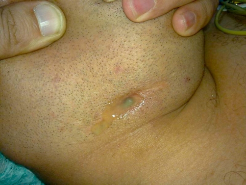
A 27-year-old Egyptian man working as a clerk presented with a discharging sinus in the right side of his neck (Figure 1). The man had a right submandibular sialadenectomy 8 years before presentation. The patient reported the sinus to appear after removing a drain which has been placed. This surgery was performed elsewhere. He also complained of pain below his chin and an increase in the amount of discharge on mastication. No history of fever. The patient reported receiving several trials of antibiotic therapy in an attempt to cure his condition. He had an X-ray done before presentation. The report of the excised gland pathological examination was not available.
Figure 1.
Discharging sinus showing copious discharge.
On examination the man was 90 kilograms body weight and 182 centimeters height, there was a healed scar of previous surgery and a discharging sinus in the right submandibular region the discharge was purulent. No palpable swellings or enlarged cervical lymph nodes were found. Bimanual examination was done and there were no palpable swelling in the floor of mouth. A provisional diagnosis of a salivary fistula was considered.
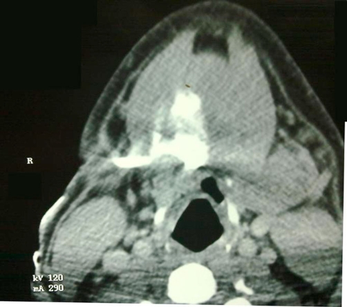
Neck X-ray showed no abnormality. A CT fistulogram was undertaken (Figure 2), which showed a blind ending track leading to cavity just beneath the right side of floor of mouth. It also confirmed excision of the right submandibular gland.
Figure 2.
CT fistulogram showing the cavity below floor of mouth.
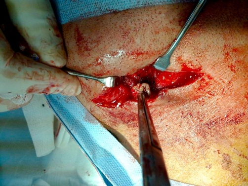
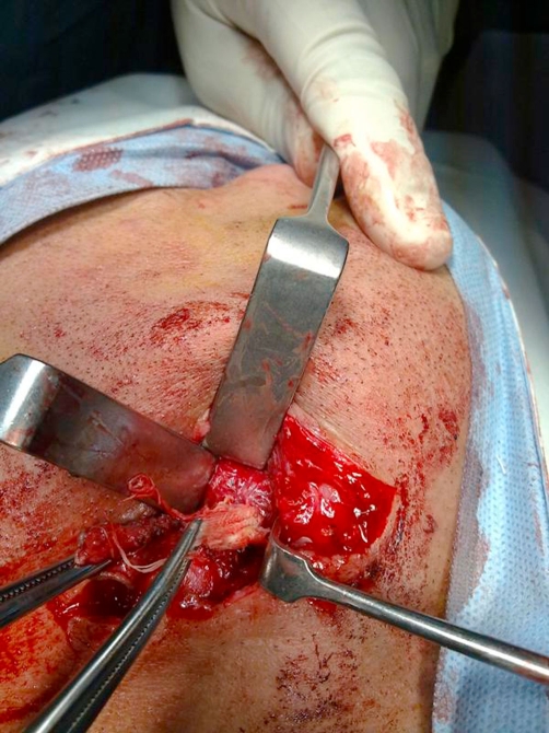
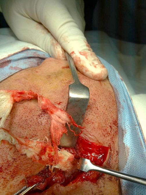
A decision to perform neck exploration was undertaken. During surgery gentle probing of the track was done then dissection was carried out revealing the sinus track passing medial the digastric muscle and up to sublingual area (Figure 3). The tracks ended into a cyst below the mandible in the area where we typically ligate and divide the submandibular duct. On opening the cyst a purulent discharge appeared and irrigation of the cavity further revealed white fabric fibers (Figure 4). With traction on these fibers two small sponges were found and removed (Figure 5). The track was then excised and the cavity and wound copiously irrigated with saline. A suction drain was inserted and wound closed. The patient had an uneventful postoperative course and primary healing was achieved.
Figure 3.
Early dissection of the well formed track.
Figure 4.
The tightly packed sponges.
Figure 5.
Extraction of the sponge.
Discussion
Retained surgical sponges in the neck are much less reported than intrabdominal ones. Nevertheless they can produce a variety of postoperative complications and can lead to diagnostic misinterpretation e.g. tumor recurrence [5]. Our patient was suspected of having a salivary fistula, another rare condition, versus a stitch sinus. The retained sponges did not show on X-ray as it contains no radio opaque marker. Although this dose not completely abolishes the problem of missed sponges, it sure allows for intraoperative radiologic screening when the accuracy of final count is in doubt, and would have helped in clenching a correct preoperative diagnosis [6].
It is well known that surgical textile materials and instruments should be counted once at the start and twice at the conclusion of surgical procedures. However, counts are not always sufficient, since most reported cases occur in spite of a normal pack count [7]. With very few exceptions, we believe that the surgeon should not leave a sponge packed behind and proceed with other operative steps, this helps to distract his attention and the blood stained sponge will be very similar to surrounding tissues. Instead the surgeon should wait until the reason for packing e.g. oozing have stopped remove the pack and proceed with other steps. Another dangerous practice is dividing swabs into 2 or more pieces to form the so-called peanut swab this helps to mess up the final count.
Conclusion
We recommend in addition to routine preoperative and postoperative counts, using sponges containing radio opaque markers. Sound surgical practice and removing packs as soon as their function have been achieved. Postoperative radiographic screening should be performed when counts are in doubt or high risk cases.
Abbreviations
- CT
Computed tomography
- RSS
retained surgical sponge
Consent
Written informed consent was obtained from the patient for publication of this case report and accompanying images. A copy of the written consent is available for review by the journal’s Editor-in-Chief.
Competing interests
The author declares that he has no competing interests.
This article is available from: http://casesjournal.com/casesjournal/article/view/6413
References
- Istvan S, Andras L, Peter S. Sudden suffocation by surgical sponge retained after a 23-year-old thoracic surgery. Am J Forensic Med Pathol. 2004;25(4):324–326. doi: 10.1097/01.paf.0000137273.73799.d6. [DOI] [PubMed] [Google Scholar]
- Zbar A, Agrawal A, Saeedi I, Utidjian M. Gossypiboma revisited: a case report and review of the literature. J R Coll Surg Edinb. 1998;43:417–418. [PubMed] [Google Scholar]
- Gawande AA, Studdert DM, Orav EJ, Brennan TA, Zinner MJ. Risk factors for retained instruments and sponges after surgery. N Engl J Med. 2003;348:229–235. doi: 10.1056/NEJMsa021721. [DOI] [PubMed] [Google Scholar]
- Yildirim S, Tarim A, Nursal TZ, Yildirim T, Caliskan K, Torer N, Karagulle E, Noyan T, Moray G, Haberal M. Retained surgical sponge (gossypiboma) after intraabdominal or retroperitoneal surgery: 14 cases treated at a single center. Langenbecks Arch Surg. 2006;391(4):390–395. doi: 10.1007/s00423-005-0581-4. [DOI] [PubMed] [Google Scholar]
- Bani-Hani KE, Gharaibeh KA, Yaghan RJ. Retained surgical sponges (gossypiboma) Asian J Surg. 2005;28(2):109–115. doi: 10.1016/s1015-9584(09)60273-6. [DOI] [PubMed] [Google Scholar]
- Burrel M, Capurro S, Arguis P, Vilana R. Sonographic appearance of a retained surgical sponge in the neck. J Clin Ultrasound. 2000;28(6):311–313. doi: 10.1002/1097-0096(200007/08)28:6<311::aid-jcu9>3.0.co;2-l. [DOI] [PubMed] [Google Scholar]
- Dux M, Ganten M, Lubienski A, Grenacher L. Retained surgical sponge with migration into the duodenum and persistent duodenal fistula. Eur Radiol. 2002;12:74–77. doi: 10.1007/s00330-002-1408-0. [DOI] [PubMed] [Google Scholar]


