Abstract
Since bone is responsive to mechanical loading, pulsating joint loading (PJL), which laterally applies oscillatory mechanical loads to joints, can be explored for preventative conditioning and therapeutic treatments. Herein the general features of PJL are reviewed, and its potential usage for sports medicine is discussed.
Summary
A recently developed pulsating-joint-loading modality is reviewed focusing on its unique features and potential usages.
Keywords: lateral loads, bone formation, wound healing, sports injury, molecular pathways
INTRODUCTION
Elite athletes frequently represent extreme cases of the need for bone strengthening therapies, and often are therefore given special attention, including extraordinary surgical interventions. For instance, tibial stress fractures (fatigue injuries) in high-level adolescent athletes are often difficult to treat (24). Similar needs are experienced by the general population, especially aging adults. Thus, the demand for bone strengthening therapies is enormous, and likely will increase as the intensity of athletic competitions escalates and as the population of developed countries ages. Bone loss due to osteoporosis, for instance, is predominantly observed in the elderly. Bone is a metabolically active tissue, and it should be possible, in principle, to devise strategies that exploit its natural anabolic and regenerative capacities. For injured athletes, geriatric patients, and astronauts options for the use of existing physical therapies, however, presently often are limited (10).
In this review we introduce a recently developed pulsating joint loading (PJL) modality and discuss its potential usage in sports medicine. PJL applies mechanical loads to a synovial joint, such as an elbow or a knee, and enhances bone formation and bone wound healing throughout long bones such as the ulna (23), the tibia (25–27), and the femur (27, 28). Target-specific therapies with PJL might offer a new dimension in physical therapies with potential advantages over pharmacological interventions. Here we review the current understanding of its efficacy and discuss its possible biophysical mechanisms as well as its potential molecular signaling features.
EXISTING APPROACHES FOR BONE-STRENGTHENING THERAPIES
Two primary traditional approaches for strengthening bone are pharmacological interventions and exercise routines (Table 1). Pharmacological agents include bisphosphonate pills, injection of parathyroid hormone, and application of selective estrogen receptor modulators. These agents offer several advantages: patient-friendly, in many cases easy to administer, and likely to be taken as prescribed for prolonged periods. Nevertheless, undesirable side effects may occur. For instance, the gastrointestinal drug-induced side effects of mucosal ulceration, gastrointestinal hemorrhage, diarrhea, and constipation have been observed (13). Only minor side effects such as occasional nausea and headache are known for the treatment with parathyroid hormone, but we do not fully understand possible long-term side effects of any of the existing drugs (15).
TABLE 1.
Examples of traditional bone-strengthening therapies
| Sample Treatment | Critique | |
|---|---|---|
| Pharmacological interventions | Bisphosphonate pill | Possible gastrointestinal side effects (13) |
| Parathyroid hormone | Promising, but frequent injections required (14) | |
| Selective estrogen receptor modulators | Possible increased risk of venous thrombosis (5) | |
| Exercise routines and mechanical loading | Rhythmic physical activity (e.g., running) | Individual variation exists (6) |
| Desensitization may develop (20) | ||
| Special needs for geriatric patients (9) | ||
| Intense athletic training (especially sport or site-specific, e.g., tennis vs swimming) | Limited effectiveness for osteoporosis (1) | |
| Routines occasionally suprathreshold intensity, and diet regulated (reviewed in Borer (4)) |
Exercise routines and mechanical stimulations include walking, jogging, running, swimming, and playing tennis as well as intense athletic training through weight lifting. Although exercise programs have long been proven to promote bone tissue growth (19), they are mostly directed towards injury prevention. Once a sports related injury happens, the active physical therapy is terminated and replaced by a procedure known as RICE (rest, ice, compression, and elevation). Few exercise programs are considered appropriate for severe injuries such as bone fractures. Furthermore, in elite athletes, or otherwise healthy young adults, rigorous exercise regimes are occasionally counterproductive by promoting stress fractures (16). Such side effects might conceivably include bone formation in unwanted areas or other negative consequences (2). Thus, there is a need to develop strategies that exploit the natural anabolic and regenerative capacities of bone which are suitable for injured athletes, geriatric patients, or astronauts with limited exercise tools.
PULSATING JOINT LOADING (PJL)
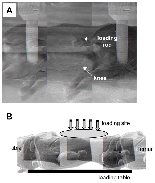
PJL in the form of knee, ankle, and elbow loading is a recently devised treatment modality, whereby bone strengthening is achieved through dynamic loads applied to a joint. Specifically, PJL loads are imposed laterally to the epiphysis of a synovial joint. Although the magnitude of loads is significantly smaller than other loading modalities such as ulna axial loading (21) and tibia four-point bending (7), PJL can induce bone formation and fracture healing in long bones. Bone histomorphometry using mice as an animal model has shown that PJL enables formation of new bone on the periosteal and endosteal surface of cortical bone in the ulna (elbow loading), tibia (knee loading and ankle loading), and femur (knee loading) (23, 25–28). In each specific joint loading modality, induction of bone formation was observed in both the metaphysis and diaphysis of long bones. Figure 1 illustrates a loading experiment using a mouse, where well-controlled pulsating loads were applied using a custom-made piezoelectric mechanical loading device to the knee (covering the epiphyses of both the femur and the tibia). Typically, loads are given at loading frequencies of 1 – 20 Hz for 3–5 min per day. For mice, 0.5 N (peak-to-peak) force is sufficient to induce significant load-driven bone formation within a week.
Fig. 1.
Configuration of the pulsating joint loading as designed for use on model laboratory animals (e.g., mouse). A. Setting up knee loading with a mouse. B. Micro computer tomography image of the mouse knee.
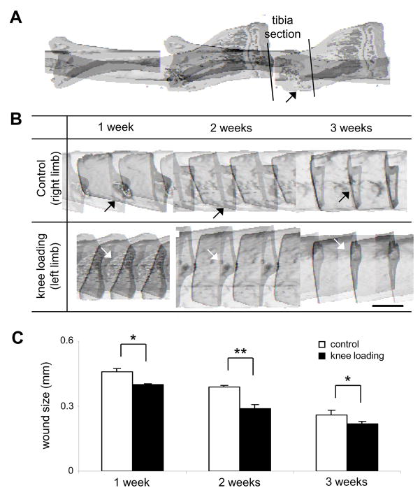
Table 2 lists the advantages of PJL over other treatment modalities. First, PJL does not require direct contact with the site of bone formation or wound healing. Since its bone-strengthening effect is exerted throughout the length of a long bone, knee loading, for instance, can accelerate bone formation in both the distal diaphysis near the knee as well as the proximal diaphysis near the femoral neck (27). Healing of a surgical wound, generated in the diaphysis of the tibia, is accelerated by loads applied at the proximal epiphysis of the tibia with knee loading (Figure 2). Second, the apparatus for clinical use is inexpensive since pulsating loads at a frequency in the order of 1 to 10 Hz can be achieved with small, commercially available portable motors. Third, the procedure is non-invasive and no surgical intervention is needed. Fourth, PJL-based therapy is likely to receive good compliance from patients because of its brief daily treatment duration (typically a few minutes) and small strains (less than 100 microstrains at the site of injury or wound healing). Fifth, PJL induces few side effects since applied loads are significantly smaller than weight-based training and no administration of chemical agents is required.
TABLE 2.
Potential advantages of pulsating joint loading as a therapeutic tool
| Feature | Comment |
|---|---|
| Might eliminate need for direct contact with injury site | Should be effective for patients with cast-immobilized wounds |
| Expected to be relatively inexpensive | Noninvasive, thus surgical intervention circumvented |
| Treatment likely to receive full compliance from patients | Requires low levels of strain, at brief intervals |
| Minimum side effects | Important for elderly patients who are taking various medications |
Fig. 2.
Micro computer tomography images of surgical wounds in the mouse tibia. A. Mouse tibia with a surgical wound. B. Healing of the surgical wound with and without knee loading. The tibia section was analyzed at 1, 2, and 3 weeks after surgery. The arrows indicate the positions of surgical wounds. Bar = 1 mm. C. Closure of the surgical wound with and without knee loading. The single and double asterisks indicate statistical significance at p < 0.05 and p < 0.01, respectively.
BIOPHYSICAL MECHANISM OF PJL
The biophysical mechanisms generated by PJL are different from most of the existing loading modalities, such as whole-body vibration (17), axial loading (21), and bending (7). Although all modalities require dynamic load application for efficient bone strengthening (8), PJL seems to eliminate the requirement for exceeding a strain threshold at the site of bone formation (25, 26, 28). This is probably because PJL effectively induces a pressure gradient throughout a long bone and this gradient establishes a remote communication link between the site of loading in the epiphysis and the site of bone formation in the metaphysis as well as in the diaphysis (30).
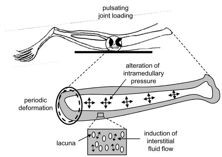
Based on several key features summarized in Table 3, the following working biophysical model is proposed: cyclic deformation of the epiphysis alters pressure in the medullary cavity and this pressure gradient induces fluid flow in the metaphysis and the diaphysis (Figure 3). Two types of fluid flow are conceivable: fluid flow in the lacuno-canalicular network in a porous bone matrix, and fluid flow in the bone medullary cavity. Flow in the lacuno-canalicular network is termed “interstitial fluid flow.” It is thought to produce dynamic flow shear to osteocytes housed in lacunae. Interstitial fluid flow is assumed to be driven by load-induced cyclic pressure alterations. Flow in the medullary cavity is thought to be induced in response to a bone wound that acts as a pressure sink. This flow is assumed to promote migration of bone-marrow derived cells including hematopoietic stem cells, mesenchymal stem cells, and endothelial progenitor cells. Their load-driven recruitment towards the site of the wound is proposed as the basis of accelerated healing.
TABLE 3.
Experimental approaches to understanding cellular and molecular basis of mechanotransduction stimulated by PJL
| Manipulation | Comment |
|---|---|
| Surgical holes drilled near site PJL | PJL effects diminished, presumably by reducing intramedullary pressure (29) |
| Fiber optical pressure sensor measurements | Suggest intramedullary pressure increased by PJL (30) |
| Biomechanical/dye transport studies | Suggest load-driven fluid flow (18) |
PJL indicates pulsating joint loading.
Fig. 3.
Schematic illustration of the proposed biophysical mechanism with knee loading. Pulsating joint loading induces periodic deformation in the epiphysis that drives alteration of intramedullary pressure in the medullary cavity and interstitial fluid flow in the lacunocanalicular network in the diaphysis.
Optimizing PJL-based therapies for sports injuries with regard to intensity and frequency, duration of rest/recovery periods may differ depending on the site and size of injury. That is, the distribution of intramedullary pressure routes might vary depending on the nature of the injury.
POTENTIAL MOLECULAR PATHWAYS INVOLVED IN PJL
In parallel with efforts directed towards elucidating the biophysical mechanisms of PJL, it is necessary to understand the molecular mechanism of action. Several cell types are likely involved in the PJL distance effects: chondrocytes in the growth plate, bone marrow-derived cells in the bone cavity, osteoblasts and osteoclasts together with cells in muscles, and the vascular system. Understanding how the activities of those diverse cell types are coordinated in response to PJL will require extensive gene expression research. Since the action of PJL is remote from the site of application, it is likely that some type of interstitial molecular transport and signaling is driven by the pressure gradient in the medullary cavity, but exact identities of biophysical mediators are yet to be understood.
Despite our lack of complete knowledge of the landscape of molecular pathways, some of the biochemical pathways activated within minutes of mechanical stimulation are known. First, mitogen-activated protein kinases (MAPKs) such as extracellular signal-regulated kinase 1/2 (ERK1/2), p38 MAPK, and Jun N-terminal kinase (JNK) are activated in osteoblasts in response to shear stress induced by fluid flow (22). Osterix, one of the critical transcription factors for bone development is induced by p38 MAP kinase. Furthermore, upregulation of type I collagen is mediated by ERK1/2 and JNK (22). Consistent with the predicted role of interstitial fluid flow, the Wnt/β-catenin pathway in osteocytes has been proposed to mediate load-driven bone formation by crosstalk with the prostaglandin pathway and a suppression of negative regulators such as Sclerostin (Sost) and Dickkopf 1(Dkk1) (3).
Second, expression of peroxisome proliferator-activated receptor gamma (PPARγ), is downregulated by mechanical stimulation (11). PPARγ is a stimulator of adipocyte proliferation and a suppressor of differentiation of osteoblasts. Therefore, its down regulation seems to favor promotion of osteogenesis over adipogenesis. Our recent in vitro experiment using mouse C57BL/6 (MC3T3 E1) osteoblast-like cells, as well as primary mesenchymal stem cells isolated from mice, shows that fluid flow at 10 dyn/cm2 for 1h significantly reduces the messenger ribonucleic acid (mRNA) level of PPARγ (unpublished observation, January 2008).
Third, previous studies have reported that expression of bone morphogenetic proteins (BMPs) and insulin-like growth factors (IGFs) are elevated in response to mechanical loading (12). The molecules are anabolic and their expression is at least in part mediated by transforming growth factor β (TGFβ) and/or phosphoinositide 3-kinase (PI3K) pathways. Taken together, molecular signaling pathways, which are potentially involved in PJL, include MAPK, PPAR, BMPs, IGFs, TGFβ, and PI3K. The detailed role of the molecules in PJL has yet to be investigated.
CONCLUSIONS AND PERSPECTIVES
In conclusion, existing animal studies support the notion that PJL offers a novel load-driven therapy for strengthening bone and accelerating healing of injured bones. Future research should be directed at evaluating the efficacy of PJL for strengthening long bones of athletes and healing of various sports-related injuries including trauma and overuse injuries in the tibia, femur, ulna, and humerus. A prototype PJL loader which could be effective for human use is being designed in our laboratory as a first attempt to develop a portable apparatus for astronauts in conditions of microgravity. However, no clinical data have been collected. In parallel with further development of a user-friendly device, evaluation of loading conditions suitable for individual sports injuries needs to be conducted.
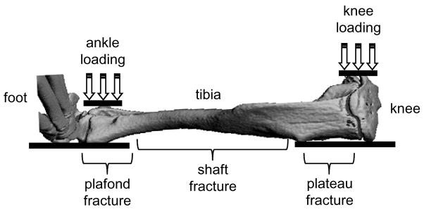
Regarding biophysical and molecular mechanisms which drive PJL effects, intriguing questions include: (a) Is the loading frequency of 2–20 Hz employed in animal studies suitable for human use? (b) Knowing that tibial stress fractures (a common form of overuse injury) are occasionally difficult to treat, which is more effective for their healing, ankle loading or knee loading (Figure 4)? (c) Does administration of either IGF or TGFβ share the same molecular pathway as application of PJL? More specifically, would simultaneous application of PJL, and, for example IGF1, be more effective than either treatment alone? (d) Is PJL effective for healing injuries not only in long bones but also in joints such as ankle sprains and knee inflammation?
Fig. 4.
Schematic illustration of ankle loading and knee loading using a tibial fracture model.
Understanding more about the physical and molecular features of bones and joints will contribute to the development of load-driven strategies for bone strengthening and injury healing.
Acknowledgments
This research was funded in part by Indiana University - Purdue University Indianapolis Research Support Fund Grant 2295058 to PZ and National Institutes of Health R01 AR52144 to HY.
References
- 1.Beaudreuil J. Nonpharmacological treatments for osteoporosis. Ann Readapt Med Phys. 2006;49:581–588. doi: 10.1016/j.annrmp.2006.05.004. [DOI] [PubMed] [Google Scholar]
- 2.Benhamou CL. Effects of osteoporosis medications on bone quality. Joint Bone Spine. 2007;74:39–47. doi: 10.1016/j.jbspin.2006.06.004. [DOI] [PubMed] [Google Scholar]
- 3.Bonewald LF, Johnson ML. Osteocytes, mechanosensing and Wnt signaling. Bone. 2008;42:606–615. doi: 10.1016/j.bone.2007.12.224. [DOI] [PMC free article] [PubMed] [Google Scholar]
- 4.Borer KT. Physical activity in the prevention and amelioration of osteoporosis in women. Sports Med. 2005;35:779–830. doi: 10.2165/00007256-200535090-00004. [DOI] [PubMed] [Google Scholar]
- 5.Cano A, Hermenegildo C, Oviedo P, Tarin JJ. The risk of cardiovascular disease in women; from estrogens to selective estrogen receptor modulators. Front Biosci. 2007;12:49–68. doi: 10.2741/2048. [DOI] [PubMed] [Google Scholar]
- 6.Daly RM. The effect of exercise on bone mass and structural geometry during growth. Med Sports Sci. 2007;51:33–49. doi: 10.1159/000103003. [DOI] [PubMed] [Google Scholar]
- 7.Gregory LS, Forwood MR. Cyclooxygenase-2 inhibition delays the attainment of peak woven bone formation following four-point bending in the rat. Calcif Tissue Int. 2007;80:176–183. doi: 10.1007/s00223-006-0170-8. [DOI] [PubMed] [Google Scholar]
- 8.Hughes JM, Novotny SA, Wetzsteon RJ, Petit MA. Lessons learned from school-based skeletal loading intervention trials: putting research into practice. Med Sport Sci. 2007;51:137–158. doi: 10.1159/000103013. [DOI] [PubMed] [Google Scholar]
- 9.Inokuchi S, Matsusaka N, Hayashi T, Shindo H. Feasibility and effectiveness of a nurse-led community exercise programme for prevention of falls among frail elderly people: a multi-centre controlled trial. J Rehabil Med. 2007;39:479–485. doi: 10.2340/16501977-0080. [DOI] [PubMed] [Google Scholar]
- 10.Iwamoto J, Takeda T. Stress fractures in athletes: review of 196 cases. J Orthop Sci. 2003;8:273–278. doi: 10.1007/s10776-002-0632-5. [DOI] [PubMed] [Google Scholar]
- 11.Kang S, Bennett CN, Gerin I, Rapp LA, Hankenson KD, Macdougald OA. Wnt signaling stimulates osteoblastogenesis of mesenchymal precursors by suppressing CAAT/enhancer-binding protein alpha and peroxisome proliferator-activated receptor gamma. J Biol Chem. 2007;282:14515–14524. doi: 10.1074/jbc.M700030200. [DOI] [PubMed] [Google Scholar]
- 12.Lau KH, Kapur S, Kesavan C, Baylink DJ. Up-regulation of the Wnt, estrogen receptor, insulin-like growth factor-I, and bone morphogenetic protein pathways in C57BL/6J osteoblasts as opposed to C3H/HeJ osteoblasts in part contributes to the differential anabolic response to fluid shear. J Biol Chem. 2006;281:9578–9588. doi: 10.1074/jbc.M509205200. [DOI] [PubMed] [Google Scholar]
- 13.Leong RW, Chan FK. Drug-induced side effects affecting the gastrointestinal tract. Expert Opin Drug Saf. 2006;5:585–592. doi: 10.1517/14740338.5.4.585. [DOI] [PubMed] [Google Scholar]
- 14.Manabe T, Mori S, Mashiba T, et al. Human parathyroid hormone (1–34) accelerates natural fracture healing process in the femoral osteotomy model of cynomolgus monkeys. Bone. 2007;40:1475–1482. doi: 10.1016/j.bone.2007.01.015. [DOI] [PubMed] [Google Scholar]
- 15.Mitlak BH. Parathyroid hormone as a therapeutic agent. Curr Opin Pharmacol. 2002;2:694–699. doi: 10.1016/s1471-4892(02)00208-4. [DOI] [PubMed] [Google Scholar]
- 16.Niva MH, Kiuru MJ, Haataja R, Pihlajamaki HK. Fatigue injuries of the femur. J Bone Joint Surg Br. 2005;87:1385–1390. doi: 10.1302/0301-620X.87B10.16666. [DOI] [PubMed] [Google Scholar]
- 17.Rubin C, Turner AS, Bain S, Mallinckrodt C, McLeod K. Anabolism: Low mechanical signals strengthen long bones. Nature. 2001;412:603–604. doi: 10.1038/35088122. [DOI] [PubMed] [Google Scholar]
- 18.Su M, Jiang H, Zhang P, Liu Y, Wang E, Hus A, Yokota H. Load-driven molecular transport in mouse femur with knee-loading modality. Ann Biomed Eng. 2006;34:1600–1606. doi: 10.1007/s10439-006-9171-z. [DOI] [PubMed] [Google Scholar]
- 19.Turner CH, Robling AG. Exercises for improving bone strength. Br J Sport Med. 2005;39:188–189. doi: 10.1136/bjsm.2004.016923. [DOI] [PMC free article] [PubMed] [Google Scholar]
- 20.Umemura Y, Ishiko T, Yamauchi T, Kurono M, Mashiko S. Five jumps per day increase bone mass and breaking force in rats. J Bone Miner Res. 1997;12:1480–1485. doi: 10.1359/jbmr.1997.12.9.1480. [DOI] [PubMed] [Google Scholar]
- 21.Warden SJ, Turner CH. Mechanotransduction in the cortical bone is most efficient at loading frequencies of 5–10 Hz. Bone. 2004;34:261–270. doi: 10.1016/j.bone.2003.11.011. [DOI] [PubMed] [Google Scholar]
- 22.Wu CC, Li YS, Haga JH, et al. Roles of MAP kinases in the regulation of bone matrix gene expressions in human osteoblasts by oscillatory fluid flow. J Cell Biochem. 2006;98:632–641. doi: 10.1002/jcb.20697. [DOI] [PubMed] [Google Scholar]
- 23.Yokota H, Tanaka SM. Osteogenic potentials with joint-loading modality. J Bone Miner Metab. 2005;23:302–308. doi: 10.1007/s00774-005-0603-x. [DOI] [PubMed] [Google Scholar]
- 24.Young AJ, McAllister DR. Evaluation and treatment of tibial stress fractures. Clin Sports Med. 2006;25:117–128. doi: 10.1016/j.csm.2005.08.015. [DOI] [PubMed] [Google Scholar]
- 25.Zhang P, Tanaka SM, Jiang H, Su M, Yokota H. Diaphyseal bone formation in murine tibiae in response to knee loading. J Appl Physiol. 2006;100:1452–1459. doi: 10.1152/japplphysiol.00997.2005. [DOI] [PubMed] [Google Scholar]
- 26.Zhang P, Sun Q, Turner CH, Yokota H. Knee loading accelerates bone healing in mice. J Bone Miner Res. 2007;22:1979–1987. doi: 10.1359/jbmr.070803. [DOI] [PMC free article] [PubMed] [Google Scholar]
- 27.Zhang P, Tanaka SM, Sun Q, Turner CH, Yokota H. Frequency-dependent enhancement of bone formation in murine tibiae and femora with knee loading. J Bone Miner Metab. 2007;25:383–391. doi: 10.1007/s00774-007-0774-8. [DOI] [PMC free article] [PubMed] [Google Scholar]
- 28.Zhang P, Su M, Tanaka SM, Yokota H. Knee loading causes diaphyseal cortical bone formation in murine femurs. BMC Musculoskel Dis. 2006;73:1–12. doi: 10.1186/1471-2474-7-73. [DOI] [PMC free article] [PubMed] [Google Scholar]
- 29.Zhang P, Yokota H. Effects of surgical holes in mouse tibiae on bone formation induced by knee loading. Bone. 2007;40:1320–1328. doi: 10.1016/j.bone.2007.01.018. [DOI] [PMC free article] [PubMed] [Google Scholar]
- 30.Zhang P, Su M, Liu Y, Hus A, Yokota H. Knee loading dynamically alters intramedullary pressure in mouse femora. Bone. 2007;40:538–543. doi: 10.1016/j.bone.2006.09.018. [DOI] [PMC free article] [PubMed] [Google Scholar]


