
Fig. 1.
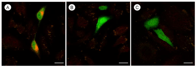
Confocal microscopic immunolabeling for the AT2 receptor antiserum in HeLa cells differentially transfected with AngII receptor subtypes. A, Immunofluorescence labeling (orange) is seen in the cytoplasm near the nucleus in cells transfected with the AT2 receptor. B and C, There is no observable immunolabeling in cells transfected with either the AT1A receptor in panel B or an empty vector plasmid in panel C. Green fluorescent protein (green) is present in all transfected cells. Scale bar=10 μm.