Abstract
Purpose
To determine by optical coherence tomography (OCT) the effect of blinking on ocular surface tear volume after instillation of artificial tears.
Design
Experimental study.
Methods
Normal human (n = 21) eyes were imaged to get dimensions of the tear film and menisci during blinking. The imaging was done immediately and 5 minutes after the instillation of 35 µl of mid-viscosity artificial tears (1.0% carboxymethylcellulose, Refresh Liquigel, Allergan, Irvine, CA). The exposed ocular surface area and the lid lengths were used to calculate the volumes.
Results
Immediately after the instillation, total tear volume was increased (P<0.001) compared to 5 minutes after the instillation with the major increases in the lower tear meniscus volume and tear film volume. After the instillation, blinking caused tear loss in total tear volume due to the decrease of the lower tear meniscus volume (P<0.05). In contrast, blinking increased the tear film volume (P<0.05). At the end of eye opening period, tear film volume decreased and lower tear meniscus volume increased significantly (P<0.05) with no significant changes in total tear volume (P>0.05). During the blink cycle immediately after the instillation, net loss was evident in tear film volume, lower tear meniscus volume and total tear volume (P<0.05).
Conclusions
Blinking plays a crucial role in distribution and removal of instilled tears. When the tear system is overloaded, the increase in blink output helps restore balance.
Blinking plays an important role in distribution and drainage of tears.1–3 The dynamics of instilled fluids into the eyes have been studied with radioscintigraphy and dye clearance methods.4–8 Recovery of tear volume occurs by active expulsion of tears from the lacrimal system. However, only the changes of total tear volume over brief periods of time were investigated previously.4–8 The impact of each blink on the tear volumes in the menisci and tear film on ocular surface remains unknown. With the advent of a newly developed, custom-built anterior segment OCT, we were able to simultaneously measure in real-time the cross-sectional areas of the upper and lower tear menisci along with tear film thickness.1,9,10 Based on eyelid lengths and ocular surface area, tear volumes in both tear menisci and the tear film can be calculated. In a previous study,1 the effect of blinking on tear volumes was reported under basal conditions and during reflex tearing. In this study, the goal was to further explore the effect of blinking on tear volume after instillation of artificial tears.
SUBJECTS AND METHODS
The same group as reported in the previous study1 continued to be tested in this study. Twenty-one young subjects (10 women and 11 men, mean age: 32.1 ± 8.7) with no history of contact lens wear or any current ocular or systemic diseases were recruited in Rochester. The study was conducted in a consulting room in which the temperature (15 to 25°C) and humidity (30 to 50%) were controlled using central air conditioning and two humidifiers as described previously.9
One eye of each subject was randomly selected and imaged using the same anterior segment OCT as described in other studies.1,9,10 In brief, after the subject completed the imaging session at baseline and delayed blink period (reported previously),1 s/he continued to be tested in this study. The subject was asked to look at an external non-illuminated target. A vertical 12 mm scan across the central cornea (apex), including the upper and lower tear menisci simultaneously, was performed. Thirty-five micro-liters of artificial tears (1.0% carboxymethylcellulose, Refresh Liquigel, Allergan, Irvine, CA) were delivered using a pipette into the lower fornix of the study eye that was immediately imaged by OCT. OCT images were acquired continuously at 8 frames per second as the subject was asked to delay each blink for as long as possible for a set of 3 blinks. To measure the length of the upper and lower eyelids and the exposed ocular surface, the selected eye of each subject was photographed with a reference scale using a digital camera mounted on another slit-lamp.
Image processing and data analysis were done at the University of Miami since two authors (JRP and JW) took positions there. To determine the total corneal thickness, including the tear film, eight OCT images corresponding to one second of time immediately before and after blinking in two consecutive blinks were processed using custom-developed software. From OCT images obtained immediately after instillation of the drop, true corneal thickness was measured, and tear film thickness was estimated by subtracting the true corneal thickness from the total thickness as described in our previous studies.9,10 The first OCT image showing both upper and lower tear menisci before and after two consecutive blinks were processed. One inter-blink interval was analyzed since two consecutive blinks defined the inter-blink interval. To determine the exposed ocular surface area, upper and lower lid lengths were measured from the two dimensional photographs of the eye using a reference scale. The values were multiplied by a factor of 1.294 to compensate for the curved ocular surface as recommended by Tiffany et al.11 The product of the tear film thickness and the exposed ocular surface area was taken as the tear film volume.12,13 The products of upper and lower eye lid lengths and cross sectional areas of upper and lower tear menisci were taken as the upper tear meniscus volume and the lower tear meniscus volume, respectively.13,14 The sum of these three volumes was taken as the total tear volume on the ocular surface.
The action of blinking and the following open eye period up to the next blink was defined as a blink cycle.15 In the present study, “blink outcome” was defined as the difference in tear volume before and after a blink. “Open eye outcome” was calculated by subtracting the tear volume at the beginning of open eye period from that at the end of open eye period. The net change during the blink cycle was the sum of blink outcome and open eye outcome.
Data analysis was conducted on a computer using Statistica software (StatSoft, Inc., Tulsa, OK). Paired t-tests were used to determine whether there were pair-wise differences (P<0.05). Pearson’s correlation was used to describe the relations among tear volumes.
RESULTS
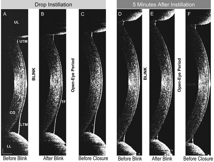
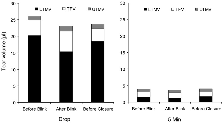
The changes due to blinking and open eye periods in each compartment were clearly visualized (Fig. 1). Immediately after the instillation, tear volume was increased (paired t-test, P<0.001) compared to 5 minutes after the instillation with all significant increases in the tear film and tear menisci (P<0.05). The major increases were found in the lower tear meniscus volume and (Fig. 2).
Figure 1. OCT images obtained during blinking.
A vertical 12 mm OCT scan was performed across the corneal apex during blinking immediately after instillation of artificial tears (Images A–C) and 5 minutes later (Images D–F). Changes in tear film (TF), upper tear meniscus (UTM) and lower tear meniscus (LTM) were noted after blinking and open-eye period. Cornea (CO), upper eyelid (UL) and lower eyelid (LL) are also shown. Bars denote to 500 µm.
Figure 2. Compartmental tear volumes of the exposed ocular surface.
Using optical coherence tomography, the tear volumes in the lower tear meniscus (LTMV), tear film (TFV) and upper tear meniscus (UTMV) were obtained immediately after the installation of artificial tears (Refresh Liquigel) and 5 minutes later. All volumes of the LTMV, TFV and UTMV were greater immediately after instillation than 5 minutes later (P<0.05). Note the volume changes in the TFV and LTMV before and after blink, indicating tear flows between the tear film and tear meniscus.
UTMV: Upper tear meniscus volume.
TFV: Tear film volume;
LTMV: Lower tear meniscus volume.
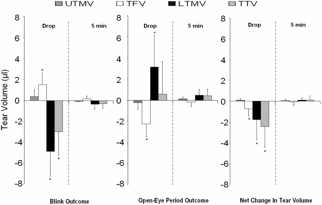
After the instillation, blinking caused tear loss in total tear volume due to the decrease of the lower tear meniscus volume (paired t-test, P<0.05, comparing tear volumes before and after blink). In contrast, blinking increased the tear film volume (P<0.05). There were no significant differences in upper tear meniscus volume with blink (Fig.3A). At the end of eye opening period, tear film volume decreased and lower tear meniscus volume increased significantly (P<0.05) with no significant changes in total tear volume (P>0.05) (Fig. 3B). During the blink cycle immediately after the instillation, net loss was evident in tear film volume, lower tear meniscus volume and total tear volume (P<0.05) (Fig. 3C). Five minutes after the instillation, no significant differences in the tear menisci and film volumes were detected. (Fig. 3A–C). Immediately after the installation of the artificial tears, the blink outcome (loss) of lower tear meniscus volume (r = −0.61, P<0.05) was negatively correlated to the total tear volume and the blink outcome (gain) of the tear film volume (r = 0.66, P<0.05) was positively related to the total tear volume.
Figure 3. Changes of tear volumes measured by OCT.
(Right) Blinking caused tear loss from total tear volume (TTV) and lower tear meniscus volume (LTMV) immediately after instillation of the artificial tears (P<0.05). In contrast, it caused the tear gain in the TFV after the instillation (P<0.05). (Middle) After the open eye period, LTMV gained volume immediately after instillation (P<0.05). TFV lost volume significantly (P<0.05) immediately after instillation. (Left) For the entire blink cycle, net loss was evident in tear film volume (TFV), LTMV, and TTV immediately after the instillation (P<0.05). Five minutes after the instillation, no significant differences in the tear menisci and film volumes were detected. Red asterisks denote to significance differences at 0.05 levels and vertical bars denote 95% confidence intervals.
UTMV: Upper tear meniscus volume.
TFV: Tear film volume;
LTMV: Lower tear meniscus volume;
TTV: Total tear volume.
DISCUSSION
We have found, in a previous study, that the tear volume in a normal tear system is 3 microliters, and that this volume is sufficient to maintain optical and physiological integrity at the ocular surface.1 This figure of the basal tear volume is slightly lower that the values reported by Mishima et al.13 The use of fluorescein in their study might have resulted in reflex tearing. With each blink, the tears are mixed and redistributed.16 Movement of the eyelids acts as a pump by compressing the canaliculi and lacrimal sac and promotes the drainage of tears.3 To keep a dynamic balance, the tear volume in the tear system must maintain a relatively steady state.13,17,18 Under normal circumstances, the drainage system itself is thought to contain a negligible volume of tears.3 This assumption is supported by our data presented in the previous study1 and the present study. At baseline, the tear volume in all of the tested compartments remained approximately the same with little gain or loss during the blink cycle.1 It appears that only a small quantity of tears in the tear film and menisci is needed for keeping the ocular surface wet during inter-blink periods. The baseline values were re-established within 5 minutes after the instillation of artificial tears (Fig. 1–2). While blinking and eye opening have little effect on normal tear volume, spreading during blinking and evaporation during the open eye period may cause minor variations in tear distribution. The mechanism of maintaining surface wetness and ocular comfort with such a small amount of tears remains a mystery. Clearly, tear quality may play a major role in this mechanism.
There is a reserve capacity in removing excessive tears out of the tear system as shown in the present and other studies.1,15,21,22 Theoretical calculations based on mathematical models of tear drainage channels demonstrated that tear output and/or drainage rate due to blinking increases by factors that ranged from 3 to 50 for overloaded tear volume compared with that at the basal condition.15 By overloading the tear film with repeated instillation of saline solution over a period of time, Sahlin and associates reported a blink output of 2 µl in the horizontal position and 4 µl in the upright position.21,22 It has been showed that blink output was significantly increased during reflex tearing due to delayed blinking1 and after the instillation of the drop. The magnitudes of the tear output due to blinking appear to be dependent on the total tear volume. More output due to blinking possibly through the drainage or other mechanism like evaporation was evident with higher tear volume. Since the major tear volume was found in the lower tear meniscus, we hypothesize that the tear volume in the lower tear meniscus may regulate the drainage system for tear removal. Further studies are needed to investigate this proposed relationship.
Although the tear film and the tear menisci are physically and functionally interconnected,9,23 they appear to respond differently to blinking under different conditions as evident in our previous study1 and the present study. When limited tears were added continuously into the system as occurs during delayed blinking, the lower tear meniscus swelled first while other compartments, including the tear film and the upper tear meniscus, remained relatively unchanged. 1 However, when a large amount of tears was added, as occurs when a drop was instilled, all compartments increased in volume, with the majority of the changes in the lower tear meniscus and tear film (Fig. 1). Under the both conditions, blinking appears to play almost no role in upper tear meniscus volume since it remained almost unchanged. However, under the latter condition with excessive tears, blinking caused the increases in both upper tear meniscus volume and tear film volume. Under both conditions, blinking caused a decrease in lower tear meniscus volume, possibly mainly due to drainage and evaporation. Clearly, blinking causes fluid redistributions among compartments and activates the drainage system to remove excessive fluid. It is not clear about the role of evaporation during such short period of the blink since no such measurement was conducted in this study. In addition to the loss of tears from the lower tear meniscus volume, the lower tear meniscus volume appears to supply fluid to the tear film and upper tear meniscus if excessive tears are present. A portion of the 4.9 µl decrease in lower tear meniscus volume after the blink was due to redistribution to the tear film and upper tear meniscus as shown by simultaneous increases in those compartments after blinking. With the limited tears available during delayed blinking tested in a previous study,1 it appears that both upper and lower tear menisci provide fluid to the tear film. Tear film volume may also depend on other sources. King-Smith et al.24 proposed that the tear film thickness may also depend on the amount of fluid under the lid, and blinking causes the deposition of the fluid. In our previous study, tear film thickness was increased when artificial tears was added into lower tear meniscus.10 However, in the present study and our previous study,1 the fluid underneath the eyelid was not investigated.
During the open eye period, tear secretion, redistribution due to gravity, evaporation, and drainage occur. This period is of much greater duration than the blink itself, and most of the tear drainage may occur in the very first instant during the opening of the lid. The tear volume during the open eye period has been reported mainly influenced by tear secretion, evaporation and absorption.16 Redistribution of tears also affects the tear volume in different compartments.16 The tear film begins to thin during the open eye period.24,25 This complex process involves mechanisms such as evaporation, dewetting, pressure-gradient flow, Marangoni flow, and gravity.25 Flow in the middle of the film has been suggested to be mediated by gravity which is proportional to the thickness of the tear film.12 Thus the change in volume of the tear film during normal blinking could be negligible because of large flow resistances in thin films.12 However in thicker tear films, gravity may play a significant role in thinning.12,26 In the present study, this was evident by significant reduction in tear film volume at the end of the open eye period after drop instillation. The tear film was also redistributed into the lower tear meniscus, most like due to fluid flow. The flow towards the lower tear meniscus appear to be aided by gravity whereas the flow towards the upper tear meniscus is against the force of gravity.12 This explains the unequal changes in upper and lower tear menisci during open eye period. With a large amount of instilled tears as tested in this study, a greater decrease of tear film volume at the end of open eye period occurred compared with that during delayed blinking.1 This indicates great fluid redistribution occurs between the tear film and tear menisci when the system is overloaded.
It appears that there is a threshold of total tear volume that is required to increase the tear film volume. Thus, tear film thickness may not change when the total tear volume is less than approximately 5 – 7 µl.1 Because normal tear volume is about 3 µl,1 it may require doubling the production of tears to increase tear film thickness, as showed during delayed blinking1 and immediately after drop instillation. Further studies are warranted to examine this viewpoint. Different effects of blinking may exist with different artificial tears with different viscosities. The mid-viscosity artificial tears were used in the present study for the demonstration of the effect of blinking when the system was overloaded. Further studies may be needed to examine the differences of the blinking effect on other artificial tears.
We have developed a novel method measuring the tear volumes with our real-time OCT and studied the effort of the blinking on artificial tears for the first time. There are some limitations in the present study. For estimating tear volume, we assumed that the tear meniscus area was similar throughout the length of the lid. In the same way, the tear film was assumed to be of uniform thickness over the entire exposed ocular surface. We were not able to measure tear film thickness at locations other than the central cornea. The variations of the corneal thickness may also introduce some errors in the measurement of the tear film thickness. Further ultra-high speed OCT with three- dimensional imaging modality may be used to image the entire tear film on ocular surface might be used to avoid the error. This modality is current not available. The values of the upper and lower eyelid lengths and the ocular surface area measured from two dimensional images were multiplied by factor 1.29411 to compensate for the curved ocular surface. These assumptions and corrections might have induced some errors in estimating the tear volume. Other possible errors associated with the OCT imaging technique have been discussed elsewhere.9,10
In conclusion, blinking plays an important role in the redistribution and removal of tears so that a dynamic balance can be maintained. When the tear system is overloaded with mid-viscosity artificial tears, the increase in blink output promotes the recovery of the tear volume to baseline. OCT is a promising method for the estimation of tear volume and its changes associated with blinking. The method may be useful in the development of new diagnostic methods for dry eye and in evaluating the efficacy of treatments for tear related eye diseases.
ACKNOWLEDGEMENT
-
Funding / Support
This study was supported by research grants from NEI (R03 EY016420), NEI core center grant P30 EY014801 to the University of Miami, Allergan, Bausch & Lomb and the grants from Research to Prevent Blindness (RPB) to the University of Rochester Eye Institute and Bascom Palmer Eye Institute.
-
Financial Disclosures
Jianhua Wang is the recipient of the research grants from NIH (R03 EY016420), Bausch & Lomb and Allergan. The authors have no proprietary interest in any materials or methods described within this article.
-
Contributions to authors
Involved in design of study (J.W.V., J.V.A.); conduct and data analysis of the study (J.W., J.R.P., J.V.A.); collection of data (J.W., J.R.P., J.V.A.); management, analysis, and interpretation of data (J.W., J.R.P., J.V.A.);
-
Statement about conformity
The research review boards of the University of Rochester and University of Miami approved this study. Each subject provided informed consent and was treated in accordance with the tenets of the Declaration of Helsinki.
-
Other acknowledgement
We thank Dr. Britt Bromberg of Xenofile Editing for providing editing services for this manuscript.
Footnotes
Publisher's Disclaimer: This is a PDF file of an unedited manuscript that has been accepted for publication. As a service to our customers we are providing this early version of the manuscript. The manuscript will undergo copyediting, typesetting, and review of the resulting proof before it is published in its final citable form. Please note that during the production process errors may be discovered which could affect the content, and all legal disclaimers that apply to the journal pertain.
References
- 1.Palakuru JR, Wang J, Aquavella JV. Effect of blinking on tear dynamics. Invest Ophthalmol Vis Sci. 2007;48:3032–3037. doi: 10.1167/iovs.06-1507. [DOI] [PubMed] [Google Scholar]
- 2.Maurice DM. The dynamics and drainage of tears. Int Ophthalmol Clin. 1973;13:103–116. doi: 10.1097/00004397-197301310-00009. [DOI] [PubMed] [Google Scholar]
- 3.Doane MG. Blinking and the mechanics of the lacrimal drainage system. Ophthalmology. 1981;88:844–851. doi: 10.1016/s0161-6420(81)34940-9. [DOI] [PubMed] [Google Scholar]
- 4.Sorensen T, Jensen FT. Methodological aspects of tear flow determination by means of a radioactive tracer. Acta Ophthalmol (Copenh) 1977;55:726–738. doi: 10.1111/j.1755-3768.1977.tb08271.x. [DOI] [PubMed] [Google Scholar]
- 5.Sorensen T, Jensen FT. Tear flow in normal human eyes. Determination by means of radioisotope and gamma camera. Acta Ophthalmol (Copenh) 1979;57:564–581. doi: 10.1111/j.1755-3768.1979.tb00504.x. [DOI] [PubMed] [Google Scholar]
- 6.Chavis RM, Welham RA, Maisey MN. Quantitative lacrimal scintillography. Arch Ophthalmol. 1978;96:2066–2068. doi: 10.1001/archopht.1978.03910060454013. [DOI] [PubMed] [Google Scholar]
- 7.Davies NM. Biopharmaceutical considerations in topical ocular drug delivery. Clin Exp Pharmacol Physiol. 2000;27:558–562. doi: 10.1046/j.1440-1681.2000.03288.x. [DOI] [PubMed] [Google Scholar]
- 8.Benedetto DA, Shah DO, Kaufman HE. The instilled fluid dynamics and surface chemistry of polymers in the preocular tear film. Invest Ophthalmol. 1975;14:887–902. [PubMed] [Google Scholar]
- 9.Wang J, Aquavella J, Palakuru J, Chung S, Feng C. Relationships between central tear film thickness and tear menisci of the upper and lower eyelids. Invest Ophthalmol Vis Sci. 2006;47:4349–4355. doi: 10.1167/iovs.05-1654. [DOI] [PubMed] [Google Scholar]
- 10.Wang J, Aquavella J, Palakuru J, Chung S. Repeated measurements of dynamic tear distribution on the ocular surface after instillation of artificial tears. Invest Ophthalmol Vis Sci. 2006;47:3325–3329. doi: 10.1167/iovs.06-0055. [DOI] [PMC free article] [PubMed] [Google Scholar]
- 11.Tiffany JM, Todd BS, Baker MR. Computer-assisted calculation of exposed area of the human eye. Adv Exp Med Biol. 1998;438:433–439. doi: 10.1007/978-1-4615-5359-5_60. [DOI] [PubMed] [Google Scholar]
- 12.Johnson ME, Murphy PJ. Temporal changes in the tear menisci following a blink. Exp Eye Res. 2006;83:517–525. doi: 10.1016/j.exer.2006.02.002. [DOI] [PubMed] [Google Scholar]
- 13.Mishima S, Gasset A, Klyce SD, Baum JL. Determination of tear volume and tear flow. Invest Ophthalmol. 1966;5:264–276. [PubMed] [Google Scholar]
- 14.Mainstone JC, Bruce AS, Golding TR. Tear meniscus measurement in the diagnosis of dry eye. Curr Eye Res. 1996;15:653–661. doi: 10.3109/02713689609008906. [DOI] [PubMed] [Google Scholar]
- 15.Zhu H, Chauhan A. A mathematical model for tear drainage through the canaliculi. Curr Eye Res. 2005;30:621–630. doi: 10.1080/02713680590968628. [DOI] [PubMed] [Google Scholar]
- 16.Zhu H, Chauhan A. A mathematical model for ocular tear and solute balance. Curr Eye Res. 2005;30:841–854. doi: 10.1080/02713680591004077. [DOI] [PubMed] [Google Scholar]
- 17.Acosta MC, Gallar J, Belmonte C. The influence of eye solutions on blinking and ocular comfort at rest and during work at video display terminals. Exp Eye Res. 1999;68:663–669. doi: 10.1006/exer.1998.0656. [DOI] [PubMed] [Google Scholar]
- 18.Paulsen FP, Schaudig U, Thale AB. Drainage of tears: impact on the ocular surface and lacrimal system. Ocul Surf. 2003;1:180–191. doi: 10.1016/s1542-0124(12)70013-7. [DOI] [PubMed] [Google Scholar]
- 19.Baxter SA, Laibson PR. Punctal plugs in the management of dry eyes. Ocul Surf. 2004;2:255–266. doi: 10.1016/s1542-0124(12)70113-1. [DOI] [PubMed] [Google Scholar]
- 20.Simmons PA, Vehige JG. Clinical performance of a mid-viscosity artificial tear for dry eye treatment. Cornea. 2007;26:294–302. doi: 10.1097/ICO.0b013e31802e1e04. [DOI] [PubMed] [Google Scholar]
- 21.Sahlin S, Laurell CG, Chen E, Philipson B. Lacrimal drainage capacity, age and blink rate. Orbit. 1998;17:155–159. doi: 10.1076/orbi.17.3.155.2757. [DOI] [PubMed] [Google Scholar]
- 22.Sahlin S, Chen E. Gravity, blink rate, and lacrimal drainage capacity. Am J Ophthalmol. 1997;124:758–764. doi: 10.1016/s0002-9394(14)71692-7. [DOI] [PubMed] [Google Scholar]
- 23.Wong H, Fatt II, I , Radke CJ. Deposition and thinning of the human tear film. J Colloid Interface Sci. 1996;184:44–51. doi: 10.1006/jcis.1996.0595. [DOI] [PubMed] [Google Scholar]
- 24.King-Smith PE, Fink BA, Hill RM, Koelling KW, Tiffany JM. The thickness of the tear film. Curr Eye Res. 2004;29:357–368. doi: 10.1080/02713680490516099. [DOI] [PubMed] [Google Scholar]
- 25.Nichols JJ, Mitchell GL, King-Smith PE. Thinning rate of the precorneal and prelens tear films. Invest Ophthalmol Vis Sci. 2005;46:2353–2361. doi: 10.1167/iovs.05-0094. [DOI] [PubMed] [Google Scholar]
- 26.Braun RJ, Fitt AD. Modelling drainage of the precorneal tear film after a blink. Math Med Biol. 2003;20:1–28. doi: 10.1093/imammb/20.1.1. [DOI] [PubMed] [Google Scholar]


