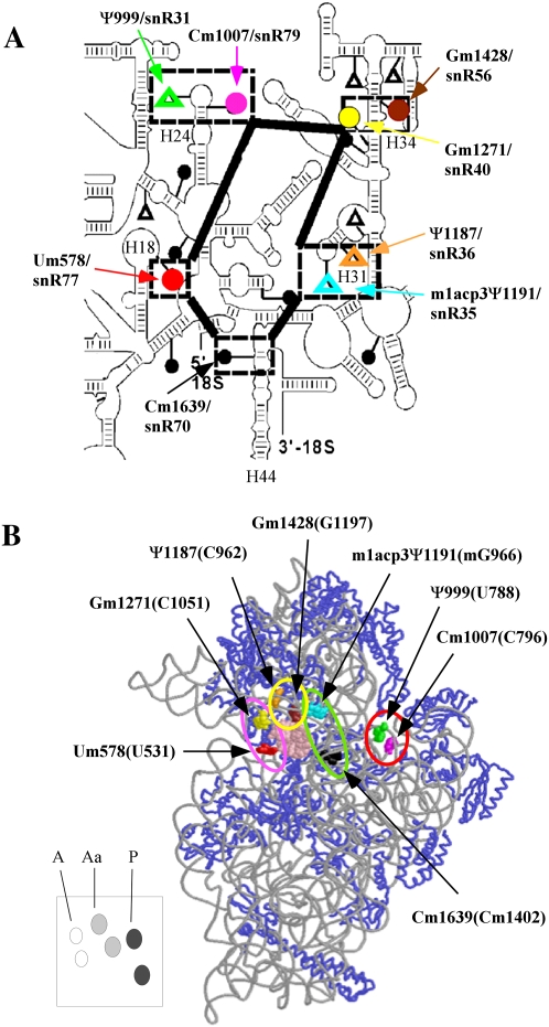
FIGURE 1.
The decoding region of yeast rRNA is rich in modifications. (A) Secondary structure of the decoding center of 18S rRNA. Modified nucleotides in the decoding center are highlighted by broken boxes, and the corresponding guide snoRNAs are also identified. Methylations (●); pseudouridines (△). (B) The 3D structure of modifications in the decoding center. Locations are deduced from the ribosome structure. The corresponding nucleotide numbers in E. coli rRNA are provided in parentheses. The system of representing modifications with colors is the same in both panels. The modifications in the A, Aa, P, and E regions are indicated by pink, yellow, green, and red circles, respectively. The lower left panel shows a schematic cartoon for positions of A-region (white), Aa-region (gray), and P-region (black) modifications. This system is used in subsequent figures to indicate modification situations.

