Abstract
Background
Body composition and fat distribution change dramatically during adolescence. Data based on longitudinal studies to describe these changes are limited. The aim of this study was to describe age-related changes in fat free–mass index (FFMI) and fat mass index (FMI), which are components of BMI, and waist circumference (WC) in participants of Project HeartBeat!, a longitudinal study of children.
Methods
Anthropometric measurements and body composition data were obtained in a mixed longitudinal study of 678 children (49.1% female, 20.1% black), initially aged 8, 11, and 14 years, every 4 months for 4 years (1991–1995). Trajectories of change from ages 8 to 18 years were measured for FFMI, FMI, and WC. Because of the small number of observations for black participants, trajectories for this group were limited to ages 8.5–15 years.
Results
Body mass index, FFMI, and WC increased steadily with age for all race–gender cohorts. However, in nonblack girls, FFMI remained constant after about age 16 years. For black boys and girls, FFMI was similar at age 8.5 years but increased more steeply for black boys by age 15 years. In girls, FMI showed an upward trend until shortly after age 14 years, when it remained constant. In boys, FMI increased between age 8 years and age 10 years, and then decreased.
Conclusions
The extent to which each component of BMI contributes to the changes in BMI depends on the gender, race, and age of the individual. Healthcare providers need to be aware that children who show upward deviation of BMI or BMI percentiles may have increases in their lean body mass rather than in adiposity.
Introduction
Overweight and obesity in children are commonly defined by CDC growth curves as a BMI greater than the 85th and 95th percentiles, respectively.1 But BMI is not necessarily a good reflection of adiposity; rather, it represents the relationship between weight and height.2–5 Amount and distribution of adiposity is important in cardiovascular disease (CVD) risk.6,7
Fat mass and fat-free mass are the components of total body mass. When stature is taken into account, these become the fat mass index (FMI) and fat free–mass index (FFMI) and represent the fat and lean components of BMI, respectively. Several reports support the use of FMI and FFMI instead of BMI for classifying the weight status of children.5,8,9 Although FMI accounts for the amount of adiposity, waist circumference (WC) is a commonly used measure of the distribution of adiposity.
Increased WC is a well recognized risk factor for CVD in adults. In children and adolescents, WC increases normally with growth. However, there are no established trajectories that enable determination of abnormal increases in WC.
Adiposity measurements differ among gender–race groups. However, trajectories for the components of BMI and WC for specific gender–race groups have not been established. It is important to create trajectories to aid in identifying populations at increased risk for developing CVD. Recognizing those children who are at greater risk will facilitate early and appropriate interventions. No attempt is made here to determine which factors affect linear growth, specifically sexual maturation, diet, and physical activity. The purpose of this paper is to establish normal trajectories for FMI, FFMI, and WC.
Methods
Study Design and Population
Project HeartBeat! is a longitudinal study designed to evaluate the changes in CVD risk factors among children and adolescents. A total of 678 boys and girls in three cohorts, aged 8, 11, and 14 years at baseline, were enrolled from The Woodlands and Conroe TX. The participants were 49.1% female, 74.6% white, 20.1% black, and 5.3% of other race/ethnicity (Table 1). Data collection began in the fall of 1991 and continued every 4 months until 1995, creating a combined cohort for ages 8–18 years.10 The study protocol was approved by the IRBs of the University of Texas Health Science Center at Houston and of Baylor College of Medicine. For each participant, informed consent or assent and parental consent were obtained. The complete design and methods of Project HeartBeat! have been described elsewhere.11
Table 1.
Selected anthropometric variables at baseline measurement: Project HeartBeat!, 1991–1995 (n=678), in kg/m2 unless otherwise noted
| Boys | ||||||
|---|---|---|---|---|---|---|
| Nonblack | Black | |||||
| Aged 8 years (n=121) | Aged 11 years (n=83) | Aged 14 years (n=75) | Aged 8 years (n=38) | Aged 11 years (n=21) | Aged 14 years (n=7) | |
| M (SD) | ||||||
| BMI | 17.2 (2.5) | 19.2 (3.4) | 20.9 (3.2) | 17.3 (3.4) | 20.8 (5.5) | 18.6 (2.9) |
| FFMI | 13.1 (1.2) | 14.2 (1.6) | 16.7 (1.7) | 13.9 (1.7) | 15.9 (2.6) | 16.5 (2.6) |
| FMI | 4.0 (1.9) | 5.0 (2.5) | 4.0 (2.1) | 3.5 (2.3) | 5.0 (3.4) | 2.1 (0.5) |
| WC (cm) | 59.7 (5.4) | 66.3 (8.2) | 72.7 (8.4) | 59.1 (7.3) | 68.3 (12.1) | 65.9 (4.9) |
|
Girls | ||||||
| Nonblack | Black | |||||
| Aged 8 years (n=114) | Aged 11 years (n=76) | Aged 14 years (n=73) | Aged 8 years (n=41) | Aged 11 years (n=17) | Aged 14 years (n=12) | |
|
M (SD) | ||||||
| BMI | 16.7 (2.1) | 18.2 (2.8) | 21.8 (3.7) | 19.4 (5.5) | 20.9 (6.9) | 27.5 (7.8) |
| FFMI | 12.5 (1.0) | 13.4 (1.1) | 15.4 (1.3) | 14.1 (2.1) | 15.1 (2.5) | 16.6 (2.4) |
| FMI | 4.2 (1.7) | 4.9 (2.3) | 5.9 (2.1) | 5.4 (3.7) | 4.6 (2.5) | 8.2 (3.2) |
| WC (cm) | 56.7 (4.8) | 62.7 (6.7) | 69.4 (7.6) | 62.1 (11.4) | 67.8 (15.1) | 74.7 (13.6) |
FFMI, fat free–mass index; FMI, fat mass index; WC, waist circumference
Data Collection
Anthropometric measures were obtained by two trained and certified technicians. Weight was measured to the nearest 0.1 kg with a balanced-beam scale; height was measured to the nearest 0.1 cm using a wall-mounted stadiometer. BMI was calculated as weight (kg)/height (m)2. Waist circumference was measured to the nearest 0.1 cm at the narrowest part of the torso using a nonelastic tape measure underneath the subject’s clothing. Skinfolds at six different sites (triceps, subscapular, midaxillary, abdomen, distal thigh, and lateral calf) were measured to the nearest 0.1 mm in full sets, then repeated two times to improve reliability.12 The three values at each site were averaged. Fat mass and fat-free mass were derived from a combination of bioelectric impedance analysis (RJL Systems bioelectric impedance analyzer BIA 101-A), performed by placing electrodes on the right arm and right foot, and anthropometry.13 Fat mass index was calculated as fat mass in (kg)/height (m)2, and FFMI was calculated as FFM in (kg)/height (m)2.
Data Analysis
Descriptive statistics (Ms and SDs) for BMI, FMI, FFMI, and WC were calculated for cohorts defined by gender, race, and age. Multilevel statistical analysis was used to calculate average age trajectories by gender and race/ethnicity for each of the measurements. A p-value of 0.05 was used as the criterion for all statistical testing. Although black participants constituted 20.1% of the total study population, the absolute numbers of observations for black participants aged 8–8.5 years and aged 15–18 years were relatively small. Accordingly, it was decided that estimates of trajectories in these age intervals might not be stable and could be misleading. Therefore, trajectories for black participants are limited to ages 8.5–15 years.14
Repeated measurements of each body composition index and WC were regressed on gender, race (nonblack/black), and age terms. For each index and WC, gender, race, a gender-by-race interaction term, three age terms (age, age2, and age3), three gender-by-age interaction terms (gender × age, gender × age 2, gender × age3), and three race-by-age interaction terms (race × age, race × age2, race × age3) were entered first into the equation. A backward-elimination procedure was used for selecting independent variables. These multilevel regression models allow construction of separate trajectories for each of the four race–gender groups from a single model using all observations simultaneously. Details of the statistical analysis have been reported previously.14 Descriptive statistical analyses were performed with SPSS version 9.0. The longitudinal modeling of trajectories of selected indices was conducted using MLwiN version 2.1.
Results
Mean baseline values for each anthropometric measure are presented in Table 1 for each age cohort by gender. In girls, FMI increased with age. In boys, FMI increased from ages 8 to 10 years, and then decreased. Mean FFMI and WC increased with age for all gender–race groups.
Multilevel linear models for BMI, FFMI, FMI, and WC are presented in Table 2. Their trajectories by gender and race are shown in Figures 1–4.
Table 2.
Estimated models for BMI, FFMI, FMI, and WC on gender, race, and age, Project HeartBeat! 1991–1995
| BMI | FFMI | FMI | WC | |||||
|---|---|---|---|---|---|---|---|---|
| Parameter | Estimate | SE | Estimate | SE | Estimate | SE | Estimate | SE |
| Fixed parameters | ||||||||
| Constant | 19.62 | 0.2561 | 15.10 | 0.117 | 4.565 | 0.160 | 68.11 | 0.560 |
| Gender (0, male; 1, female) | 0.0206ns | 0.3651 | −0.671 | 0.166 | 0.577 | 0.229 | −3.041 | 0.799 |
| Race (0, nonblack; 1 black) | 0.9153ns | 0.5677 | 0.812 | 0.252 | −0.220ns | 0.366 | −0.148ns | 1.249 |
| Gender × race | 2.167 | 0.7664 | 0.773 | 0.327 | 1.372 | 0.518 | 5.551 | 1.695 |
| Age(years)a | 0.7491 | 0.0488 | 0.877 | 0.031 | −0.141 | 0.037 | 2.555 | 0.115 |
| Age2 | — | 0.00728 | 0.036 | 0.005 | −0.030 | 0.006 | −0.007ns | 0.018 |
| Age3 | — | 0.00163 | −0.010 | 0.001 | 0.004 | 0.001 | −0.023 | 0.004 |
| Gender × age | 0.1873 | 0.0662 | −0.194 | 0.042 | 0.362 | 0.050 | −0.330 | 0.157 |
| Gender × age2 | −0.0203 | 0.00994 | −0.048 | 0.007 | 0.031 | 0.008 | −0.090 | 0.024 |
| Gender × age3 | — | 0.00225 | 0.003 | 0.001 | −0.008 | 0.002 | 0.003ns | 0.006 |
| Race × age | 0.1173ns | 0.0892 | 0.013ns | 0.058 | 0.023ns | 0.068 | −0.038ns | 0.212 |
| Race × age2 | — | 0.0138 | 0.015ns | 0.009 | −0.017ns | 0.011 | −0.033ns | 0.034 |
| Race × age3 | 0.01053 | 0.00338 | 0.006 | 0.002 | 0.009 | 0.003 | 0.030 | 0.008 |
| Between-subjects variances/covariance | ||||||||
| Constant | 17.91 | 1.004 | 3.660 | 0.212 | 6.727 | 0.386 | 84.759 | 4.803 |
| Age/constant | 1.077 | 0.1184 | 0.415 | 0.036 | 0.026ns | 0.047 | 4.786 | 0.581 |
| Age | 0.3514 | 0.0239 | 0.135 | 0.010 | 0.149 | 0.011 | 1.705 | 0.124 |
| Within-subjects variance | ||||||||
| Error | 0.4092 | 0.00883 | 0.221 | 0.005 | 0.332 | 0.007 | 3.010 | 0.065 |
FFMI, fat free–mass index; FMI, fat mass index; WC, waist circumference; ns, not significant at the level of ≥0.05
Age centered at 12.1 years (mean age of 678 participants)
Figure 1.
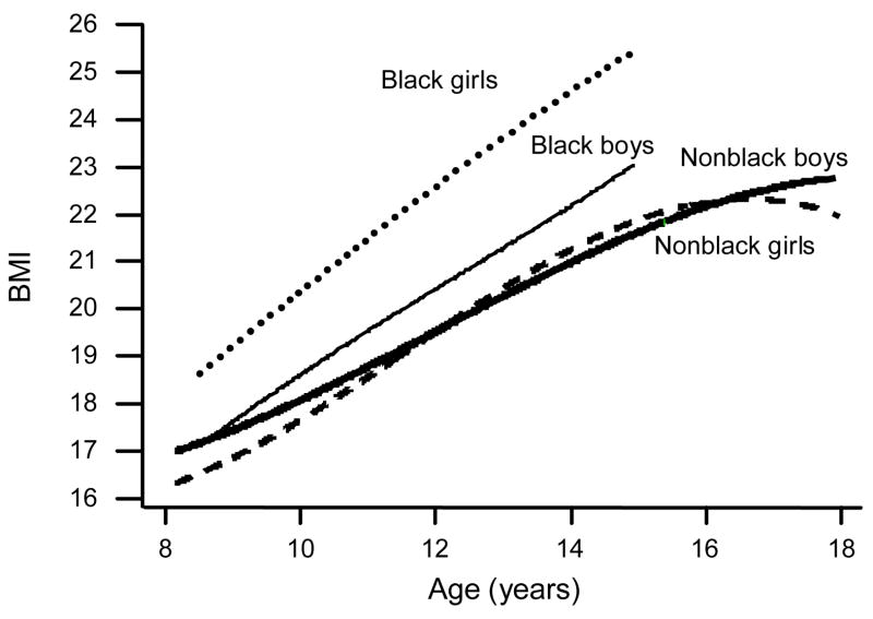
Gender- and race-specific trajectories of BMI, Project HeartBeat!, 1991–1995 (n=678)
Figure 4.
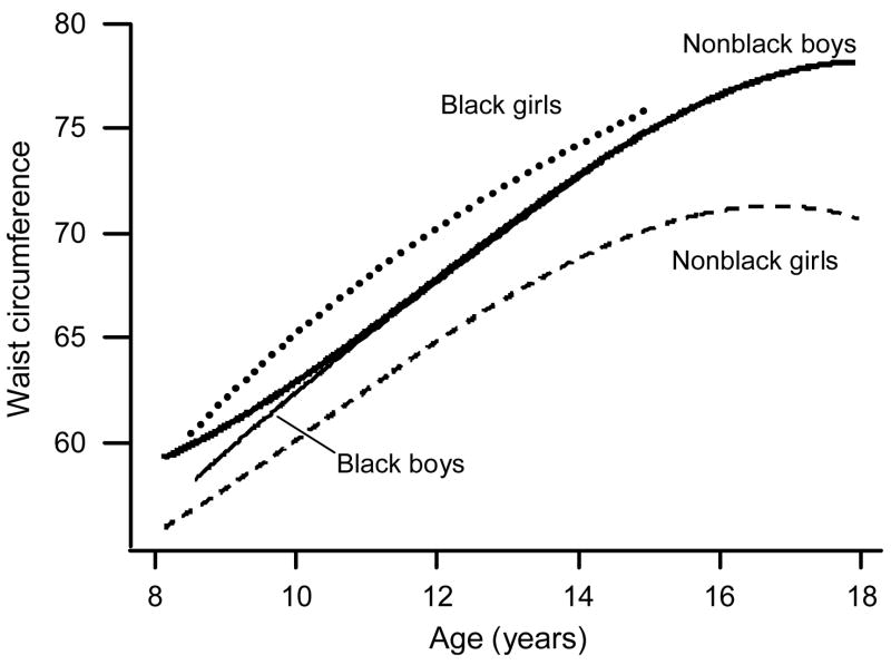
Gender- and race-specific trajectories of waist circumference, Project HeartBeat!, 1991–1995 (n=678)
Body Mass Index
Average BMI values increased steadily with age, except in nonblack girls, in whom BMI showed no increase after age 16.5 years and even showed a slight decrease after age 17 years (Figure 1). In nonblack boys, BMI values increased from 17.0 kg/m2 at age 8 years to ~22.7 kg/m2 at age 18 years. In black boys, BMI was similar to that for nonblack boys aged 8.5 years (17.1 kg/m2) but increased to 23.0 kg/m2 by age 15 years. The BMI values were consistently higher in black girls at all ages than in other gender–race groups.
Fat Mass Index
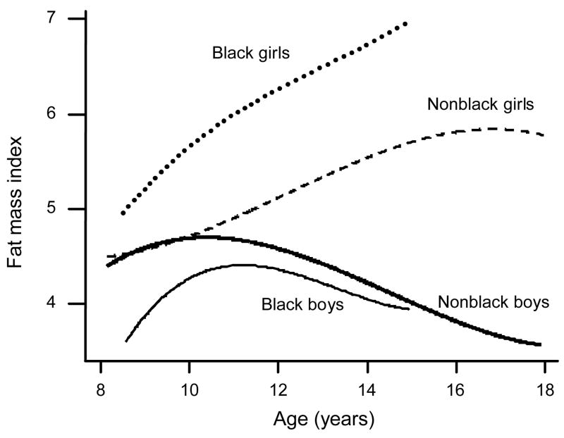
Age patterns of change in FMI differed markedly by gender. In nonblack girls and boys, the trajectory began at ~4.4 kg/m2 at age 8 years and then diverged, with nonblack girls’ FMI increasing consistently until reaching a plateau at 5.8 kg/m2 at about age 17 years, and nonblack boys’ FMI decreasing consistently after a subtle peak of ~4.7 kg/m2 at age 10 years. Average FMI of 5.0 kg/m2 for black girls aged 8.5 years was higher than that for nonblack boys and girls (~4.5 kg/m2), and it continued to increase, to ~7.0 kg/m2 at age 15 years. Black boys had a low FMI of 3.6 kg/m2 at age 8.5 years, which rose to only 4.4 kg/m2 at age 11 years, and then declined slightly until age 15 years.
Fat Free–Mass Index
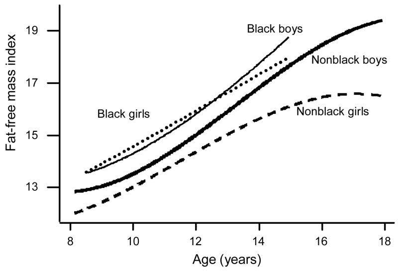
Trajectories for FFMI were dramatically different from those for FMI (Figure 3). The average FFMI increased steadily with age, except for an apparent plateau in nonblack girls aged 17 years (Figure 2). In nonblack boys, FFMI increased from 12.9 kg/m2 at age 8 years to approximately 19.3 kg/m2 at age 18 years. Among nonblack children, average FFMI values were consistently higher for boys than for girls at all ages. The FFMI was similar for black boys and girls at age 8.5 years (~13.6 kg/m2) but increased more steeply for black boys to ~18.8 kg/m2 by age 15 years. The FFMI demonstrated a similar pattern in black and nonblack boys, but values were higher for black boys at all ages. Average FFMI values were consistently higher among blacks than nonblacks.
Figure 3.
Gender- and race-specific trajectories of fat mass index, Project HeartBeat!, 1991–1995 (n=678)
Figure 2.
Gender- and race-specific trajectories of fat free–mass index, Project HeartBeat!, 1991–1995 (n=678)
Waist Circumference
Waist circumference increased steadily with age, except for a plateau at ~71 cm after age 16 years for nonblack girls (Figure 4). Trajectories for nonblack boys and for black boys and girls followed a similar pattern, increasing steadily for the age range studied, except that the trajectory started ~2 cm lower for black boys at age 8.5 years (~58 cm) and ended slightly higher for black girls at age 15 years (~76 cm). The trajectory for nonblack girls was consistently lower, by ~2–10 cm, than those for the other gender–race subgroups.
Discussion
This analysis provides trajectories for FMI, FFMI, and WC for black and nonblack adolescents. The trajectories varied remarkably by age and gender and to a lesser extent by race. There were gender and age differences in the measurements after age 10 years, with a strong divergence between boys and girls in the growth pattern of FMI; boys decreased in FMI with age, and girls continued to increase in FMI. This finding was similar to those from other studies,8,15,16 except in one,8 FMI values in boys did not decrease until after age 12 years.
Although BMI growth pattern and rate were similar for boys and girls, the BMI components differed greatly between genders, particularly during adolescence. In both genders, BMI was composed predominantly of lean mass. For boys, the age-related increase in BMI was concurrent with a large increase in FFMI and a decrease in FMI, which may explain the changes in boys’ physiques as they become more lean and muscular during adolescence. These results were similar to those of other studies.3,5,15,17 Demerath et al.3 reported that white boys aged 13–18 years experience an increase in BMI percentile that may be accompanied by a uniform increase in the FFM component across the range of BMI percentiles; further, older boys increase faster in FFM than younger boys and experience a decrease in percent body fat, even in the overweight range.
Waist circumference trajectories differed by gender and race. Black girls had a larger WC compared with other gender–race groups at all ages, similar to the findings in another report.18 Waist circumference did not differ between black and nonblack boys, which supports findings19 of no differences in the rate of visceral and subcutaneous abdominal adiposity between gender groups. Of the four adiposity trajectories presented in the current study, WC showed the fewest differences among the four gender–race groups and generally was most similar to BMI and FFMI, except that black boys had a steeper increase in FFMI. An explanation for the increase in WC is that it represents the measurement for truncal fat, including muscles and bones without differentiation of visceral and subcutaneous fat, and the increase for boys may represent a greater increase in FFM than in fat mass.
The trajectories for all measurements varied remarkably by age and gender and to a lesser extent by race. In the study cohort, black girls, at all ages, had higher values of FMI, BMI, and WC than other race–gender groups, likely making them the highest risk group for CVD and its risk factors. In a study of healthy multi-ethnic children aged 5–18 years,7 distribution of lean and fat mass was similar to the current findings. The current findings are also in agreement with national data indicating that BMI values for black girls are higher than those for nonblack girls during this age period.20 The analyses did not seek to determine the etiology of excess fat mass development in black girls, but they clearly demonstrate the excess fat mass that occurs in adolescents, which likely increases their risk for development of CVD risk factors and CVD as adults. Some possible mechanisms for the differences between black girls and other youth in this study are physiologic, genetic, cultural, and socioeconomic.
Our findings support the recommendation17 to use FFMI and FMI in determining the deeper meaning of BMI. These two measures of adiposity allow independent evaluation and assessment of the contribution of FFM and fat mass relative to body size.4 Calculation of FFMI and FMI in the context of BMI enable the clinician to identify children with normal BMI and excess adiposity (large FMI) to initiate intervention. Similarly, they enable identification of children who are overweight as a result of a large FFMI for primary prevention counseling.
Standards for FFMI, FMI, and WC need to be established. The current findings contribute to the understanding of FFMI, FMI, and WC. A possible weakness of the findings is that FFMI and FMI calculations require measurement of percent body fat. This measurement can be done using bioelectric impedance analysis or dual x-ray absorptiometry. Neither of these measures is readily available in the majority of outpatient pediatric clinics. Another limitation of the current study is that the population was predominantly white with few black children, and there were no other race/ethnic groups for comparison. Finally, the participants were at a variety of pubertal stages, which may account for significant differences in trajectories between boys and girls and between races.
Conclusion
Each component of the adiposity index contributes to the changes in BMI and depends on the gender, race, and age of the individual. Trajectories were created for FMI, FFMI, and WC, which were not previously defined for children and adolescents. Additionally, black girls have the highest values on each measure of adiposity, but they also have a lot of lean mass compared to white boys and girls. Standards for FMI, FFMI, and WC should be available to identify individuals at higher risk for having excess adiposity. Intervention strategies should be developed to target high-risk populations, especially black girls.
Acknowledgments
The authors gratefully acknowledge the contribution of each Project HeartBeat! participant and family. The researchers recognize the support of the Conroe Independent School District and the generous support of The Woodlands Corporation. Funding for the study was provided by the National Heart, Lung, and Blood Institute through Cooperative Agreement U01-HL-41166 and by the CDC through the Southwest Center for Prevention Research (U48/CCU609653).
The findings and conclusions in this report are those of the authors and do not necessarily represent the views of the CDC.
No financial disclosures were reported by the authors of this paper.
Footnotes
Publisher's Disclaimer: This is a PDF file of an unedited manuscript that has been accepted for publication. As a service to our customers we are providing this early version of the manuscript. The manuscript will undergo copyediting, typesetting, and review of the resulting proof before it is published in its final citable form. Please note that during the production process errors may be discovered which could affect the content, and all legal disclaimers that apply to the journal pertain.
References
- 1.National Center for Health Statistics. CDC growth charts: United States. 2000 www.cdc.gov. [PubMed]
- 2.Franklin MF. Comparison of weight and height relations in boys from 4 countries. Am J Clin Nutr. 1999;70(1):157S–162S. [PubMed] [Google Scholar]
- 3.Demerath EW, Schubert CM, Maynard LM, et al. Do changes in body mass index percentile reflect changes in body composition in children? Data from the Fels Longitudinal Study. Pediatrics. 2006;117(3):e487–95. doi: 10.1542/peds.2005-0572. [DOI] [PubMed] [Google Scholar]
- 4.Freedman DS, Ogden CL, Berenson GS, Horlick M. Body mass index and body fatness in childhood. Curr Opin Clin Nutr Metab Care. 2005;8(6):618–23. doi: 10.1097/01.mco.0000171128.21655.93. [DOI] [PubMed] [Google Scholar]
- 5.Maynard LM, Wisemandle W, Roche AF, Chumlea WC, Guo SS, Siervogel RM. Childhood body composition in relation to body mass index. Pediatrics. 2001;107(2):344–50. doi: 10.1542/peds.107.2.344. [DOI] [PubMed] [Google Scholar]
- 6.Kuk JL, Katzmarzyk PT, Nichaman MZ, Church TS, Blair SN, Ross R. Visceral fat is an independent predictor of all-cause mortality in men. Obesity (Silver Spring) 2006;14(2):336–41. doi: 10.1038/oby.2006.43. [DOI] [PubMed] [Google Scholar]
- 7.Wiklund P, Toss F, Weinehall L, et al. Abdominal and gynoid fat mass are associated with cardiovascular risk factors in men and women. J Clin Endocrinol Metab. 2008;93(11):4360–6. doi: 10.1210/jc.2008-0804. [DOI] [PubMed] [Google Scholar]
- 8.Freedman DS, Wang J, Maynard LM, et al. Relation of BMI to fat and fat-free mass among children and adolescents. Int J Obes (Lond) 2005;29(1):1–8. doi: 10.1038/sj.ijo.0802735. [DOI] [PubMed] [Google Scholar]
- 9.Nakao T, Komiya S. Reference norms for a fat-free mass index and fat mass index in the Japanese child population. J Physiol Anthropol Appl Human Sci. 2003;22(6):293–8. doi: 10.2114/jpa.22.293. [DOI] [PubMed] [Google Scholar]
- 10.Mueller WH, Harrist RB, Doyle SR, Ayars CL, Labarthe DR. Body measurement variability, fatness, and fat-free mass in children 8, 11, and 14 years of age: Project HeartBeat! Am J Hum Biol. 1999;11(1):69–78. doi: 10.1002/(SICI)1520-6300(1999)11:1<69::AID-AJHB7>3.0.CO;2-T. [DOI] [PubMed] [Google Scholar]
- 11.Labarthe DR, Nichaman MZ, Harrist RB, Grunbaum JA, Dai S. Development of cardiovascular risk factors from ages 8 to 18 in Project HeartBeat! Study design and patterns of change in plasma total cholesterol concentration. Circulation. 1997;95(12):2636–42. doi: 10.1161/01.cir.95.12.2636. [DOI] [PubMed] [Google Scholar]
- 12.Lohman TG, Roche AF, Martorell R. Anthropometric standardization reference manual. Champaign IL: Human Kinetics Press; 1988. [Google Scholar]
- 13.Guo SM, Roche AF, Houtkooper L. Fat-free mass in children and young adults predicted from bioelectric impedance and anthropometric variables. Am J Clin Nutr. 1989;50(3):435–43. doi: 10.1093/ajcn/50.3.435. [DOI] [PubMed] [Google Scholar]
- 14.Dai S, Labarthe DR, Grunbaum JA, Harrist RB, Mueller WH. Longitudinal analysis of changes in indices of obesity from age 8 years to age 18 years. Project HeartBeat! Am J Epidemiol. 2002;156(8):720–9. doi: 10.1093/aje/kwf109. [DOI] [PubMed] [Google Scholar]
- 15.Hattori K, Tahara Y, Moji K, Aoyagi K, Furusawa T. Chart analysis of body composition change among pre- and postadolescent Japanese subjects assessed by underwater weighing method. Int J Obes Relat Metab Disord. 2004;28(4):520–4. doi: 10.1038/sj.ijo.0802593. [DOI] [PubMed] [Google Scholar]
- 16.Chumlea WC, Siervogel RM, Roche AF, Webb P, Rogers E. Increments across age in body composition for children 10 to 18 years of age. Hum Biol. 1983;55(4):845–52. [PubMed] [Google Scholar]
- 17.Wells JC, Coward WA, Cole TJ, Davies PS. The contribution of fat and fat-free tissue to body mass index in contemporary children and the reference child. Int J Obes Relat Metab Disord. 2002;26(10):1323–8. doi: 10.1038/sj.ijo.0802077. [DOI] [PubMed] [Google Scholar]
- 18.Dekkers JC, Podolsky RH, Treiber FA, Barbeau P, Gutin B, Snieder H. Development of general and central obesity from childhood into early adulthood in African American and European American males and females with a family history of cardiovascular disease. Am J Clin Nutr. 2004;79(4):661–8. doi: 10.1093/ajcn/79.4.661. [DOI] [PubMed] [Google Scholar]
- 19.Huang TT, Johnson MS, Figueroa-Colon R, Dwyer JH, Goran MI. Growth of visceral fat, subcutaneous abdominal fat, and total body fat in children. Obes Res. 2001;9(5):283–9. doi: 10.1038/oby.2001.35. [DOI] [PubMed] [Google Scholar]
- 20.Ogden CL, Carroll MD, Curtin LR, McDowell MA, Tabak CJ, Flegal KM. Prevalence of overweight and obesity in the United States, 1999–2004. JAMA. 2006;295(13):1549–55. doi: 10.1001/jama.295.13.1549. [DOI] [PubMed] [Google Scholar]


