Abstract
In a 71-year-old female with evolving anterior wall myocardial infarction, coronary angiography revealed a monocoronary artery which arose from the right sinus of Valsalva. Originating from a short common trunk, the left main stem showed a thrombotic lesion that occluded the left anterior descending coronary artery while the circumflex artery was obstructed. Intracoronary administration of abciximab, followed by stenting of the transition between the left anterior descending coronary artery and the main stem, and final kissing balloon inflation of the bifurcation resulted in an excellent angiographic result and favourable clinical outcome. (Neth Heart J 2009;17:274-6.)
Keywords: monocoronary artery, single coronary artery, primary PCI, myocardial infarction, congenital anomaly, stent
In a very small number of patients, the entire coronary tree originates from one single common trunk or a monocoronary artery.1 In general, the presence of such anomalies does not result in symptoms. Nevertheless, such patients may develop symptomatic atherosclerotic coronary disease and even myocardial infarction. Percutaneous coronary interventions (PCI) in monocoronary arteries are technically challenging procedures,2 which may be even more complex in the setting of cardiogenic shock, as described in the present case report.
A 71-year-old woman was referred to our centre with progressive chest pain for seven hours and anterolateral ST-segment depression. Her medical history was notable for hypertension and obesity. Initial physical examination showed no significant abnormalities. Cardiac markers were normal. Intravenous metoprolol and nitroglycerine were administered in an attempt to medically alleviate the ischaemia, but the chest pain and electrocardiographic abnormalities did not improve. We therefore decided to conduct emergency coronary angiography 30 minutes after admission. During transfer to the catheterisation suite, her haemodynamic status deteriorated and the patient developed cardiogenic shock.
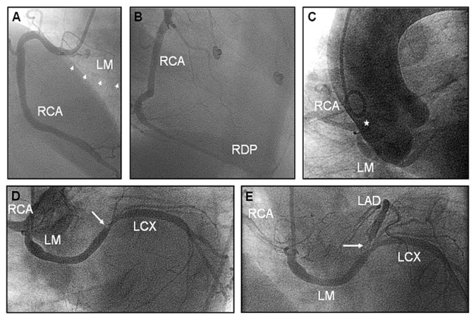
Angiography of the right coronary artery did not show any stenosis (figures 1A and B). We could not engage the ostium of the left coronary artery at its common anatomical position. Aortography, which was performed to find the left coronary artery (LCA), demonstrated a large coronary artery (common trunk) that originated from the right sinus of Valsalva. This vessel divided shortly after its origin into the right coronary artery and the left main coronary artery (figure 1C). Selective angiography of the left coronary artery demonstrated a thrombus in the left main stem, resulting in an occlusion of the left anterior descending artery and an obstruction of the left circumflex artery (figure 1D).
Figure 1.
Coronary angiography and aortography. Angiography of the right coronary artery (RCA) revealed no stenosis but wall irregularities in the right descending posterior (RDP) artery (A, B). Aortography (C) showed the common coronary trunk of a monocoronary artery (marked by star), arising from the right coronary cusp and dividing into the RCA and left main stem (LM). Retrospectively, the left main stem was hazily visible during angiography of the RCA (A, arrowheads). Just proximal to the bifurcation of the LM into left anterior descending (LAD) and left circumflex (LCX) coronary arteries, a thrombus (D, arrowhead) occluded the LAD while the LCX was obstructed. Slow TIMI 2 flow was restored in the LAD after passing the lesion with a guidewire (E).
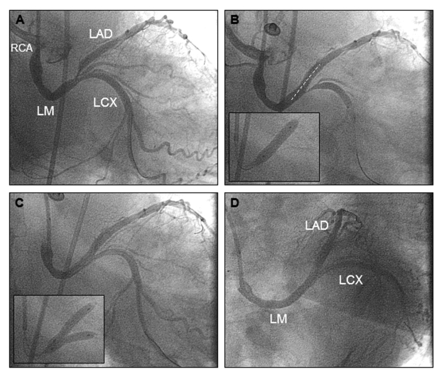
The lesion could be passed by floppy guidewires and slow flow (TIMI 2) was restored in the previously occluded left anterior descending artery. Shortly after intracoronary injection of a bolus of abciximab, the thrombus resolved. Following predilatation with a 2.5/12 mm balloon catheter at 12 atm, the transition from the left main stem to the left descending artery was stented using a 4.0/18 mm bare metal stent at 16 atm (figure 2B). Angiography revealed a good result in the left anterior descending artery with TIMI 3 flow, but worsening of the obstruction in the proximal circumflex artery – most likely the result of plaque shift. Final kissing balloon inflation with two 3.5/20 mm balloon catheters, both at 14 atm (figure 2C, insert), led to an excellent angiographic result in both vessels (figures 2C and D).
Figure 2.
Percutaneous intervention in left main stem of monocoronary artery. The thrombus was resolved after intracoronary injection of a bolus of abciximab with a remaining stenosis of the left main bifurcation (A). Stenting of the left anterior descending artery (B, insert) resulted in a good result in the left main stem and anterior descending artery but worsened the proximal left circumflex artery (B). Final kissing balloon inflation (C, insert) resulted in an excellent angiographic result (C, D).
At the end of the procedure, her haemodynamic status normalised. The further in-hospital course was uneventful, and the patient was asymptomatic at 30-day follow-up.
Comment
This case of primary PCI in a monocoronary artery type RII-C according to Lipton's classification is a rare procedure.3 Such anomalies have an incidence of no more than 1-2/10,000 coronary angiographies.1
Primary PCI procedures in monocoronary arteries often represent technically challenging cases4,5 with an increased risk of procedural failure or unfavourable clinical outcome. The increased risk and technical challenge of such cases sometimes trigger surgical treatment,2 which may be avoided in cases with comparable clinical syndromes but no such (unusual) anatomy.
In the literature, only a few cases of primary PCI in monocoronary arteries have been described. Most reports concerned either different vessels in slightly different anatomical settings4 or completely different anatomical settings.5 The only report on a patient with a basically identical anatomy and clinical scenario ended up with successful emergency coronary bypass surgery because of failed coronary angioplasty.2
In the present case we have demonstrated that with this anatomy a primary PCI of the left main stem is feasible, even in the presence of a partial thrombotic occlusion of the main stem with haemodynamic compromise. However, we faced two technical difficulties. First, it was necessary to recognise the aberrant anatomy and find the left coronary artery. Second, we used a guiding catheter which is quite uncommon for engagement of the left coronary artery and we had to adjust our standard angiographic projections to the different anatomy of the left main bifurcation.
In a different and much rarer type of monocoronary artery, i.e. in the form of solitary dominant coronary vessel such as recently described by our group,6 ostial or proximal thrombotic occlusion of the vessel is generally fatal and the patient often does not reach the catheterisation laboratory. Mid or distal lesions in such solitary dominant vessels, however, may represent a unique technical challenge or may be inaccessible for PCI.
Thus, the present case demonstrates that in monocoronary arteries type RII-C (Lipton's classification) primary PCI of left main stem lesions may be feasible with a good angiographic result and favourable clinical outcome.
References
- 1.Yamanaka O, Hobbs RE. Coronary artery anomalies in 126,595 patients undergoing coronary arteriography. Cathet Cardiovasc Diagn 1990;21:28-40. [DOI] [PubMed] [Google Scholar]
- 2.Geyik B, Ozeke O, Deveci B, Maden O, Senen K. Single coronary artery presenting with cardiogenic shock due to acute myocardial infarction. Int J Cardiovasc Imaging 2006;22:5-7. [DOI] [PubMed] [Google Scholar]
- 3.Lipton MJ, Barry WH, Obrez I, et al. Isolated single coronary artery: diagnosis, angiographic classification and clinical significance. Radiology 1979;130:39-47. [DOI] [PubMed] [Google Scholar]
- 4.Raddino R, Pedrinazzi C, Zanini G, Leonzi O, Robba D, Chieppa F, et al. Percutaneous coronary angioplasty in a patient with anomalous single coronary artery arising from the right sinus of Valsalva. Int J Cardiol 2006;112:e60-e62. [DOI] [PubMed] [Google Scholar]
- 5.Braun MU, Stolte D, Rauwolf T, Strasser RH. Single coronary artery with anomalous origin from the right sinus Valsalva. Clin Res Cardiol 2006;95:119-21. [DOI] [PubMed] [Google Scholar]
- 6.Hartmann M, Verhorst PM, von Birgelen C. Isolated “superdominant” single coronary artery: a particularly rare coronary anomaly. Heart 2007;913:687. [DOI] [PMC free article] [PubMed] [Google Scholar]


