
Figure 5. Conservation of the Spt4-Spt5 Interface.
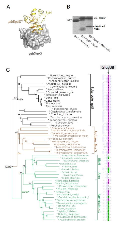
(A) Model of Archaeal NusG bound to RpoE″ : Archaeal RpoE″-NusG complex was modeled by superimposing the pfuRpoE″ structure and pfuNusG model onto the Spt4-Spt5NGN structure. The pfuNusG model is shown as a white surface; yellow, Spt4; orange, pfuRpoE″. (B) GST Pull-down assay of Methanocaldococcus jannaschii RpoE″-NusG interaction. (C) A sequence-based phylogeny derived from an alignment of NGN domain in Spt5/NusG homologues. Organism names are color-coded according to the domain of life. Alignment of Glu338 for the acid-dipole interaction is shown at right. Bootstrap support is indicated for major braches.