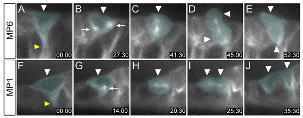
Fig. 2. Time-lapse imaging of sequential MP delamination and division.
Images in sagittal view, internal (basal) up, from time-lapse imaging of an (A–E) MP6 division and an (F–J) MP1 division. GFP fluorescence was visualized in sim-Gal4 UAS tau-GFP embryos during stage 11. Time is displayed as minutes:seconds. Relevant cells in each panel are pseudocolored. (A) Prior to division, the MP6 (white arrowhead) delaminates from the apical surface and takes on a triangular shape. The tip of the retracting cell is indicated by the yellow arrowhead. (B–D) During mitosis, (B) the centrosomes (arrows) move toward opposite poles, (C) the spindle fibers have an apical-basal orientation, and (D) the MP6 divides (arrowheads) along this axis. (E) Two MP6 neurons (arrowheads) are produced. (F) The MP1 (white arrowhead) delaminates from the apical surface, also acquiring a triangular shape (retraction point, yellow arrowhead). (G) The centrosomes (arrow) can be seen just before they separate and begin their migration. (H) The MP1 spindle maintains an orientation perpendicular to the apical-basal axis. (I,J) Cytokinesis results in the formation of two MP1 neurons (arrowheads).

