Abstract
There currently is great interest in translating findings about the importance of nitric oxide (NO) in vascular biology to the Clinical arena. The bioactivity of endothelium-derived NO can readily be assessed in human subjects as vasodilation of conduit arteries or increased flow, which reflects vasodilation of resistance vessels. This chapter provides an update on the available noninvasive methodology to assess endothelium-dependent vasodilation in human subjects.
Background
A vast body of work has emphasized the importance of endothelium-derived nitric oxide (NO) in vascular biology, and there currently is great interest in translating these findings to the clinical practice. Because it is a potent vasodilator, the bioavailability of endothelium-derived NO can readily be evaluated in human subjects by measuring changes in arterial diameter and blood flow. Human studies have shown that impaired endothelium dependent vasodilation is associated with the presence of atherosclerosis and recognized cardiovascular disease risk factors. Many interventions that reduce cardiovascular risk also restore endothelium-dependent vasodilation toward normal. Importantly, prospective studies have shown that the presence of impaired endothelium-dependent vasodilation in the coronary or peripheral circulation identifies patients with increased risk for future cardiovascular disease events (Widlansky et al., 2003). In a review of methods for measurement of NO-dependent vasodilation in humans. Vita (2002) described invasive methods for the study of the coronary and peripheral circulations and use of two-dimensional ultrasound to study flow-mediated dilation of the brachial artery. Since that time, additional noninvasive approaches have emerged for study of NO-dependent control of vascular tone in the coronary circulation, central aorta, and peripheral microcirculation. In this chapter, we briefly mention advances in the previously described methods (Vita, 2002) and then describe these newer methods for assessment of NO-dependent vasodilation in humans (Table I).
TABLE I.
Noninvasive methods for study of NO-dependent vasodilation in human subjects
| Method | Vascular bed | Stimulus for NO release | Advantages | Disadvantages |
|---|---|---|---|---|
| Vascular ultrasound | Conduit brachial artery | Flow produced by reactive hyperemia | Well established | Operator dependent |
| Flow-mediated dilation (FMD) | Demonstrated prognostic Value | Low signal/noise ratio | ||
| Correlates with coronary circulation | Lack of standardized methodology | |||
| Pulse amplitude tonometry (PAT) | Small arteries in the fingertip | Flow produced by reactive hyperemia | Automatic | Clinical relevance not established |
| Rapid results | Minimal information about response to interventions | |||
| Correlates with coronary circulation | ||||
| Venous occlusion | Forearm resistance vessels | Flow produced by reactive hyperemia | Stronger correlation with risk factors than FMD | Low reproducibility |
| Plethysmography | Can be measured by several different techniques | Operator dependent | ||
| Arterial compliance | Central aorta or brachial artery | None | More reproducible and stable overtime | Only partially NO dependent |
| Pulse wave analysis | Low operator dependence | Relates to arterial structure and vascular tone | ||
| Transthoracic Doppler | Left anterior descending coronary artery | Adenosine or adenosine triphosphate | More clinically relevant circulation | Only partially NO dependent |
| Highly operator dependent | ||||
| Lack of standardized methodology |
Studies of the Coronary Circulation
Invasive Studies
A number of studies have examined NO-dependent vasodilation in the coronary circulation of patients undergoing cardiac catheterization. These studies assess changes in coronary artery diameter or coronary blood flow during intraarterial agonist infusion using quantitative coronary angiography and intracoronary Doppler, respectively (Vita, 2002). Though generally extremely safe, these studies have the potential to produce major complications such as coronary thrombosis or death. Because of their invasive nature, they are not well suited for repeated studies in the same individual or for the study of relatively low-risk populations. Despite these limitations, studies of the coronary circulation are the most clinically relevant for coronary artery disease. In particular, studies have shown that abnormalities of NO-dependent vasodilation in the coronary circulation are associated with increased risk for cardiovascular disease events (Halcox et al., 2002; Schachinger et al., 2000; Schindler et al., 2003; Suwaidi et al., 2000; Targonski et al., 2003).
Noninvasive Studies
Given its clinical relevance, it would be desirable to obtain information about endothelium-dependent vasodilation of the coronary circulation in a noninvasive manner. Several studies have used transthoracic Doppler echocardiography to assess coronary blood flow reserve in left anterior descending (LAD) circulation. This method involves obtaining Doppler flow signals of the distal LAD using an acoustic window near the midclavicular line in the fourth and fifth intercostals spaces. Signals are recorded at baseline and then after 2 min of intravenous adenosine triphosphate (ATP) infusion (140 µg/kg/min) to increase coronary blood flow. Changes in coronary blood flow are expressed as the ratio of ATP-induced to basal coronary flow velocity (Olsuka et al., 2001). Coronary “flow reserve” measured in this manner is acutely impaired by passive cigarette smoking and improved by interventions known to improve endothelium-dependent vasodilation (Hirata et al., 2004; Otsuka et al., 2001).
The methodology is limited as a test of endothelial vasodilator function, however, because the response to ATP is only partially dependent on endothelium-derived NO. Furthermore, systemic ATP infusion lowers blood pressure and increases heart rate, which may alter coronary blood flow independently of endothelial function. The technique is highly operator dependent. No published studies have evaluated the effect of inhibitors of NO synthase (NOS) on the response. Thus, the technique has potential as a noninvasive method for assessing NO-dependent and NO-independent vasodilation in the human coronary circulation, but further studies are required before it can be generally accepted. A number of other noninvasive methodologies also show promise for examination of endothelium-dependent changes in coronary blood flow, including magnetic resonance imaging and positron emission tomography but currently are not in use for this purpose.
Studies of the Arm and Hand
Invasive Studies
In light of the difficulty of studying the coronary circulation, many investigators have turned to the study of NO-dependent vasodilation in peripheral arteries. As reviewed by Vita (2002), studies of tins type involve infusion of various vasoactive drugs into the brachial artery and measurement of vasodilation as changes in forearm blood flow using venous occlusion plethysmography or changes in radial artery diameter using high-resolution vascular ultrasound (Creager et al., 1990; Lieberman et al., 1996). The clinical relevance of these studies is predicated on the assumption that many cardiovascular disease risk factors are systemic in nature and have parallel effects in different vascular beds. This assumption is strongly supported by studies showing that an impaired blood flow response to acetylcholine and other endothelium-dependent vasodilators is associated with increased risk for cardiovascular disease events (Fichtlscherer et al., 2004; Heitzer et al., 2001; Perticone et al., 2001). Despite their clinical relevance, these studies require insertion of an arterial catheter, which reduces their applicability to the general population. Thus, the methodology remains extremely useful for studying selected populations and examining mechanisms of vascular dysfunction. However, there continues to be great interest in noninvasive methods to examine No-dependent vasodilation in the periphery.
Noninvasive Studies: Flow-Mediated Dilation
A widely used noninvasive method to assess endothelial vasomotor function is brachial artery flow-mediated dilation as assessed by ultrasound (Corretti et al., 2002; Vita, 2002). In these studies, reactive hyperemia is induced by cuff occlusion of the arm, and changes in arterial diameter are measured using high-resolution ultrasound (Corretti et al., 2002). Flow-mediated dilation measured in this fashion depends on NO synthesis (Lieberman et al., 1996), correlates with endothelial vasomotor function in the coronary circulation (Anderson et al., 1995), and is reduced in the setting of traditional risk factors for coronary artery disease (Benjamin et al., 2004). In addition, impaired brachial artery flow-mediated dilation predicts short-term and long-term risk for cardiovascular disease events in patients with advanced atherosclerosis (Gokce et al., 2002) and in patients with hypertension (Modena et al., 2002).
Despite its clinical relevance, ultrasound-based studies have a number of limitations. The technique is technically demanding, and changes in brachial diameter produced by hyperemic flow (0.1–0.6 mm) are close to the limit of detection of ultrasound. Reproduciability depends greatly on image quality, and the technique requires time-consuming off-line image analysis. For these reasons, investigators have sought new techniques that are faster and simpler to perform.
Noninvasive Studies: Pulse Amplitude Tonometry
One emerging method is known as fingertip pulse amplitude tonometry (PAT). Studies are performed using a commercially available device (Endo-PAT 2000, Itamar Medical, Ltd.) that records the pulse amplitude in fingertip at baseline and during reactive hyperemia. Hyperemia induces flow-mediated dilation within the fingertip and increases pulse amplitude. Simultaneous recordings are made from the contralateral finger and are used to adjust for changes in sympathetic tone and other systemic effects that might affect the signal during cuff occlusion and the hyperemic phase. Proprietary software provides further adjustment based on an empiric regression equation to account for baseline pulse amplitude, although the importance of making this adjustment remains unproven. The net response is expressed as the “reactive hyperemia PAT index.” A preliminary study demonstrated that the increase in pulse amplitude is blocked, in part, by intraarterial infusion of monomethyl-l-arginine (l-NMMA), confirming that it depends in part on NO synthesis (Gerhard-Herman et al., 2002). Interestingly, the reactive hyperemia PAT index has been reported to correlate with brachial artery flow-mediated dilation in the arm and is inversely related to risk factors and the presence of coronary artery disease (Kuvin et al., 2003). The response also correlates with endothelial function in the coronary circulation (Bonettiet al., 2004). Finally, the response improves after enhanced external counter pulsation therapy, an intervention known to improve peripheral artery endothelia] function (Bonetti et al., 2003).
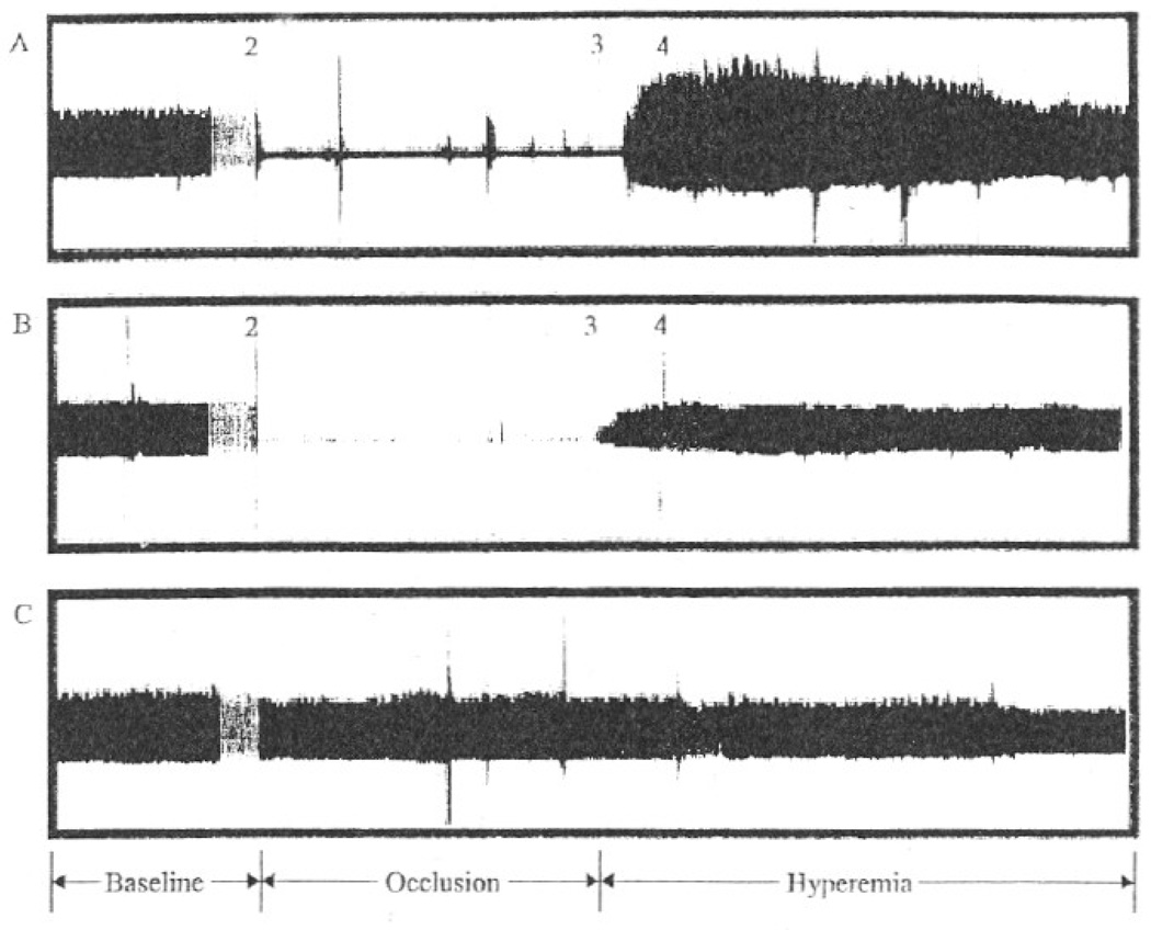
In our laboratory at Boston University School of Medicine, PAT and brachial ultrasound studies are done simultaneously using a single cuff occlusion to generate a period of reactive hyperemia, which stimulates flow-mediated dilation of both the conduit brachial artery and the small arteries in the finger. The PAT signals are recorded using thimble-shaped pneumatic probes that are placed on the index fingers of each hand. Patients lie supine with both wrists supported on foam blocks to allow the fingers to hang in an unsupported manner. The inflation pressure of the finger cuff is set to the diastolic pressure or 80 mm Hg (whichever is lower). Pulse recordings are made before cuff inflation and during the 1-min period beginning 1 min after 5-min cuff occlusion of the arm with the cuff placed on the upper arm. Figure 1 displays signals from a healthy subject and a subject with coronary artery disease. In a group of 252 unselected patients undergoing study of vascular function from our laboratory, the mean (±SD) reactive hyperemia PAT ratio was 2.2 ± 0.74 (range 1.23–5.69) with a highly skewed distribution. A prior study demonstrated that among patients referred for evaluation of chest pain, the reactive hyperemia PAT ratios were 1.31 ± 0.11 and 1.62 ± 0.47 for patients with and without exercise induced myocardial ischemia, respectively. We calculate that a sample size of 29 subjects per group would be required to detect a difference between groups of this magnitude with 80% power (alpha = 0.05) using log-transformed values for the reactive hyperemia/PAT ratio. These results suggest that clinically important differences between study groups can be detected using this methodology in studies with samples sizes that are similar to those needed for study of brachial artery flow-mediated dilation (Vita, 2002). Overall, PAT appears to be a promising new methodology, but much work needs to be done to confirm its relation to other measures of NO-dependent vasodilation and to cardiovascular disease.
FIG. 1.
Pulse amplitude recorded in the index finger with the Endo-PAT 2000 device (Itamar Medical, Ltd.) before, during, and after cuff occlusion of the arm, as described in the text. (A) The response from a healthy individual with no risk factor. (B) The very blunted response in an individual with coronary artery disease. (C) The response in the contralateral finger not subject to cuff occlusion, which demonstrates that the signal remains stable over time. The PAT ratio is calculated at baseline and between 1 and 2 min after cuff release. Reproduced, with permission, from Kuvin et al. (2003).
Noninvasive Studies: Extent of Reactive Hyperemia
Reactive hyperemia is the transient increase in limb blood flow that occurs after a period of limb occlusion and reflects ischemia-induced production of a variety of vasodilators, including adenosine and hydrogen ions that locally act on microvessels. A portion of the hyperemic response also depends on NO, possibly stimulated by local increases in shear stress during hyperemic flow. l-NNMA infusion blunts both the peak and the net hyperemic response in the forearm (Meredith et al., 1996). Many investigators had suggested that reactive hyperemia is unaffected by cardiovascular disease. However, other studies have emphasized that reactive hyperemia is reduced in the setting of risk factors (Hayoz et al., 1995; Higashi et al., 2001; Mitchell et al., 2004b) or coronary artery disease (Lieberman et al., 1996), particularly the NO-dependent portion of the response (Higashi et al., 2001). Reactive hyperemia also correlates inversely with systemic markers of inflammation, including C-reactive protein, interleukin-6, and the soluble form of intercellular adhesion molecule-1 (Vita et al., 2004). Reactive hyperemia is the stimulus for brachial artery flow-mediated dilation, and we observed that a reduction in this stimulus accounts for much of the observed impairment in flow-mediated dilation observed in the setting of systemic risk factors (Mitchell et al., 2004b). These findings suggest that noninvasive measures of flow can be used to assess reactive hyperemia as a clinically relevant correlate of endothelial vasomotor function.
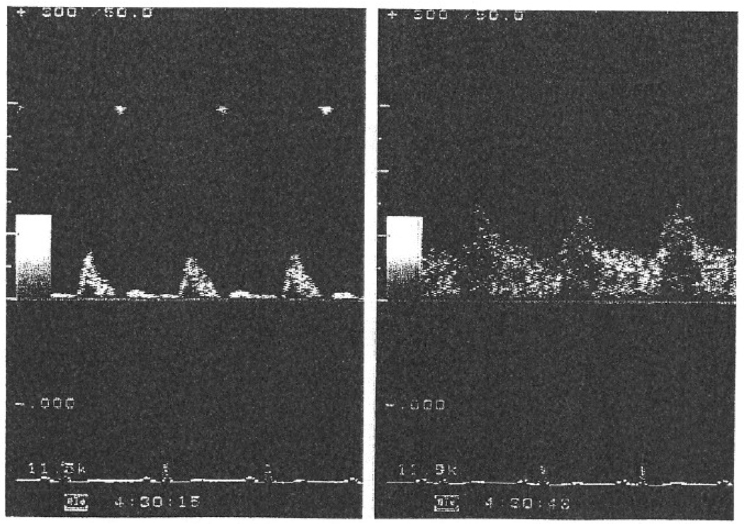
We take two approaches to assessing reactive hyperemia. First, we use Doppler ultrasound to record flow signals from the brachial artery at baseline and for 15 s after cuff release after 5-min occlusion of the upper arm (Vita, 2002). Typical flow signals are displayed in Fig. 2. The peak hyperemic response is typically observed within two or three beats after cuff release. Images are digitized on-line, and we measure the average flow velocity (area under the curve) for the peak cardiac cycle using one of several image analysis software packages (Brachial Analyzer, Medical Imaging Applications, Iowa City, IA). Table II presents reference values from a cohort of 503 healthy subjects studied in our laboratory. Many investigators express hyperemic flow as the ratio of peak to baseline flow, but a recent study suggests that the hyperemic flow velocity and hyperemic shear stress (calculated from the velocity, brachial artery diameter, and assumed values for blood viscosity) correlate most strongly with cardiovascular disease risk factors and prevalent cardiovascular disease (Mitchell et al., 2004b).
FIG. 2.
Representative Doppler recordings from the brachial artery at baseline (left) and immediately after cuff release (right) reflecting hyperemic flow.
TABLE II.
Mean values with 95% confidence intervals for brachial artery flowa
| Age (y) | Sample size | Hyperemia volume flow ratio | Baseline flow velocity (cm/s) | Hypermia flow velocity (cm/s) |
|---|---|---|---|---|
| <30 | n = 173 | 8.0 (6.0–10.1) | 12.0 (11.1–13.0) | 79.5 (75.2–83.7) |
| 30–39 | n = 125 | 7.3 (6.6–8.0) | 11.4 (10.3–12.5) | 78.0 (72.2–83.9) |
| 40–49 | n = 121 | 6.9 (6.1–7.6) | 11.9 (10.7–13.0) | 77.6 (72.0–82.3) |
| 50–59 | n = 56 | 6.7 (5.6–7.7) | 11.4 (9.9–13.0) | 74.1 (64.6–83.4) |
| ≥50 | n = 37 | 5.1 (3.3–6.9) | 12.2 (9.9–14.6) | 63.5 (50.1–76.2) |
Displayed are mean values and 95% confidence intervals according to age. Results Shown are for subjects without clinical history of coronary artery disease, peripheral vascular disease, diabetes mellitus, or hypertension.
A second method to assess reactive hyperemic uses venous occlusion plethysmography to measure forearm blood flow before and after cuff release (Higashi et al., 2001). Blood flow measurements are made using a mercury-in-silastic strain gauge, upper arm and wrist cuffs, and a computerized plethysmograph (Hokanson, Inc.) (Vita, 2002). During these studies, the upper arm venous occlusion cuff is inflated to 40 mm Hg (or adjusted to optimize the tracing), and circulation to the hand is excluded by inflation of the wrist cuff to suprasystolic pressure before initiation of flow measurements. At least five measurements are made and averaged at baseline, and a recording is made every 20 s after cuff release for 2 min. Although this methodology has limited ability to “capture” the peak flow response, it provides a reproducible approach to examine the entire hyperemic response.
Noninvasive Studies: Pulse Wave Analysis of Arterial Stiffness
There is great interest in examining arterial stiffness as a surrogate marker of atherosclerosis (Cohn et al., 2004). A number of approaches can be used, including simple assessment of arterial pulse pressure measured by blood pressure cuff, pulse wave contour analysis assessed by tonometry, ultrasound visualization of arterial distensibility (calculated from the change in arterial diameter in relation to changes in blood pressure), and examination of pulse wave velocity. In regard to pulse wave velocity, a number of studies have shown that carotid-femoral pulse-wave velocity relates to cardiovascular disease risk factors and risk for future cardiovascular disease events (Cohn et al., 2004). Whereas structural components of the arterial wall are major determinants of arterial stiffness, there is growing recognition that there also is a dynamic component of arterial stiffness that depends in part on arterial tone and endothelial release of NO. In support of the possibility, Wilkinson et al. (2002a,b)have observed that several measures of arterial stiffness are increased after systemic l-NMMA.
In our laboratory, we use applanation tonometry to assess vascular stiffness with a device developed at Cardiovascular Engineering, Inc. (Holliston, MA). Subjects lie quietly in a supine position, and pulse recordings are made from the carotid artery, brachial artery, radial artery, and femoral artery. Distances between recording sites are measured, and the pressures are calibrated using the brachial cuff pressure. Pulse recordings are gated using the electrocardiogram R-wave, and pulse wave velocity and the time of reflected waves are determined by blinded investigators. Reference values for a healthy, risk factor-free cohort were published this year (Mitchell et al., 2004a). In some studies, we also made ultrasound recordings of flow and diameter of the left ventricular outflow tract, allowing us to calculate characteristic impendence, a variable that relates to stiffness of the proximal aorta (Mitchell et al., 2002). One study demonstrated significant correlations between these measures of arterial stiffness and endothelial function (Nigam et al., 2003), but further study will be required to define the precise contribute of endothelium-derived NO to arterial stiffness in different disease states.
Conclusions
The methodology for the study of NO-dependent vasodilation in intact humans continues to evolve. Many of the techniques are well established and have proven useful to study mechanisms of impaired NO bioavailability in atherosclerosis and related disease states and to evaluate potential therapies for these conditions. We suggest that some or all of these methods could be used clinically to assess cardiovascular risk or to guide risk-reduction therapy in individual patients. However, a great deal of work remains to be done to determine the clinical utility of these techniques.
Acknowledgments
A Program Project Grant (HL60886), a Specialized center of Research Grant (HL55993), and the Boston Medical center General Clinical Research Center (M01RR00533) provided support for portions of this work.
References
- Anderson TJ, Uehata A, Gerhard MD, Meredith JT, Knab S, Delagrange D, Leiberman E, Ganz P, Creager MA, Yeung AC, Selwyn AP. Close relation of endothelial function in the human coronary and peripheral circulations. J. Am. Coll. Cardiol. 1995;26:1235–1241. doi: 10.1016/0735-1097(95)00327-4. [DOI] [PubMed] [Google Scholar]
- Benjamin EJ, Larson MG, Keyes MJ, Mitchell GF, Vasan RS, Keaney JF, Jr, Lehman B, Fan S, Osypiuk E, Vita JA. Clinical correlates and heritability of endothelial function in the community: The Framingham Heart Study. Circulation. 2004;109:613–619. doi: 10.1161/01.CIR.0000112565.60887.1E. [DOI] [PubMed] [Google Scholar]
- Bonetti PO, Barsness GW, Keelan PC, Schnell TL, Pumper GM, Holmes DR, Stuart TH, Lerman A. Enhanced external counterpulsation improves endothelial function in patients with coronary artery disease. J. Am. Coll. Cardiol. 2003;41:370A. doi: 10.1016/s0735-1097(03)00329-2. (abstract) [DOI] [PubMed] [Google Scholar]
- Bonetti PO, Pumper GM, Higano ST, Holmes DR, Kuvin JT, Lerman A. Noninvasive identification of patients with early coronary atherosclerosis by assessment of digital reactive hyperemia. J. Am. Coll. Cardiol. 2004;44:2137–2141. doi: 10.1016/j.jacc.2004.08.062. [DOI] [PubMed] [Google Scholar]
- Cohn JN, Quyyumi AA, Hollenberg NK, Jamerson KA. Surrogate markers for cardiovascular disease: Functional markers. Circulation. 2004;109:IV31–IV46. doi: 10.1161/01.CIR.0000133442.99186.39. [DOI] [PubMed] [Google Scholar]
- Corretti MC, Anderson TJ, Benjamin EI, Celermajer D, Charbonneau F, Creager MA, Deanfield J, Drexler H, Gerhard-Herman M, Herrington D, Vallance P, Vita J, Vogel R. Guidelines for the ultrasound assessment of endothelial-dependent flow-mediated vasodilation of the brachial artery. A report of international Brachial Artery Reactivity Task Force. J. Am. Coll. Cardiol. 2002;39:257–265. doi: 10.1016/s0735-1097(01)01746-6. [DOI] [PubMed] [Google Scholar]
- Creager MA, Cooke JP, Mendelsohn ME, Gallagher SJ, Coleman SM, Loscalzo J, Dzau VJ. Impaired vasodilation of forearm resistance vessels in hypercholesterolemic humans. J. Clin. Invest. 1990;86:228–234. doi: 10.1172/JCI114688. [DOI] [PMC free article] [PubMed] [Google Scholar]
- Fichtlscherer S, Breuer S, Zeiher AM. Prognostic value of systemic endothelial dysfunction in patients with acute coronary syndromes: Further evidence for the existence of the “vulnerable” patient. Circulation. 2004;110:1926–1932. doi: 10.1161/01.CIR.0000143378.58099.8C. [DOI] [PubMed] [Google Scholar]
- Gerhard-Herman M, Hurley S, Mitra D, Creager MA, Ganz P. Assessment of endothelial function (nitric oxide) at the tip of a finger. Circulation. 2002;106:11–170. (abstract) [Google Scholar]
- Gokce N, Keaney JF, Jr, Menzoian JO, Watkins M, Hunter L, Duffy SJ, Vita JA. Risk stratification for post operative cardiovascular events via noninvasive assessment of endothelial function. Circulation. 2002;105:1567–1572. doi: 10.1161/01.cir.0000012543.55874.47. [DOI] [PubMed] [Google Scholar]
- Halcox JP, Schenke WH, Zalos G, Mincemoyer R, Prasad A, Waclawiw MA, Nour KR, Quyynmi AA. Prognostic value of coronary vascular endothelial dysfunction. Circulation. 2002;106:653–658. doi: 10.1161/01.cir.0000025404.78001.d8. [DOI] [PubMed] [Google Scholar]
- Hayoz D, Weber R, Rutschmann B, Darioli R, Burnier M, Waeber B, Brunner HR. Postischemic blood flow response in hypercholesterolemic patients. Hypertension. 1995;26:497–502. doi: 10.1161/01.hyp.26.3.497. [DOI] [PubMed] [Google Scholar]
- Heitzer T, Schlinzig T, Krohn K, Meinertz T, Munzel T. Endothelial dysfunction, oxidative stress, and risk of cardiovascular events in patients with coronary artery disease. Circulation. 2001;104:2673–2678. doi: 10.1161/hc4601.099485. [DOI] [PubMed] [Google Scholar]
- Higashi Y, Sasaki S, Nakagawa K, Matsuura H, Kajiyama G, Oshima T. A noninvasive measurement of reactive hyperemia that can be used to assess resistance artery endothelial function in human. Am. J. Cardiol. 2001;87:121–125. doi: 10.1016/s0002-9149(00)01288-1. [DOI] [PubMed] [Google Scholar]
- Hirata K, Shimada K, Watanabe H, Otsuka R, Tokai K, Yoshiyama M, Homma S, Yoshikawa J. Black tea increases coronary flow velocity reserve in healthy male subjects. Am J. Cardiol. 2004;93:1384–1388. doi: 10.1016/j.amjcard.2004.02.035. A6. [DOI] [PubMed] [Google Scholar]
- Kuvin JT, Patel AR, Sliney KA, Pandian GP, Sheffy J, Schnall RP, Karas RH, Udelson JE. Assessment of peripheral vascular endothelial function with finger arterial pulse wave amplitude. Am. Heart. J. 2003;146:168–174. doi: 10.1016/S0002-8703(03)00094-2. [DOI] [PubMed] [Google Scholar]
- Lieberman EH, Gerhard MD, Uehata A, Selwyn AP, Ganz P, Yeung AC, Creager MA. Flow-induced vasodilation of the human brachial artery is impaired in patients <40 years of age with coronary artery disease. Am. J. Cardiol. 1996;78:1210–1214. doi: 10.1016/s0002-9149(96)00597-8. [DOI] [PubMed] [Google Scholar]
- Meredith IT, Currie KE, Anderson TJ, Roddy MA, Ganz P, Creager MA. Postischemic vasodilation in human forearm is dependent on endothelium-derived nitric oxide. AJP Heart Circ. Physiol. 1996;270:H1435–H1440. doi: 10.1152/ajpheart.1996.270.4.H1435. [DOI] [PubMed] [Google Scholar]
- Mitchell GF, Izzo JL, Jr, Lacourciere Y, Ouellet JP, Neutel J, Qian C, Kerwin LJ, Block AJ, Pfeffer MA. Omapatrilat reduces pulse pressure and proximal aortic stiffness in patients with systolic hypertension: Results of the conduit hemodynamics of omapatrilat international research study. Circulation. 2002;105:2955–2961. doi: 10.1161/01.cir.0000020500.77568.3c. [DOI] [PubMed] [Google Scholar]
- Mitchell GF, Parise H, Benjamin EJ, Larson MG, Keyes MJ, Vita JA, Vasan RS, Levy D. Changes in arterial stiffness and wave reflection with advancing age in healthy men and women: The Framingham Heart Study. Hypertension. 2004a;43:1239–1245. doi: 10.1161/01.HYP.0000128420.01881.aa. [DOI] [PubMed] [Google Scholar]
- Mitchell GF, Parise H, Vita JA, Larson MG, Warner E, Kenney JF, Jr, Keyes MJ, Levy D, Vasan RS, Benjamin EJ. Local shear stress and brachial artery flow-mediated dilation: The Framingham Heart Study. Hypertension. 2004b;44:134–139. doi: 10.1161/01.HYP.0000137305.77635.68. [DOI] [PubMed] [Google Scholar]
- Modena MG, Bonetti L, Coppi F, Bursi F, Rossi R. Prognostic role of reversible endothelial dysfunction in hypertensive postmenopausal women. J. Am. Coll. Cardiol. 2002;40:505–510. doi: 10.1016/s0735-1097(02)01976-9. [DOI] [PubMed] [Google Scholar]
- Nigam A, Mitchell GF, Lambert J, Tardif JC. Relation between conduit vessel stiffness (assessed by tonometry) and endothelial function (assessed by flow-mediated dilatation) in patients with and without coronary heart disease. Am. J. Cordial. 2003;92:395–399. doi: 10.1016/s0002-9149(03)00656-8. [DOI] [PubMed] [Google Scholar]
- Otsuka R, Watanabe H, Hirata K, Tokai K, Muro T, Yoshiyama M, Takeuchi K, Yoshikawa J. Acute effects of passive smoking on the coronary circulation in healthy young adults. JAMA. 2001;286:436–441. doi: 10.1001/jama.286.4.436. [DOI] [PubMed] [Google Scholar]
- Perticone F, Ceravolo R, Pujia A, Ventura G, Iacopino S, Scozzafava A, Ferraro A, Chello M, Mastroroberto P, Verdecchia P, Schillaci G. Prognostic significance of endothelial dysfunction in hypertensive patients. Circulation. 2001;104:191–196. doi: 10.1161/01.cir.104.2.191. [DOI] [PubMed] [Google Scholar]
- Schachinger V, Britten MB, Zeiher AM. Prognostic impact coronary vasodilator dysfunction on adverse long-term outcome of coronary heart disease. Circulation. 2000;101:1899–1906. doi: 10.1161/01.cir.101.16.1899. [DOI] [PubMed] [Google Scholar]
- Schindler TH, Hornig B, Buser PT, Olschewski M, Magosaki N, Pfisterer M, Nitzsche EU, Solzbach U, Just H. Prognostic value of abnormal vasoreactivity of epicardial coronary arteries to sympathetic stimulation in patient with normal coronary angiograms. Arteriosel. Thromb Vase. Biol. 2003;23:495–501. doi: 10.1161/01.ATV.0000057571.03012.F4. [DOI] [PubMed] [Google Scholar]
- Suwaidi JA, Hamasaki S, Higano ST, Nishimmra RA, Holmes DR, Lerman A. Long-term follow-Up of patients with mild coronary artery disease and endothelial dysfunction. Circulation. 2003;101:948–954. doi: 10.1161/01.cir.101.9.948. [DOI] [PubMed] [Google Scholar]
- Targonski PV, Bonetti PO, Pumper GM, Higano ST, Holmes DR, Jr, Lerman A. Coronary endothelial dysfunction is associated with an increased risk of cerebrovascular events. Circulation. 2003;107:2805–2809. doi: 10.1161/01.CIR.0000072765.93106.EE. [DOI] [PubMed] [Google Scholar]
- Vita JA. Nitric oxide-dependent vasodilation in human subjects. Methods Enzymol. 2002;359:186–200. doi: 10.1016/s0076-6879(02)59183-7. [DOI] [PubMed] [Google Scholar]
- Vita JA, Keaney JF, Jr, Larson MG, Keyes MJ, Massro JM, Lipinska L, Lehman B, Fan S, Osypink E, Wilson PWF, Vasan RS, Mitchell GF, Benjamin EJ. Brachial artery vasodilator function and systemic inflammation in the Framingham Offspring Study. Circulation. 2004;110:3604–3609. doi: 10.1161/01.CIR.0000148821.97162.5E. [DOI] [PubMed] [Google Scholar]
- Widlansky ME, Gokce N, Keaney JF, Jr, Vita JA. The clinical implication of endothelial dysfunction. J. Am. Coll. Cardiol. 2003;42:1149–1160. doi: 10.1016/s0735-1097(03)00994-x. [DOI] [PubMed] [Google Scholar]
- Wilkinson IB, MacCallum H, Cockcroft JR, Webb DJ. Inhibition of basal nitric oxide synthesis increases aortic augmentation index and pulse wave velocity in vivo. Br. J. Clin. Pharmacol. 2002a;53:189–192. doi: 10.1046/j.1365-2125.2002.1528adoc.x. [DOI] [PMC free article] [PubMed] [Google Scholar]
- Wilkinson IB, Qasem A, McEniery CM, Webb DJ, Avolio AP, Cockcroft JR. Nitric oxide regulates local arterial distensibility in vivo. Circulation. 2002b;105:213–217. doi: 10.1161/hc0202.101970. [DOI] [PubMed] [Google Scholar]


