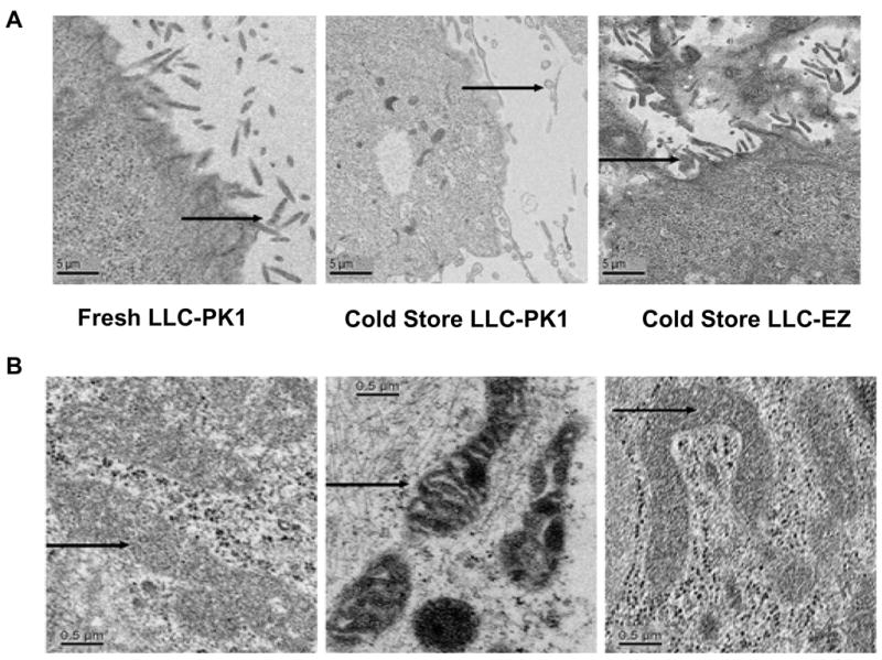
Figure 5.

Transmission electron microscopy (TEM) of fresh LLC-PK cells before cold storage (left), LLC-PK cells after 8 hrs of cold storage (middle), and TEM of ezrin over expressing LLC-EZ cells after 8 hrs of cold storage preservation (right). Microvilli details of the plasma membrane are featured in panel-A and show loss of brush border after cold storage in LLC-PK cells that is largely prevented in cells that over express ezrin. Arrows show cell microvilli. Similarly, mitochondria (panel-B) are shrunken, electron dense, and some show signs of cristalysis after 8 hrs of cold storage in control LLC-PK1 cells (B, middle). However, the mitochondrial changes are much milder in the ezrin over expressing LLC-EZ cells after cold storage (B, right). Many of their mitochondria appear normal with fewer electron dense structures. The mitochondrial structural preservation associated with ezrin over expression is consistent with the changes observed in mitochondrial function in these cells. Arrows show mitochondria.
