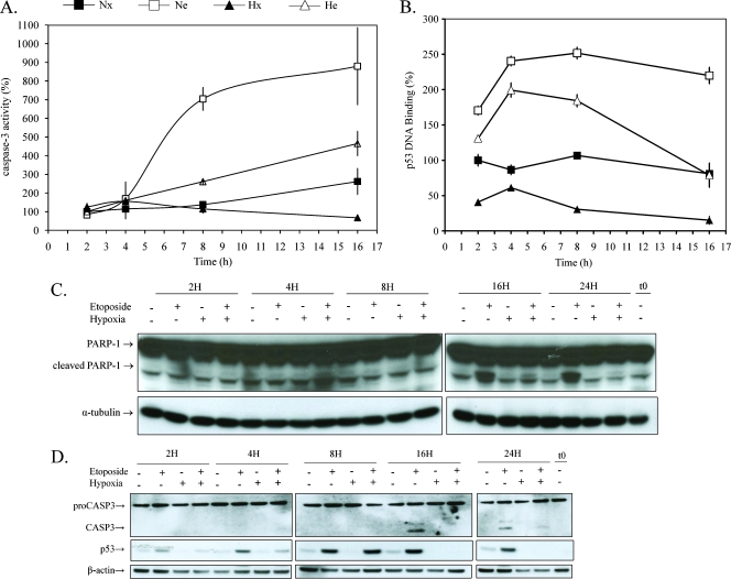
Figure 1.
Effect of hypoxia and/or etoposide on caspase 3 activity and p53 regulation. HepG2 cells were incubated under normoxic (Nx; 21% O2) or hypoxic (Hx; 1% O2) conditions with (Ne-He; 50 µM) or without etoposide (Nx-Hx) for increasing periods from 2 to 16 hours or from 2 to 24 hours. (A) Caspase 3 activity was assayed by measuring fluorescence intensity associated to free AFC released from the cleavage of caspase 3 substrate Ac-DEVD-AFC. Data are expressed in percentages of caspase 3 activity in control cells (Nx) as means ± 1 SD (n = 3). (B) p53 DNA binding activity was assessed by colorimetric assay. Data are expressed in percentage of DNA binding in control cells (Nx) and correspond to the mean value ± 1 SD from three independent experiments. (C and D) PARP-1, caspase 3 (CASP3), and p53 were detected in total cell extracts by Western blot analysis, using specific antibody. α-Tubulin and β-actin were used to assess the total amount of proteins loaded on the gel. t0 = time zero.

