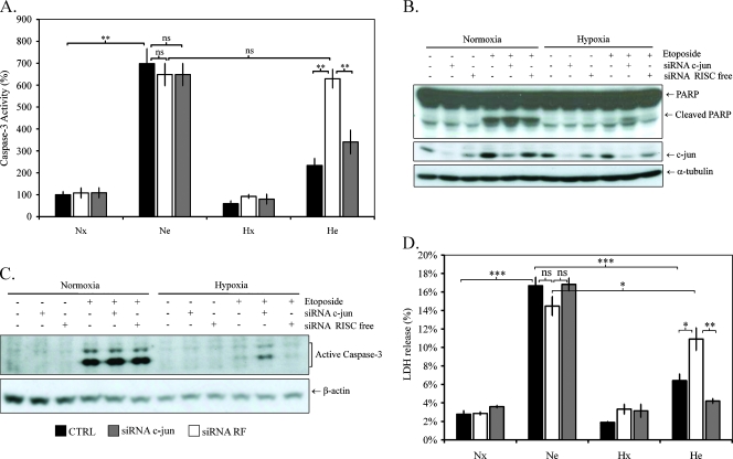
Figure 5.
Effect of c-jun silencing on hypoxia-induced resistance to etoposide-induced apoptosis. HepG2 cells were transfected with 50 nM c-jun siRNA or negative control siRNA for 24 hours. Eight hours later, cells were incubated under normoxic (Nx; 21% O2) or hypoxic (Hx; 1% O2) conditions with (Ne-He; 50 µM) or without etoposide (Nx-Hx) for 16 hours (A, B, C) or 40 hours (D). (A) Caspase 3 activity was assayed by measuring fluorescence intensity associated to free AFC released from the cleavage of the caspase 3 substrate Ac-DEVD-AFC. Results are normalized by fluorescence intensity of control cells (Nx) and are expressed in percentages as means ± 1 SD (n = 3). (B and C) Total extracts were analyzed by Western blot analysis for PARP-1 and active caspase 3 using specific anti-PARP-1 and active caspase 3 antibodies. c-jun protein level was assessed using specific anti-c-jun antibody, and α-tubulin was used to assess the total amount of proteins loaded on the gel. (D) LDH release was measured after 40 hours of incubation. Results are presented in percentages of LDH release in control cells (Nx) as means ± 1 SD (n = 3). Statistical analysis was carried out with the Student's t test. ns indicates nonsignificant; * .05 > P > .01; ** .01 > P > .001; ***P < .001.

