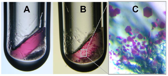
Fig. 1.

Growth of bacteriorhodopsin crystals within a monoolein-based lipidic cubic phase matrix in a glass vial. (A) The 10 μl crystallization experiment was set up in a glass vial as lined out in 2.2.1.1. At first the purple bacteriorhodopsin is distributed homogeneously in the lipidic cubic phase. The bottom of the 3 mm diameter tube is filled with solid Sørensen salt. (B) Bacteriorhodopsin microcrystals formed within 1 month. (C) Schematic close-up of purple bacteriorhodopsin crystals with sizes up to ca. 100 μm along their longest dimension.
