

Abstract
Guided by nuclear magnetic resonance (NMR) binding assays and computational docking studies, a series of 5, 5′ substituted Apogossypol derivatives was synthesized that resulted in potent pan-active inhibitors of anti-apoptotic Bcl-2 family proteins. Compound 8r inhibits the binding of BH3 peptides to Bcl-XL, Bcl-2, Mcl-1 and Bfl-1 with IC50 values of 0.76, 0.32, 0.28 and 0.73 μM, respectively. The compound also potently inhibits cell growth of human lung cancer and BP3 human B-cell lymphoma cell lines with EC50 values of 0.33 and 0.66 μM, respectively. Compound 8r shows little cytotoxicity against bax−/−bak−/− cells, indicating that it kills cancers cells via the intented mechanism. The compound also displays in vivo efficacy in transgenic mice in which Bcl-2 is overexpressed in splenic B-cells. Together with its improved chemical, plasma and microsomal stability relative to compound 2 (Apogossypol), compound 8r represents a promising drug lead for the development of novel apoptosis-based therapies for cancer.
Introduction
Programmed cell-death (apoptosis) plays critical roles in the maintenance of normal tissue homeostasis, ensuring a proper balance of cell production and cell loss.1, 2 Defects in the regulation of programmed cell death promote tumorgenesis, and also contribute significantly to chemoresistance.3, 4 B-cell lymphoma/leukemia-2 (Bcl-2) family proteins are central regulators of apoptosis.5–7 In humans, six anti-apoptotic members of the Bcl-2 family have been identified and characterized thus far, including Bcl-2, Bcl-XL, Mcl-1, Bfl-1, Bcl-W and Bcl-B. Over-expression of anti-apoptotic Bcl-2 family proteins occurs in many human cancers and leukemias, and therefore these proteins are very attractive targets for the development of novel anticancer agents.8–11 Members of the Bcl-2 family proteins also include pro-apoptotic effectors such as Bak, Bax, Bad, Bim and Bid. Anti-apoptotic and pro-apoptotic Bcl-2 family proteins dimerize and negate each other’s functions.3 Structural studies revealed the presence of a deep and relatively large hydrophobic crevice on the surface of anti-apoptotic Bcl-2 family proteins that binds the BH3 dimerization domain (an α-helical region) of pro-apoptotic family members.10 Thus, molecules that mimic the BH3 domain of pro-apoptotic proteins induce apoptosis and/or abrogate the ability of anti-apoptotic Bcl-2 proteins to inhibit cancer cell death.
We and others have reported that the natural product 1 (Gossypol) (Figure 1A) is a potent inhibitor of Bcl-2, Bcl-XL and Mcl-1, functioning as a BH3 mimetic.12–17 Compound 1 is currently in phase II clinical trials, displaying single-agent antitumor activity in patients with advanced malignancies.14, 17, 18 In mice studies, compound 1 displays some toxicity and off target effects likely due to two reactive aldehyde groups, which are important for targeting other cellular proteins such as dehydrogenases, for example. Our previous molecular docking studies, however, suggested that these two reactive groups are not essential for the compound to bind to Bcl-2 proteins, hence we designed compound 2 (Apogossypol) (Figure 1A), that lacks the aldehydes. In agreement with our predicted docked structure, compound 2 retains activity against anti-apoptotic Bcl-2 family proteins in vitro and in cells.19 Recently, we further compared the efficacy and toxicity in mice of compounds 1 and 2. Our preclinical in vivo data show that compound 2 has superior efficacy and markedly reduced toxicity compared to 1.20 We also evaluated the single-dose pharmacokinetic characteristics of compound 2 in mice. Compound 2 displayed superior blood concentrations over time compared to compound 1, due to slower clearance.21 These observations indicate that compound 2 is a promising lead compound for cancer therapy.
Figure 1.

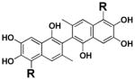
(A) Structure of compound 1, 2 and 3. (B) Structure of 5, 5′ substituted compound 2 derivatives. (C) and (D), Molecular docking studies. Stereo views of docked structures of (C) compound 2 and (D) compound 8r into Bcl-2 (PDB ID:1YSW).
Recently, we reported the separation and characterization of atropoisomers of compound 2.22 These studies revealed that the racemic compound 2 is as effective as its individual isomers.22 We further reported the synthesis and evaluation of 5, 5′ ketone substituted compound 2 derivatives. Among these derivatives, compound 3 (BI79D10)23 displayed improved in vitro and in vivo efficacy compared to compound 2 (Figure 1A and 1B). However, contrary to what we observed with compound 2, compound 3 displayed also mild GI toxicity in mice. The observed toxicity in compound 3 may be attributable to relatively active ketone groups.23 Based on these premises, in this current work, we focused our attention on preparing and evaluating activities of novel 5, 5′ substituted compound 2 derivatives which further replace the reactive ketone groups with more druggable amide and alkyl groups (Figure 1B).
Results and Discussion
We have recently reported that compound 2 is a promising inhibitor of Bcl-XL and Bcl-2 with improved in vivo efficacy and reduced toxicity compared to compound 1.12, 19, 20 Molecular docking studies of compound 2 into the BH3 binding groove in Bcl-2 24, 25 (Figure 1C) suggest that 2 forms two hydrogen bonds with residues Arg 143 and Tyr 105 in Bcl-2 through the 1 and 1′ hydroxyl groups, respectively. Compound 2 is also involved in hydrogen bonding interactions with Trp 141 and Tyr 199 in Bcl-2 through the 6′ hydroxyl group on naphthalene ring. According to this model, the isopropyl group on the left naphthalene ring inserts into the first hydrophobic pocket (P1) in Bcl-2 (Figure 1C), while the isopropyl group on the right naphthalene ring inserts into the second hydrophobic pocket (P2) (Figure 1C). Hence, the analysis of the predicted binding models indicates that while the overall core structure of compound 2 fits rather well into BH3 binding groove of Bcl-2, the two isopropyl groups do not apparently fully occupy the hydrophobic pockets P1 and P2. Therefore, a library of 5, 5′ substituted compound 2 derivatives (Figure 1B) that replace the isopropyl groups with suitable substituents was designed with the aim of deriving novel molecules that could occupy the hydrophobic pockets on Bcl-2 more efficiently.
A synthetic route (Scheme 1) was developed to introduce a variety of amide derivatives at the 5, 5′ positions. Compound 1 was treated with NaOH solution at 90°C to provide compound 2, which was readily methylated by dimethyl sulfate in the presence of potassium carbonate to afford compound 4.26 Reaction of compound 4 with TiCl4 followed by dichloromethyl methyl ether at room temperature resulted in loss of the isopropyl groups and simultaneous bisformylation to give the aldehyde compound 5.26 The aldehyde groups of compound 5 were converted to carboxylic acid 6 by mild oxidation with sodium hypochlorite.27 The carboxylic acid 6 was then coupled with a variety of commercially available amines in the presence of 1-ethyl-3-(3′-dimethylaminopropyl)carbodiimide (EDCI) at room temperature to give compound 7.28 Subsequent demethylation of the compound 7 using boron tribromide afforded compound 8.27 The synthesis of 5, 5′ alkyl substituted compound 2 derivatives was outlined in Scheme 2. Compound 5 was treated with different Grignard or lithium reagents to afford a secondary alcohol 9, which was oxidized to give the phenone 10 by pyridinium chlorochromate. Triethylsilane reduced phenone 10 to alkyl compound 1129 followed by subsequent demethylation using boron tribromide to afford compound 12 (Scheme 2). Compounds 13 and 14 (Scheme 3), with only hydrogen atom or carboxylic acid at 5, 5′ positions, were synthesized to explore if substitution at 5, 5′ position is important for enhancing biological activities. Compound 13 was synthesized by treating compound 4 with concentrated sulfuric acid to lose isopropyl group.26 The resulting product and compound 6 was then treated individually with boron tribromide to give compounds 13 and 14, respectively (Scheme 3).
Scheme 1.

Reagents and conditions: (a) NaOH, H2O, reflux; (b) H2SO4; (c) DMS, K2CO3; (d) TiCl4, Cl2CHOCH3, rt; (e) HCl, H2O (f) NaClO2, H2O2, KH2PO4, CH3CN, rt; (g) HCl, H2O; (h) EDCI, NH2R, HOBT, rt; (i) BBr3, CH2Cl2 (j) HCl, H2O.
Scheme 2.

Reagents and conditions: (a) RMgBr or RLi, rt; (b) NH4Cl, H2O; (c) Pyridinium chlorochromate, CH2Cl2, rt; (d) Et3SiH, TFA or Pd/C, H2; (e) BBr3; (f) HCl, H2O.
Scheme 3.

Reagents and conditions: (a) H2SO4, rt; (b) H2O; (c) BBr3, CH2Cl2; (d) HCl, H2O.
The synthesized 5, 5′ substituted compound 2 derivatives were first screened by one-dimensional 1H nuclear magnetic resonance spectroscopy (1D-1H NMR) binding assays against Bcl-XL, as we reported previously (Table 1 and 2).30 Active compounds in 1D-1H NMR binding assays were then selected and evaluated in Isothermal Titration Calorimetry assays (ITC), competitive fluorescence polarization assays (FPA) and cell viability assays (Tables 1–3). A group of compounds (8r, 8q, 8m) displayed high binding affinity to Bcl-XL in these assays (Table 1, 8r Figure 2A). The most potent compound induced significant chemical shift changes in active site methyl groups (region between −0.38 and 0.42 ppm) in the one-dimensional 1H-NMR spectra of Bcl-XL (Figure 2A) and also has an IC50 value of 0.76 μM in the FP displacement assays, which is 5 times more effective than 2 (Table 3 and Supplementary Figure 2A). To further confirm the results obtained by the NMR binding data and the FP assays, we further evaluated the binding affinity of compound 8r for Bcl-XL using ITC assay (Table 3 and Supplementary Figure 1). In agreement with NMR binding and FPA data, compound 8r displayed potent binding affinity to Bcl-XL with a Kd value of 0.11 μM, which is 15 times more potent than compound 2 (Kd = 1.7 μM) in the same assay. Consistent with NMR binding, FPA, and ITC data, compound 8r displayed efficacy in inhibiting growth of PC3ML cells, which express high levels of Bcl-XL. The EC50 value of 8r is 1.7 μM, hence 6-fold more potent than 2 (EC50 = 10.4 μM). Compounds (8j-8k and 8p-8s) displayed similar binding affinity as 8r for Bcl-XL in these assays with average IC50 value of 2.8 μM (Table 1).
Table 1.
Evaluation of 5, 5′ substituted compound 2 derivatives using a combination of 1D 1H-NMR binding assays and cell viability assays
 |
EC50 (μM) | ||||||
|---|---|---|---|---|---|---|---|
| Compound | R = | 1D-1H NMRa* | RS11846c* | H1299b* | H460b* | PC3MLb* | BP3c* |
| 1 | +++ | 4.2 | 6.0 | 3.0 | 3.1 | 1.42 | |
| 2 |  |
++ | 5.0 | 3.4 | 3.5 | 10.4 | 4.7 |
| 14 |  |
− | >30 | >30 | >30 | >30 | >30 |
| 8a | 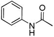 |
+ | 15.1 | 8.0 | 3.5 | 15.1 | NDd* |
| 8b | 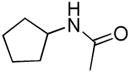 |
+ | 13.7 | 8.0 | 8.5 | 12.2 | ND |
| 8c | 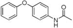 |
+ | 10.8 | 17.0 | 10.1 | 8.5 | ND |
| 8d | 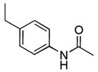 |
+ | 4.7 | 5.0 | 4.1 | 8.3 | ND |
| 8e | 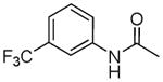 |
+ | 12.6 | 28.7 | 16.7 | 12.2 | ND |
| 8f | 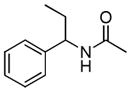 |
+++ | 4.2 | ND | 1.5 | ND | ND |
| 8g | 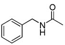 |
+ | 9.3 | 3.6 | 4.6 | 13.7 | ND |
| 8h |  |
+++ | 4.8 | 3.1 | 2.9 | 10.2 | ND |
| 8i |  |
+++ | 7.3 | 5.2 | 3.3 | 8.3 | ND |
| 8j |  |
++ | 3.0 | 3.6 | 1.4 | 0.7 | 1.5 |
| 8k |  |
+++ | 5.5 | 3.2 | 0.50 | 5.0 | 1.5 |
| 8l | 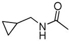 |
+ | >10 | ND | ND | ND | ND |
| 8m | 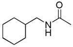 |
+++ | 4.9 | 4.8 | 0.41 | 3.7 | 0.90 |
| 8n | 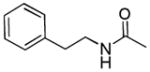 |
++ | 3.1 | 3.6 | 0.55 | 3.9 | 0.72 |
| 8o | 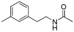 |
+ | 5.6 | ND | 0.70 | ND | ND |
| 8p | 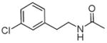 |
+++ | 4.6 | 7.8 | 0.99 | 3.2 | 0.41 |
| 8q | 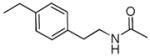 |
+++ | 3.0 | 0.36 | 0.40 | 1.7 | 0.2 |
| 8r | 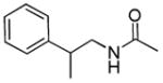 |
++ | 5.8 | 3.2 | 0.33 | 1.7 | 0.66 |
| 8s | 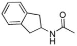 |
++ | 4.1 | 7.1 | 0.94 | 3.0 | 1.1 |
| 8t |  |
++ | 5.1 | ND | 1.3 | ND | 0.70 |
4-point-rating scale: +++, Very Active; ++, Active; +, Mild; −, Weak
Compounds against cell line using ATP-LITE assay
Compounds against cell line using Annexin V-FITC and propidium iodide assay
ND: not determined
Table 2.
Evaluation of 5, 5′ substituted compound 2 derivatives using a combination of 1D 1H-NMR binding assays and cell viability assays
 |
EC50 (μM) | ||||||
|---|---|---|---|---|---|---|---|
| Compound | R = | 1D-1H NMRa* | RS11846c* | H1299b* | H460b* | PC3MLb* | BP3c* |
| 2 |  |
++ | 5.0 | 3.4 | 3.5 | 10.4 | 4.7 |
| 13 | H | − | 24.5 | 13.4 | >10 | >10 | NDD* |
| 12a |  |
+ | 8.4 | 1.2 | 3.2 | 6.7 | ND |
| 12b |  |
+ | 4.8 | 1.8 | 1.1 | 5.2 | ND |
| 12c |  |
+ | 4.5 | 10.9 | 1.8 | 11.2 | ND |
| 12d |  |
+ | 3.2 | 1.28 | 1.2 | 8.3 | ND |
| 12e |  |
++ | 9.8 | 0.58 | 0.92 | 2.4 | 4.14 |
4-point-rating scale: +++: Very Active; ++: Active; +: Mild; −: Weak
Compounds against cell line using ATP-LITE assay
Compounds against cell line using Annexin V-FITC and propidium iodide assay
ND: not determined
Table 3.
Cross-activity of selected 5, 5′ substituted compound 2 derivatives against Bcl-XL, Bcl-2, Mcl-1 and Bfl-1.
| Compound | IC50 (μM) FPA | Kd (μM) ITC | |||
|---|---|---|---|---|---|
| IC50 | Bcl-XL | Bcl-2 | Mcl-1 | Bfl-1 | Bcl-XL |
| 2 | 3.7 | 4.3 | 2.6 | >10 | 1.7 |
| 12e | 3.5 | 0.48 | 0.83 | 5.0 | 0.41 |
| 8m | 1.1 | 0.71 | 0.78 | 2.0 | 0.85 |
| 8n | 0.80 | 0.15 | 0.30 | 0.55 | ND |
| 8p | 6.3 | 4.4 | 3.2 | NDa* | ND |
| 8q | 0.93 | 0.67 | 0.59 | 1.3 | 0.12 |
| 8j | 0.8 | 0.70 | 1.1 | ND | ND |
| 8k | 0.27 | 0.49 | 0.23 | 0.40 | 0.11 |
| 8r | 0.76 | 0.32 | 0.28 | 0.73 | 0.11 |
| 8s | 0.85 | 0.70 | 0.35 | 0.67 | ND |
ND = Not determined
Figure 2.

(A) NMR binding studies. Aliphatic region of the 1H-NMR spectrum of Bcl-XL (25 μM, black) and Bcl-XL in the presence of compound 8m (200 μM, grey), 8q (200 μM, blue), and 8r (200 μM, red). (B) Fluorescence polarization-based competitive binding curves of compound 8m (solid squares), 8q (solid up triangle), 8r (solid down triangle) and 2 (solid dots) using Bcl-2. (C) Inhibition of cell growth by compound 8m (red square), 8q (green triangle), 8r (blue diamond), 8p (dark triangle) and 2 (dark dots) in the H460 human lung cell line. Cells were treated for 3 days and cell viability was evaluated using ATP-LITE assay. (D) Mouse embryonic fibroblast cells with wild-type (MEF/WT; blue bars) or bax−/−bak−/− double knockout (red bars) genotypes were treated with various 5, 5′ substituted compound 2 derivatives at 10 μM and apoptosis was monitored by Annexin V-FITC assays.
In addition to Bcl-XL, other members of the Bcl-2 family are known to play critical roles in tumor cell survival.31, 32 Therefore, we further evaluated the binding properties and specificity of selected 5, 5′ substituted compound 2 derivatives against Bcl-2, Mcl-1 and Bfl-1 using FP assays (Table 3 and Figure 2B). In these assays, compound 8r inhibited Bcl-2, Mcl-1 and Bfl-1 with IC50 values of 0.32, 0.28 and 0.73 μM, respectively, values that correspond to approximately 10 times higher potency thanwhat observed with compound 2 in similar FP assays (Figure 2B, Table 3 and Supplementary Figure 2A). Compound 8r was further evaluated against H460, H1299 and BP3 cell lines, which express high levels of Bcl-2, Mcl-1 and Bfl-1, respectively (Table 1).32–34 Consistent with FPA data, compound 8r displayed significant efficacy in inhibiting growth of H460 and BP3 cells, with IC50 values of 0.33 μM and 0.66 μM, repectively, which are approximately 7–10 times more potent than what observed with compound 2 against the same cell lines (Table 1, 8r Figure 2C and Supplementary Figure 3A). Molecular docking studies with compound and Bcl-2 (Figure 1D) suggest that 2-phenylpropyl groups at 5, 5′ positions could insert deeper into hydrophobic pockets (P1 and P2) in Bcl-2, hence occupying these regions more efficiently compared to the isopropyl groups of compound 2 (Figure 1B). In addition, the carbonyl group on the right naphthalene ring also formed an additional hydrogen bond with residue Tyr199. Other 5, 5′ substituted derivatives, such as compounds 12e, 8n, 8p, 8q, 8k also displayed strong pan-active inhibitory properties against Bcl-2, Mcl-1 and Bfl-1. The most potent derivative, compound 8q, inhibits Bcl-2, Mcl-1 and Bfl-1 with IC50 values of 0.67, 0.59 and 1.3 μM (Figure 2B and Table 3), respectively, in FP assays. In agreement with these data, the compound showed potent cell growth inhibitory activity against H460, H1299 and BP3 cell lines, with IC50 values of 0.40, 0.36 and 0.20 μM, respectively (Table 1, Figure 2C and Supplementary Figure 3A).
As anticipated by our initial docking studies with compound 2, the synthesized derivatives reveals that substitution at 5, 5′ position with larger hydrophobic groups result in improved binding affinity to the anti-apoptotic Bcl-2 family proteins tested. Consisted to these observations, compounds 13 and 14 (Table 1 and 2), with only hydrogen atoms or carboxylic acid groups on 5, 5′ positions, displayed weak or no inhibition in all reported in vitro and cellular assays (Table 1 and 2). A closer look at the emerging SAR for the synthesized 5, 5′ amide substituted derivatives further indicates that longer and flexible hydrophobic groups display higher potency than small, short and rigid hydrophobic groups. Replacement of the methylcyclopropane (8l) or the cyclopentyl (8b) groups by the longer methylcyclohexyl group (8m) significantly increased cell inhibition potency. Also, compounds (8n-8s) having phenethyl groups at 5, 5′ positions displayed potent cell activity in the H460 and PC3ML cell lines with average EC50 values of 0.64 μM and 2.6 μM, respectively, while related compounds (8a-8e) having only phenyl groups displayed relatively weaker cell activity with average EC50 values of 8.6 μM and 11.3 μM, respectively (Table 1) against the same cell lines.
These observations are corroborated by our computational docking studies suggesting that longer and flexible groups may insert deeper into the P1 and P2 pockets (Figure 1C and 1D).
We further explored the SAR of the 5, 5′ alkyl substituted derivatives. Overall we observed the similar trend that suggests longer, hydrophobic groups to improve potency. Compounds 12a and 12b with isobutyl and isopentyl groups displayed improved activity compared to 2 with isopropyl groups. Similarly, compound 12e with phenethyl groups is more active than compound 12d with benzyl groups (Table 2).
Next, we profiled the activity of the compounds in cells and compared their cell killing ability with their reported levels of Bcl-2 proteins. For example, the lung cancer H460 cell line has been studied by several groups with respect to sensitivity to Bcl-2 antagonists.33–35 In our studies, however, we also introduce the BP3 cell line to profile compounds’ activity. The BP3 cell line originates from a human diffuse large B-cell lymphoma (DLBCL) overexpressing Bfl-1. The mRNA ratio of Bfl-1, Bcl-XL and Mcl-1 is approximately 10:3:1.32 However, we determined that BP3 cells express high levels of both Bfl-1 and Mcl-1 by Western blot analysis (Supplementary Table 1). In agreement with these observations, the potent dual Bcl-XL and Bcl-2 antagonist, compound 15 (ABT-737) 24 displayed no cytotoxic activity against BP3 cell lines presumably because this molecule is not effective against Mcl-1 and Bfl-1 (Supplementary Figure 2B).24, 31, 36 By comparison, our most active compounds 8q and 8r, targeting Bfl-1 and Mcl-1, showed submicromolar cell growth inhibitory activity against BP3 cells with IC50 values of 0.20 and 0.66 μM, respectively (Table 1). We also evaluated the ability of 5, 5′ derivatives to induce apoptosis of the human lymphoma RS11846 cell line, which expresses high levels of Bcl-2 and Bcl-XL. For these assays, we used Annexin V-FITC and propidium iodide (PI) double staining, followed by flow-cytometry analysis (Table 1). Most of synthesized derivatives effectively induced apoptosis of the RS11846 cells in a dose-dependent manner (Table 1 and Supplementary Figure 3B). In particular, compounds 8q, 8r and 8n are effective with EC50 values ranging from 3.0 to 5.8 μM, which is consistent with previous results obtained for human PC3ML and H460 cancer cell lines. Again, the negative control compounds 13 and 14 induced little or no apoptosis of the RS11846 cell line.
We next explored whether 5, 5′ substituted compound 2 derivatives had cytotoxicity against wild type mouse embryonic fibroblast cells (wt-MEFs) and transformed Bax/Bak double knockout MEF cells (DKO/MEFs) in which anti-apoptotic Bcl-2 family proteins lack a cytoprotective phenotype.37, 38 The most potent pan-active Bcl-2 compound 2 derivatives, namely compounds 8m, 8q, 8r, 8k, 8p, induced nearly complete cell death (80–90%) of wt-MEFs at 10 μM (Figure 2D). However, at the same concentration they are ineffective in killing DKO/MEFs measured by the same FITC-Annexin V/PI assay (Figure 2D). These data strongly suggest that cytotoxic activities of those compound 2 derivatives are largely dependant on the Bcl-2 pathway. In contrast, compound 1 seems equally effective in killing both wt MEFs and MEF/DKOs at 10 μM (Figure 2D), suggesting that other possible killing mechanisms not related to Bcl-2 inhibition are induced by this compound. Due to the lack of the reactive aldehydes of compound 1, compound 2 showed improved selectivity against Bcl-2 proteins hence reduced cytotoxicity against MEF/DKOs (Figure 2D). Recently, Vogler et al.38 based on an extensive sets of validation studies on a panel of reported Bcl-2 antagonists, including compounds 1 and 2, concluded that only the Abbott compound ABT-737 shows remarkable selectivity for Bcl-2 and Bcl-xL. However, among the compounds examined, Vogler et al. also concluded that it is possible that analogs of compounds 1 and 2 may have the potential for becoming selective Bcl-2 antagonists.38 This conclusion is now corroborated by our studies that resulted in more potent pan-active Bcl-2 antagonists.
Compound 2 is a polyphenol scaffold with six hydroxyl groups on the naphthalene ring (Figure 1A) which can be oxidized to quinones. We previously stabilized 2 by co-crystalizing it with ascorbic acid.22 Compound 2 can also be stabilized by introducing electron withdrawing groups, such as carbonyl groups on the naphthalene rings, which slow oxidation and other side reactions. The chemical stability of our compounds (8m, 8q, 8r, 8k, 8p, 12e) was evaluated as solid powders at room temperature. Compound stability was monitored using a combination of HPLC and LC-MS. Overall, 5, 5′ amide substituted compound 2 derivatives show superior chemical stability compared to 2 (Figure 3A). In particular, 8r and 8q were only 10% degraded after 60 days at room temperature while 2 is almost 80% decomposed under the same condition in the absence of ascorbic acid. Compound 12e having phenethyl groups at the 5, 5′ positions are also less stable than amide compounds, presumably due to the lack of electron withdrawing groups.
Figure 3.

Characterization of compounds in vitro and in vivo. (A) Chemical stability of 5, 5′-substituted compound 2 derivatives when left at room temperature in powder form: 8m (red dot), 8p (green square), 8q (purple dot), 8r (blue triangle), 8k (pink dot), 12e (dark dot), 2* (compound 2 with ascorbic acid, dark square) and 2 (pure 2, dark triangle). Chemical stability was evaluated in the air for 60 days at room temperature. The stability was monitored using a combination of HPLC and LCMS. (B) Effects of 5, 5′ substituted compound 2 derivatives on shrinkage of Bcl-2 mouse spleen at a single intraperitoneal injection dose of 0.072 mmol/kg. All shrinkage data are percentage of maximum reduction of mice spleen size. (C) % Weight loss in mice induced by single ip injection of various amount of compound 8r. (D) Effects of compound 8r at 42 mg/kg (0.06 mmol/kg) on reduction of spleen weight of six Bcl-2 mice treatment with a single intraperitoneal injection. Data shown as means ± S.E. (n =6). P < 0.0001.
To test the pharmacological properties of 5, 5′ substituted compound 2 derivatives, we determined their in vitro plasma stability, microsomal stability, and cell membrane permeability (Table 4). From these studies, we could conclude that our synthesized compounds displayed superior plasma and microsomal stability compared to 2 (Table 4). Compounds 8r and 8m only degraded 4% and 11%, respectively, after 1 h incubation in rat plasma while 2 degraded 47% under the same conditions. In addition, compounds 8r and 8m degraded by 24% and 10%, respectively, after 1 h incubation in rat microsomal preparations, while 2 degraded by 36% under the same conditions (Table 4). Compounds 8r and 8m also showed similar or improved cell membrane permeability compared to 2.
Table 4.
Plasma stability, microsomal stability, and cell permeability of selected 5, 5′ substituted compound 2 derivatives.
| Compound | Plasma stability (T = 1 h) | Microsomal Stability (T = 1 h) | Cell Permeability |
|---|---|---|---|
| 2 | 53% | 64% | −7.16 |
| 12e | 80% | 89% | −6.61 |
| 12c | 81% | 75% | −6.27 |
| 8n | 63% | 60% | −6.49 |
| 8m | 89% | 90% | −6.67 |
| 8p | NDa* | 90% | −7.71 |
| 8q | 94% | 87% | −8.15 |
| 8r | 96% | 76% | −7.51 |
| 8k | 94% | 71% | −7.92 |
ND = Not determined
Hence, using a combination of NMR-based binding assays, FP assays, ITC assays, cytotoxicity assays and preliminary in vitro ADME data, we selected compounds to be tested in subsequent in vivo studies using a Bcl-2 transgenic mouse model. B-cells of the B6 transgenic mice overexpress human Bcl-2 and accumulate in the spleen of mice. The spleen weight is used as an end-point for assessing in vivo activity as we have determined that the spleen weight is highly consistent in age- and sex-matched Bcl-2-transgenic mice, varying by only ±2% among control Bcl2 mice.20 We first screened the in vivo activities of compounds such as 8r and 8q side by side with 1 and 2 in Bcl-2 transgenic mice with a single intraperitoneal (ip) injection at 72 μmol/kg. In agreement with the in vitro data, tested 5, 5′ amide substituted compound 2 derivatives displayed superior in vivo activity compared to 1 and 2 (Figure 3B). In particular, compounds (8r, 8k and 8p) induced more than 40% spleen weight reduction compared to only ≤20% by 1 and 2. Since the maximum spleen shrinkage would be no more than 50% in this experimental model,20 these compounds induced near maximal (85–95%) biological activity, while 1 and 2 induced ≤40% of maximum reduction in spleen weight at the same dose. The negative control, compound 13, displayed no activity in transgenic mice, as expected. Overall, the 5, 5′ alkyl substituted compound 2 derivatives tested (12c and 12e) displayed lower in vivo activity compared to 5, 5′ amide substituted compound 2 derivatives. However, the 5, 5′ alkyl substituted compound 2 derivatives show no significant signs of toxicity at 72 μmol/kg and even at 120 μmol/kg while 5, 5′ amide substituted compound 2 derivatives show evidence of toxicity at the 72 μmol/kg (Supplementary Table 2). In particular, mice treated with compound 8r had more apparent signs of GI toxicity at the 72 μmol/kg dose (50 mg/kg).
To balance the toxicity verse efficacy of compound 8r, we next explored the maximum tolerated dose (MTD) of 8r using a group of five mice. Mice were treated with a single dose of 100, 75, 50 and 25 mg/kg (ip) and observed for a period of 14 days monitoring morbidity (body weight loss) and mortality. All mice were alive after 14 days. The maximum weight loss was observed at the fifth day, with 80–100% recovery after 14 days (Figure 3C). The mice dosed at 25 mg/kg showed slightly weight loss, while the mice dosed at 50 mg/kg displayed <15% weight loss. Therefore the MTD of compound 8r is likely between 25 mg to 50 mg/kg. We next evaluated the in vivo activity and toxicity of the compound 8r in groups of six mice each at a dose of 42 mg/kg (60 μmol/kg). Consistent with the single mouse experiment, compound 8r treatment resulted in a significant (~70%) reduction of spleen weight (P<0.0001) compared to the control group of six mice (Figure 3D). All mice tolerated the treatment well, with only mild signs of GI toxicity. The average weight loss of mice was 7.8% during the course of this study of compound 8r.
Conclusions
In summary, a library of 5, 5′ substituted compound 2 derivatives was synthesized and evaluated in a variety of in vitro and in vivo assays. The most potent compound, 8r, was found to bind to Bcl-xL, Bcl-2, Mcl-1 and Bfl-1 with IC50 values of 760 nM, 320 nM, 280 nM and 730 nM, respectively. The compound also potently inhibited growth in cultures of the PC3ML, H460, H1299 and BP3 cancer cell lines, which express Bcl-XL, Bcl-2, Mcl-1 and Bfl-1, respectively, with EC50 values in the submicromolar to nanomolar range. Compound 8r effectively induced apoptosis of the RS11846 human lymphoma cell line in a dose-dependent manner and show little cytotoxicity against Bax/Bak double knockout mouse embryonic fibroblast cells in which anti-apoptotic Bcl-2 family proteins lack a cytoprotective phenotype. Hence, compound 8r is much more effective against Bcl-2 proteins in in vitro assays resulting in increased cytotoxicity against cancer cells compared to compounds 1 and 2, and descreased cytotoxicity against Bax/Bak MEF/DKO cells, suggesting that owe to its increased affinity for the Bc-2 proteins, off target effects that characterize cell death induced by compounds 1 and 2, respectively, are much less prominent in this compound. Finally, compound 8r showed favorable chemical stability, in vitro ADME properties, and superior in vivo efficacy compared to 2 in Bcl-2 transgenic mice in which Bcl-2 is overexpressed in B-cells. Considering the critical roles of anti-apoptotic Bcl-2 family proteins in tumorgenesis and chemoresistance, and the potent inhibitory activity of 8r against anti-apoptotic Bcl-2 family proteins, we speculate that this compound represents a viable drug candidate for the development of novel apoptosis-based cancer therapies.
Experimental Section
General Synthetic Procedures
Unless otherwise indicated, all reagents and anhydrous solvents (CH2Cl2, THF, diethyl ether, etc) were obtained from commercial sources and used without purification. All reactions were performed in oven-dried glassware. All reactions involving air or moisture sensitive reagents were performed under a nitrogen atmosphere. Silica gel or reverse phase chromatography was performed using prepacked silica gel or C-18 cartridges (RediSep), repectively. All final compounds were purified to > 95% purity, as determined by a HPLC Breeze from Waters Co. using an Atlantis T3 3 μM 4.6 mm × 150 mm reverse phase column. Compounds for in vivo studies were purified to ≥ 98% purity using preparative HPLC. The eluant was a linear gradient with a flow rate of 1 mL/min from 50% A and 50% B to 5% A and 95% B in 15 min followed by 5 min at 100% B (Solvent A: H2O with 0.1% TFA; Solvent B: ACN with 0.1% TFA). Compounds were detected at λ = 254 nm. 1H NMR spectra were recorded on Varian 300 or Bruker 600 MHz instruments. Chemical shifts are reported in ppm (d) relative to 1H (Me4Si at 0.00 ppm). Coupling constant (J) are reported in Hz throughout. Mass spectral data were acquired on Shimadzu LCMS-2010EV for low resolution, and on an Agilent ESI-TOF for high resolution.
1,1′,6,6′,7,7′-Hexahydroxy-5,5′-diisopropyl-3,3′-dimethyl-2,2′-binaphthyl-8,8′-dicarboxaldehyde (1)
Compound 1 is commercially available from Yixin Pharmaceutical Co. HPLC purity 99.0%, tR = 12.50 min.
5,5′-Diisopropyl-1,1′,6,6′,7,7′-hexahydroxy-3,3′-dimethyl-2,2′-binaphthalene (2)
The compound 1 (5 g, 8.65 mmol) in 50 mL of 40% NaOH was heated under nitrogen at 90 °C for 3.5 h in the dark. The reaction mixture was cooled and poured slowly onto ice (300 mL) and concentrated H2SO4 (35 mL) mixture to form white precipitation. The precipitation was filtered, washed with water and dried to afford 3.8 g of compound 2 (95%) as a white solid. 1H NMR (300 MHz, CDCl3) δ 7.61 (s, 2H), 7.50 (s, 2H), 5.93 (s, 2H), 5.27 (s, 2H), 5.13 (s, 2H), 3.88 (m, 2H), 2.12 (s, 6H), 1.55 (d, J = 5.5 Hz, 12H). HPLC purity 99.2%, tR = 13.12 min. HRMS calcd for C28H30O6 463.2115 (M + H), found 463.2108.
5,5′-Diisopropyl-1,1′,6,6′,7,7′-hexamethoxy-3,3′-dimethyl-2,2′-binaphthalene (4)
The compound 2 (3.8 g, 8.21 mmol) was dissolved into acetone (200 mL). K2CO3 (23.9 g, 206.7 mmol) and dimethyl sulfate (16.3 mL, 206.7 mmol) were added and the reaction mixture was refluxed under nitrogen for 24 h. The solid was collected by filtration and washed using acetone and water and dried to yield 4.2 g of compound 4 as white solid (93%). 1H NMR (300 MHz, CDCl3) 7.83 (s, 2H), 7.43 (s, 2H), 3.98 (m, 8H), 3.94 (s, 6H), 3.57 (s, 6H), 2.20 (s, 6H), 1.56 (s, 12H).
1,1′,6,6′,7,7′-Hexamethoxy-3,3′-dimethyl-2,2′-binaphthalene-5,5′-dicarboxaldehyde (5)
To a solution of compound 4 (1.6 g, 2.93 mmol) in dry methylene chloride (40 mL) at 0 °C was added titanium tetrachloride (14.3 g, 75.5 mmol). After addition was completed, the dark red solution was stirred an additional 15 min at 0 °C. Dichloromethyl methyl ether (2.93 g, 25.5 mmol) was added dropwise over 15 min, and the reaction mixture was stirred at ambient temperature under nitrogen for 12 h. The reaction mixture was poured onto ice and the resulting aqueous layer was extracted twice with methylene chloride. The combined organic fractions were washed with water and brine, dried over MgSO4, and concentrated to give dark red oil. The oil was chromatographed (acetonitrile/methylene chloride) followed by trituration of crude product with diethyl ether to afford compound 5 (0.60 g, 40%) as yellow solid. 1H NMR (300 MHz, CDCl3) 10.84 (s, 2H), 8.93 (s, 2H), 7.82 (s, 2H), 4.10 (s, 6H), 4.03 (s, 6H), 3.48 (s, 6H), 2.22 (s, 6H).
1,1′,6,6′,7,7′-Hexamethoxy-3,3′-dimethyl-2,2′-binaphthalene-5,5′-dicarboxylic acid (6)
Compound 5 (6.6 g, 12.7 mmol) was dissolved in 40 mL of acetonitrile and 40 mL of THF in an ice bath. Sodium dihydrogen phosphate (876 mg, 6.35 mmol), 30% hydrogen peroxide (2.6 mL, 25.4 mmol) were added. Sodium chlorite (4.14 g, 45.8 mmol) dissolved in 20 mL of water was added. The reaction mixture was stirred overnight at room temperature and then poured onto 100 g of ice with 30 mL of 6M HCl. The solution was extracted with ether (3 × 100 mL). The ether extracts were washed with brine, dried over magnesium sulfate and filtered. Evaporation of the solvent in vacuo and the residue was purified by C-18 column chromatography (H2O/Acetonitrile) to give 5.9 g (85%) of compound 6 as a red solid. 1H NMR (600 MHz, CD3OD) δ 8.0 (s, 2H), 7.68 (s, 2H), 4.1 (s, 6H), 4.06 (s, 6H), 3.54 (s, 6H), 2.21 (s, 6H).
1,1′,6,6′,7,7′-Hexamethoxy-3,3′-dimethyl-N5,N5′-bis(2-phenylpropyl)-2,2′-binaphthyl-5,5′-dicarboxamide (7r)
Compound 6 (500 mg, 0.907 mmol), EDCI (522 mg, 2.72 mmol) and HOBT (244 mg, 1.81 mmol) were dissolved in 15 mL of dry CH2Cl2 and stirred at room temperature for 10 min under nitrogen atmosphere. 2-phenyl-1-propanamine (0.30 mL, 2.09 mmol) and Et3N (0.51 mL, 3.7 mmol) were added and the reaction mixture was stirred at room temperature for 24 h. The mixture was then poured onto 50 mL of water and the solution was extracted with CH2Cl2 (3 × 100 mL). The ether extracts were washed with water and brine, dried over magnesium sulfate and filtered. Evaporation of the solvent in vacuo and the residue was purified by silica chromatography to give 320 mg (45%) of compound 7r as a yellow solid. 1H NMR (600 MHz, CD3OD) δ 7.56 (s, 2H), 7.37 (m, 8H), 7.22 (m, 4H), 3.98 (s, 6H), 3.85 (s, 6H), 3.77 (m, 2H), 3.62 (m, 2H), 3.55 (s, 3H), 3.53 (s, 3H), 3.20 (m, 2H), 2.01 (s, 3H), 2.00 (s, 3H), 1.38 (s, 3H), 1.39 (s, 3H).
1,1′,6,6′,7,7′-Hexahydroxy-3,3′-dimethyl-N5,N5′-bis(2-phenylpropyl)-2,2′-binaphthyl-5,5′-dicarboxamide (8r)
0.45 mL of BBr3 solution (1.18 g, 4.73 mmol) was added dropwise into a solution of compound 7 (310 mg, 0.40 mmol) in 20 mL of anhydrous CH2Cl2 at −78 °C. Stirring was continued at −78 °C for 1 h, 0 °C for 1 h, and ambient temperature for 1 h. 50 grams of ice containing 10 mL of 6M HCl was added to the mixture and stirred for 1 h at room temperature. The aqueous layer was extracted with dichloromethane (3 × 50 mL). The combined organic layer was washed with water, brine and dried over MgSO4. The solvent was concentrated in vacuo and the residue was purified using C-18 column chromatography (H2O/Acetonitrile) to give 200 mg of compound 8r (72%) as white-yellow solid. 1H NMR (600 MHz, CD3OD) δ 7.56 (s, 2H), 7.37 (d, J = 6.0 Hz, 4H), 7.32 (t, J1 = J2 = 7.2 Hz, 4H), 7.20 (t, J1 = J2 = 7.2 Hz, 2H), 7.07 (s, 1H), 6.99 (s, 1H), 3.76-3.60 (m, 4H), 3.19 (m, 2H), 1.90 (d, J = 3.0 Hz, 3H), 1.88 (d, J = 3.0 Hz, 3H), 1.41 (m, 6H); HPLC purity 99.4%, tR = 8.71 min. HRMS calcd for C42H40N2O8 701.2857 (M + H), found 701.2864.
Following above mentioned procedure and the appropriate starting materials and reagents used; compounds 8a-8t were synthesized.
1,1′,6,6′,7,7′-Hexahydroxy-3,3′-dimethylN5,N5′-diphenyl-2,2′-binaphthyl-5,5′-dicarboxamide (8a)
Yield, 75%; 1H NMR (600 MHz, CD3OD) δ 7.77 (d, J = 7.8 Hz, 4H), 7.63 (s, 2H), 7.38 (t, J1 = 7.8 Hz, J2 = 7.2 Hz, 4H), 7.28 (s, 2H), 7.16 (t, J1 = 7.8 Hz, J2 = 7.2 Hz, 2H), 2.01 (s, 6H). HPLC purity 98.0%, tR = 5.95 min. HRMS calcd for C36H28N2O8 617.1918 (M + H), found 617.1912.
N5,N5′-Dicyclopentyl-1,1′,6,6′,7,7′-hexahydroxy-3,3′-dimethyl-2,2′-binaphthyl-5,5′-dicarboxamide (8b)
Yield, 76%; 1H NMR (600 MHz, CD3OD) δ 7.57 (s, 2H), 7.19 (s, 2H), 4.46 (m, 2H), 2.09 (m, 4H), 1.97 (s, 6H), 1.80 (m, 4H), 1.69 (m, 8H). HPLC purity 98.1%, tR = 4.23 min. HRMS calcd for C34H36N2O8 601.2544 (M + H), found 601.2531.
1,1′,6,6′,7,7′-Hexahydroxy-3,3′-dimethylN5,N5′-bis(4-phenoxyphenyl)-2,2′-binaphthyl-5,5′-dicarboxamide (8c)
Yield, 65%; 1H NMR (600 MHz, CD3OD) δ 7.78 (d, J = 8.4 Hz, 4H), 7.64 (s, 2H), 7.35 (t, J1 = 7.8 Hz, J2 = 7.8 Hz, 4H), 7.28 (s, 2H), 7.08 (m, 2H), 7.02 (m, 8H), 2.01 (s, 6H). HPLC purity 99.0%, tR = 12.09 min. HRMS calcd for C48H36N2O10 801.2443 (M + H), found 801.2425.
N5,N5′-Bis(3-ethylphenyl)-1,1′,6,6′,7,7′-hexahydroxy-3,3′-dimethyl-2,2′-binaphthyl-5,5′-dicarboxamide (8d)
Yield, 69%; 1H NMR (600 MHz, CD3OD) δ7.63 (s, 4H), 7.60 (d, J = 7.8 Hz, 2H), 7.28 (m, 4H), 7.02 (d, J = 7.8 Hz, 2H), 2.68 (q, J1 = J2 = 8.4 Hz, 4H), 2.01 (s, 6H), 1.28 (t, J1 = J2 = 8.4 Hz, 6H). HPLC purity 99.5%, tR = 10.27 min. HRMS calcd for C40H36N2O8 673.2544 (M + H), found 673.2537.
1,1′,6,6′,7,7′-Hexahydroxy-3,3′-dimethylN5,N5′-bis(3-(trifluoromethyl)phenyl)-2,2′-binaphthyl-5,5′-dicarboxamide (8e)
Yield, 69%; 1H NMR (600 MHz, CD3OD) δ 8.26 (s, 2H), 7.96 (d, J = 7.8 Hz, 2H), 7.65 (s, 2H), 7.57 (t, J1 = J2 = 6.6 Hz, 2H), 7.44 (d, J = 7.2 Hz, 2H), 7.27 (s, 2H), 2.01 (s, 6H). HPLC purity 99.0%, tR = 13.52 min. HRMS calcd for C38H26F6N2O8 751.1511 (M + H), found 751.1506.
1,1′,6,6′,7,7′-Hexahydroxy-3,3′-dimethylN5,N5′-bis(1-phenylpropyl)-2,2′-binaphthyl-5,5′-dicarboxamide (8f)
Yield, 70%; 1H NMR (600 MHz, CD3OD) δ 7.50 (m, 4H), 7.31 (m, 6H), 7.09 (s, 2H), 6.95 (s, 2H), 5.09 (m, 2H), 1.88 (m, 6H), 1.09 (m, 6H). HPLC purity 99.3%, tR = 8.09 min. HRMS calcd for C40H40N2O8 701.2857 (M + H), found 701.2867.
N5,N5′-Dibenzyl-1,1′,6,6′,7,7′-hexahydroxy-3,3′-dimethyl-2,2′-binaphthyl-5,5′-dicarboxamide (8g)
Yield, 78%; 1H NMR (600 MHz, CD3OD) δ 7.58 (s, 2H), 7.54 (d, J = 7.2 Hz, 4H), 7.36 (t, J1 = J2 = 7.2 Hz, 4H), 7.27 (t, J1 = J2 = 7.2 Hz, 2H), 7.16 (s, 2H), 4.70 (s, 4H), 1.91 (s, 6H). HPLC purity 99.0%, tR = 4.92 min. HRMS calcd for C38H32N2O8 645.2231 (M + H), found 645.2237.
1,1′,6,6′,7,7′-Hexahydroxy-3,3′-dimethylN5,N5′-bis(3-methylbenzyl)-2,2′-binaphthyl-5,5′-dicarboxamide (8h)
Yield, 75%; 1H NMR (600 MHz, CD3OD) δ 7.58 (s, 1H), 7.36 (s, 2H), 7.30 (d, J = 7.8 Hz, 2H), 7.23 (t, J1 = 7.8 Hz, J2 = 7.2 Hz, 2H), 7.16 (s, 2H), 7.08 (d, J = 7.2 Hz, 2H), 4.65 (t, J1 = J2 = 15.0 Hz, 4H), 2.36 (s, 6H), 1.91 (s, 6H). HPLC purity 99.0%, tR = 6.22 min. HRMS calcd for C40H36N2O8 673.2544 (M + H), found 673.2536.
N5,N5′-Bis(3-chlorobenzyl)-1,1′,6,6′,7,7′-hexahydroxy-3,3′-dimethyl-2,2′-binaphthyl-5,5′-dicarboxamide (8i)
Yield, 70%; 1H NMR (600 MHz, CD3OD) δ 7.59 (d, J = 4.2 Hz, 4H), 7.46 (d, J = 7.2 Hz, 2H), 7.35 (t, J1 = J2 = 7.2 Hz, 2H), 7.28 (d, J = 7.2 Hz, 2H), 7.15 (s, 2H), 4.68 (s, 4H), 1.93 (s, 6H). HPLC purity 98.0%, tR = 6.71 min. HRMS calcd for C38H30N2O8 713.1452 (M + H), found 713.1426.
1,1′,6,6′,7,7′-Hexahydroxy-3,3′-dimethylN5,N5′-bis(2,4,6-trimethylbenzyl)-2,2′-binaphthyl-5,5′-dicarboxamide (8j)
Yield, 70%; 1H NMR (600 MHz, CD3OD) δ 7.54 (s, 2H), 7.18 (s, 2H), 6.87 (s, 4H), 4.70 (s, 4H), 2.46 (s, 12H), 2.22 (s, 6H), 1.91 (s, 6H). HPLC purity 98.8%, tR = 10.45 min. HRMS calcd for C44H44N2O8 729.3170 (M + H), found 729.3167.
N5,N5′-Bis(1-(4-chlorophenyl)ethyl)-1,1′,6,6′,7,7′-hexahydroxy-3,3′-dimethyl-2,2′-binaphthyl-5,5′-dicarboxamide (8k)
Yield, 73%; 1H NMR (600 MHz, CD3OD) δ 7.53 (m, 6H), 7.35 (m, 4H), 7.09 (s, 2H), 6.95 (s, 2H), 5.33 (m, 2H), 1.91 (s, 3H), 1.86 (s, 3H), 1.56 (m, 3H), 1.54 (m, 3H). HPLC purity 99.5%, tR = 8.73 min. HRMS calcd for C40H34N2O8 741.1765 (M + H), found 741.1763.
N5,N5′-Bis(cyclopropylmethyl)-1,1′,6,6′,7,7′-hexahydroxy-3,3′-dimethyl-2,2′-binaphthyl-5,5′-dicarboxamide (8l)
Yield, 70%; 1H NMR (600 MHz, CD3OD) δ 7.58 (s, 2H), 7.26 (s, 2H), 3.36 (m, 4H), 1.97 (s, 6H), 1.18 (m, 2H), 0.57 (d, J = 8.1 Hz, 4H), 0.37 (m, 4H). HPLC purity 98.5%, tR = 3.95 min. HRMS calcd for C32H32N2O8 573.2231 (M + H), found 573.2214.
N5,N5′-Bis(cyclohexylmethyl)-1,1′,6,6′,7,7′-hexahydroxy-3,3′-dimethyl-2,2′-binaphthyl-5,5′-dicarboxamide (8m)
Yield, 80%; 1H NMR (600 MHz, CD3OD) δ 7.58 (s, 2H), 7.22 (s, 2H), 3.32 (m, 4H), 1.96 (s, 6H), 1.79 (d, J = 7.2 Hz, 4H), 1.71 (d, J = 8.4 Hz, 4H), 1.39-1.08 (m, 14H). HPLC purity 99.5%, tR = 9.24 min. HRMS calcd for C38H44N2O8 657.3170 (M + H), found 657.3169.
1,1′,6,6′,7,7′-Hexahydroxy-3,3′-dimethylN5,N5′-diphenethyl-2,2′-binaphthyl-5,5′-dicarboxamide (8n)
Yield, 80%; 1H NMR (600 MHz, CD3OD) δ 7.58 (s, 2H), 7.36 (d, J = 7.2 Hz, 4H), 7.31 (t, J1 = J2 = 7.2 Hz, 4H), 7.21 (t, J1 = J2 = 7.2 Hz, 2H), 7.09 (s, 2H), 3.74 (m, 4H), 3.01 (t, J1 = J2 = 7.2 Hz, 4H), 1.92 (s, 6H). HPLC purity 99.2%, tR = 5.89 min. HRMS calcd for C40H36N2O8 673.2544 (M + H), found 673.2536.
1,1′,6,6′,7,7′-Hexahydroxy-3,3′-dimethylN5,N5′-bis(3-methylphenethyl)-2,2′-binaphthyl-5,5′-dicarboxamide (8o)
Yield, 76%; 1H NMR (600 MHz, CD3OD) δ 7.57 (s, 2H), 7.23 (d, J = 7.8 Hz, 4H), 7.11 (d, J = 7.8 Hz, 4H), 7.06 (s, 2H), 3.80 (m, 4H), 2.96 (t, J1 = J2 = 7.2 Hz, 4H), 2.29 (s, 6H), 1.90 (s, 6H), 1.40 (s, 4H). HPLC purity 98.0%, tR = 7.83 min. HRMS calcd for C40H40N2O8 701.2857 (M + H), found 701.2859.
N5,N5′-Bis(3-chlorophenethyl)-1,1′,6,6′,7,7′-hexahydroxy-3,3′-dimethyl-2,2′-binaphthyl-5,5′-dicarboxamide (8p)
Yield, 70%; 1H NMR (600 MHz, CD3OD) δ 7.57 (s, 2H), 7.39 (s, 2H), 7.30 (t, J1 = 7.2 Hz, J2 = 6.6 Hz, 4H), 7.21 (d, J = 6.6 Hz, 2H), 7.03 (s, 2H), 3.79 (m, 2H), 3.70 (m, 2H), 3.00 (m, 4H), 1.91(s, 6H). HPLC purity 98.5%, tR = 7.72 min. HRMS calcd for C40H34N2O8 741.1765 (M + H), found 741.1769.
N5,N5′-Bis(4-ethylphenethyl)-1,1′,6,6′,7,7′-hexahydroxy-3,3′-dimethyl-2,2′-binaphthyl-5,5′-dicarboxamide (8q)
Yield, 75%; 1H NMR (600 MHz, CD3OD) δ 7.58 (s, 2H), 7.26 (d, J = 7.8 Hz, 4H), 7.15 (d, J = 7.8 Hz, 4H), 7.10 (s, 2H), 3.75 (m, 2H), 3.70 (m, 2H), 2.98 (t, J1 = J2 = 7.2 Hz, 4H), 2.60 (q, J1 = 7.8 Hz, J2 = 7.2 Hz, 4H), 1.91 (s, 6H), 1.20 (t, J1 = 7.8 Hz, J2 = 7.2 Hz, 6H). HPLC purity 99.7%, tR = 11.44 min. HRMS calcd for C44H44N2O8 729.3170 (M + H), found 729.3175.
N5,N5′-Bis(2,3-dihydro-1H-inden-2-yl)-1,1′,6,6′,7,7′-hexahydroxy-3,3′-dimethyl-2,2′-binaphthyl-5,5′-dicarboxamide (8s)
Yield, 72%; 1H NMR (600 MHz, CD3OD) δ 7.57 (s, 2H), 7.24 (s, 4H), 7.19 (s, 2H), 7.14 (s, 4H), 4.94 (m, 2H), 3.42 (m, 4H), 3.07 (m, 4H), 1.94 (s, 6H). HPLC purity 98.5%, tR = 6.66 min. HRMS calcd for C42H36N2O8 697.2544 (M + H), found 697.2541.
N5,N5′-Bis(4-chlorophenethyl)-1,1′,6,6′,7,7′-hexahydroxy-3,3′-dimethyl-2,2′-binaphthyl-5,5′-dicarboxamide (8t)
Yield, 75%; 1H NMR (600 MHz, CD3OD) δ 7.57 (s, 2H), 7.35 (d, J = 7.8 Hz, 4H), 7.30 (d, J = 7.8 Hz, 4H), 7.02 (s, 2H), 3.76 (m, 2H), 3.71 (m, 2H), 2.99 (t, J1 = J2 = 6.6 Hz, 4H), 1.93 (s, 6H). HPLC purity 98.7%, tR = 8.12 min. HRMS calcd for C40H34N2O8 741.1765 (M + H), found 741.1765.
1,1′,6,6′,7,7′-Hexamethoxy-3,3′-dimethyl-5,5′-diphenethyl-2,2′-binaphthalene (11e)
To a freshly benzylmagnesium chloride (5.4 mmol) solution at room temperature was added a solution of 5 (1.0 g, 1.93 mmol) in anhydrous tetrahydrofuran (15 mL) and the reaction mixture was stirred at this temperature for 12 h. The reaction mixture was poured onto saturated ammonium chloride solution and the aqueous layer was extracted twice with diethyl ether, washed with brine and dried over MgSO4. Filtration followed by evaporation of the ether gave yellow oil. The solution of yellow oil in dry methylene chloride (10 mL) was added into pyridinium chlorochromate (2.6 g, 12.1 mmol) in dry methylene chloride (12 mL). The reaction mixture was stirred at ambient temperature for 4 h and was filtrated through celite. The filtrate was chromatographed to afford 0.3 g of 10e (22%). 1H NMR (600 MHz, CDCl3) δ 7.54 (s, 2H), 7.32 (m, 10H), 7.14 (s, 2H), 4.29 (s, 4H), 4.02 (s, 6H), 3.96 (s, 6H), 3.49 (s, 6H), 2.02 (s, 6H). To a solution of compound 10e (170 mg, 0.29 mmol) in 10 mL TFA was added 0.6 mL of triethylsilane dropwise. The solution was stirred overnight at room temperature and concentrated in vacuo followed by silica gel column chromatography to give compound 11e as colorless oil (140 mg, 90%). 1H NMR (600 MHz, CDCl3) δ 7.67 (s, 2H), 7.44 (s, 2H), 7.35 (s, 8H), 7.25 (s, 2H), 4.04 (s, 6H), 3.95 (s, 6H), 3.60 (s, 6H), 3.41 (m, 4H), 3.02 (m, 4H), 2.18 (s, 6H).
3,3′-Dimethyl-5,5′-diphenethyl-2,2′-binaphthyl-1,1′,6,6′,7,7′-hexaol (12e)
0.27 mL of BBr3 solution (0.72 g, 2.88 mmol) was added dropwise into a solution of 11e (200 mg, 0.30 mmol) in 8 mL of anhydrous CH2Cl2 at −78 °C. Stirring was continued at −78 °C for 1 h, 0 °C for 1 h, and ambient temperature for 1 h, respectively. 100 grams of ice containing 10 mL of 6M HCl was added to the mixture and stirred for 1 h at room temperature. The aqueous layer was extracted with dichloromethane (3 × 50 mL). The combined organic layer was washed with water, brine and dried over MgSO4. The solvent was concentrated in vacuo and the residue was purified by C-18 column chromatography (H2O/Acetonitrile) to give 128 mg of compound 12e (75%) as orange solid. 1H NMR (600 MHz, CDCl3) δ 7.52 (s, 2H), 7.44 (s, 2H), 7.30 (m, 10H), 5.35 (s, OH, 4H), 5.17 (s, OH, 2H), 3.37 (t, J1 = J2 = 6.6 Hz, 4H), 3.03 (t, J1 = J2 = 6.6 Hz, 4H), 2.13 (s, 6H). HPLC purity 99.5%, tR = 14.93 min. HRMS calcd for C38H34O6 587.2428 (M + H), found 587.2425.
Following above mentioned procedure and the appropriate starting materials and reagents used; compounds 12a–12e were synthesized.
5,5′-Diisobutyl-3,3′-dimethyl-2,2′-binaphthyl-1,1′,6,6′,7,7′-hexaol (12a)
Yield, 80%; 1H NMR (600 MHz, CD3OD) δ 7.44 (s, 2H), 7.34 (s, 2H), 2.95 (d, J = 7.2 Hz, 4H), 2.14 (m, 2H), 2.05 (s, 6H), 1.02 (d, J = 6.0 Hz, 6H), 1.00 (d, J = 6.0 Hz, 6H). HPLC purity 98.5%, tR = 13.19 min. HRMS calcd for C30H34O6 491.2428 (M + H), found 491.2429.
5,5′-Diisopentyl-3,3′-dimethyl-2,2′-binaphthyl-1,1′,6,6′,7,7′-hexaol (12b)
Yield, 79%; 1H NMR (600 MHz, CD3OD) δ 7.42 (s, 2H), 7.34 (s, 2H), 3.04 (t, J1 = J2 = 5.4 Hz, 4H), 2.05 (s, 6H), 1.74 (m, 2H), 1.55 (m, 4H), 1.05 (d, J = 3.6 Hz, 6H), 1.04 (d, J = 3.6 Hz, 6H). HPLC purity 99.5%, tR = 15.56 min. HRMS calcd for C32H38O6 519.2741 (M + H), found 519.2739.
5,5′-Bis(cyclopentylmethyl)-3,3′-dimethyl-2,2′-binaphthyl-1,1′,6,6′,7,7′-hexaol (12c)
Yield, 78%; 1H NMR (600 MHz, CD3OD) δ 7.41 (s, 2H), 7.37 (s, 2H), 3.06 (d, J = 7.2 Hz, 4H), 2.36 (m, 2H), 2.03 (s, 6H), 1.72 (m, 8H), 1.50 (m, 8H). HPLC purity 99.5%, tR = 16.93 min. HRMS calcd for C34H38O6 543.2741 (M + H), found 543.2739.
5,5′-Dibenzyl-3,3′-dimethyl-2,2′-binaphthyl-1,1′,6,6′,7,7′-hexaol (12d)
Yield, 72%; 1H NMR (600 MHz, (CD3)2SO) δ 9.81 (s, 2H), 8.64 (s, 2H), 7.76 (s, 2H), 7.39 (s, 2H), 7.24 (m, 10H), 7.10 (m, 2H), 4.28 (dd, J1 = 15.0 Hz, J2 = 19.8 Hz, 4H), 1.94 (s, 6H). HPLC purity 97.5%, tR = 10.64 min. HRMS calcd for C36H30O6 559.2115 (M + H), found 559.2112.
3,3′-Dimethyl-2,2′-binaphthyl-1,1′,6,6′,7,7′-hexaol (13)
The compound 4 (2.8 g, 5.1 mmol) was added in portions to 36 mL of concentrated sulfuric acid. The solution was vigorous stirring for 50 min at room temperature. The reaction mixture was poured onto ice and water mixture. The solution was extracted with chloroform and the organic layer was washed with water, dilute ammonium hydroxide, and brine, dried, and concentrated under vacuum. Purification by flash chromatography (10% acetonitrile in CH2Cl2) followed by recrystallization from benzene/methanol afforded 1.2 g white solid as methylated compound 13. 0.27 mL of BBr3 solution (0.72 g, 2.88 mmol) was added dropwise into a solution of the above white solid (87 mg, 0.19 mmol) in 8 mL of anhydrous CH2Cl2 at −78 °C. Stirring was continued at −78 °C for 1 h, 0 °C for 1 h, and ambient temperature for 1 h, respectively. 100 grams of ice containing 10 mL of 6M HCl was added to the mixture and stirred for 1 h at room temperature. The aqueous layer was extracted with dichloromethane (3 × 50 mL). The combined organic layer was washed with water, brine and dried over MgSO4. The solvent was concentrated in vacuo and the residue was purified by C-18 column chromatography (H2O/Acetonitrile) to give 57 mg of compound 13 (80%) as white solid. 1H NMR (600 MHz, CD3OD) δ 7.46 (s, 2H), 7.11 (s, 2H), 7.02 (s, 2H), 1.97 (s, 6H). HPLC purity 99.0%, tR = 3.46 min. HRMS calcd for C22H18O6 379.1176 (M + H), found 379.1168.
Following above demethylation procedure, compound 14 was synthesized.
1,1′,6,6′,7,7′-Hexahydroxy-3,3′-dimethyl-2,2′-binaphthyl-5,5′-dicarboxylic acid (14)
Yield, 70%; 1H NMR (600 MHz, CD3OD) δ 8.29 (s, 2H), 7.83 (s, 2H), 2.04 (s, 6H). HPLC purity 99.4%, tR = 2.56 min. HRMS calcd for C24H18O10 467.0973 (M + H), found 467.0964.
Molecular Modeling
Molecular modeling studies were conducted on a Linux workstation and a 64 × 3.2-GHz CPUs Linux cluster. Docking studies were performed using the crystal structure of Bcl-2 in complex with a benzothiazole BH3 mimetic ligand (Protein Data Bank code 1YSW).24, 25 The ligand was extracted from the protein structure and was used to define the binding site for small molecules. Compound 2 and its derivatives were docked into the Bcl-2 protein by the GOLD 39 docking program using ChemScore 40 as the scoring function. The active site radius was set at 10 Å and 10 GA solutions were generated for each molecule. The GA docking procedure in GOLD 39 allowed the small molecules to flexibly explore the best binding conformations whereas the protein structure was kept static. The protein surface was prepared with the program MOLCAD 41 as implemented in Sybyl (Tripos, St. Louis) and was used to analyze the binding poses for the studied small molecules.
Fluorescence Polarization Assays (FPAs)
A Bak BH3 peptide (F-BakBH3) (GQVGRQLAIIGDDINR) was labeled at the N-terminus with fluorescein isothiocyanate (FITC) (Molecular Probes) and purified by HPLC. For competitive binding assays, 100 nM GST–Bcl-XL ΔTM protein was preincubated with the tested compound at varying concentrations in 47.5 μL PBS (pH=7.4) in 96-well black plates at room temperature for 10 min, then 2.5 μL of 100 nM FITC-labeled Bak BH3 peptide was added to produce a final volume of 50 μL. The wild-type and mutant Bak BH3 peptides were included in each assay plate as positive and negative controls, respectively. After 30 min incubation at room temperature, the polarization values in millipolarization units 42 were measured at excitation/emission wavelengths of 480/535 nm with a multilabel plate reader (PerkinElmer). IC50 was determined by fitting the experimental data to a sigmoidal dose-response nonlinear regression model (SigmaPlot 10.0.1, Systat Software, Inc., San Jose, CA, USA). Data reported are mean of three independent experiments ± standard error (SE). Performance of Bcl-2 and Mcl-1 FPA are similar. Briefly, 50 nM of GST-Bcl-2 or -Mcl-1 were incubated with various concentrations of compound 2, or its 5, 5′ substituted derivatives for 2 min, then 15 nM FITC-conjugated-Bim BH3 peptide 43 was added in PBS buffer. Fluorescence polarization was measured after 10 min.
Cell Viability and Apoptosis Assays
The activity of the compounds against human cancer cell lines (PC3ML, H460, H1299, RS11846) was assessed by using the ATP-LITE assay (PerkinElmer). All cells were seeded in either 12F2 or RPMI1640 medium with 5 mM L-glutamine supplemented with 5% fetal bovine serum (Mediatech Inc.), penicillin and streptomycin (Omega). For maintenance, cells were cultured in 5% FBS. Cells plated into 96 well plates at varying initial densities depending on doubling time. H460 and H1299 plated at 2000 cells/well, A549 and PC3 at 3000 cells/well, and RS118456S at 10,000 cells/well. Compounds were diluted to final concentrations with 0.1% DMSO. Prior to dispensing compounds onto cells, fresh 5% media was placed into wells. Administration of compounds occurred 24 hours after seeding into the fresh media. Cell viability was evaluated using ATP-LITE reagent (PerkinElmer) after 72 hours of treatment. Data were normalized to the DMSO control-treated cells using Prism version 5.01 (Graphpad Software).
The apoptotic activity of the compounds against RS11846 cells was assessed by staining with Annexin V- and propidium iodide (PI). Lymphoma cell line, RS11846, was cultured in RPMI 1640 medium (Mediatech Inc., Herndon, VA 20171) containing 10% fetal bovine serum (Mediatech Inc., Herndon, VA 20171) and Penicillin/Streptomycin (Mediatech Inc., Herndon, VA 20171). Cells were cultured with various concentrations of 5, 5′ substituted compound 2 derivatives for 1 – 2 days. The percentage of viable cells was determined by FITC-Annexin V- and propidium iodide (PI)-labeling, using an Apoptosis Detection kit (BioVision Inc.), and analyzing stained cells by flow cytometry (FACSort; Bectin-Dickinson, Inc.; Mountain View, CA). Cells that were annexin-V-negative and PI-negative were considered viable.
The apoptotic activity of the compounds, such as 8r and 8q against mouse embryonic fibroblast wild-type cells (wt-MEFs) and mouse embryonic fibroblast BAX/Bak double knockout cells (DKO/MEFs) was assessed by staining with Annexin V- and propidium iodide (PI). Wild-type MEFs and DKO/MEFs were seeded in 24-well plate at a seeding density of half a million per well (in 1 ml of DMEM medium supplemented by 10% FCS). Next day, compound was added to wild-type and DKO cells at final concentration of 0, 2.5, 5.0, 7.5 and 10 μM. On the following day, floating cells were pooled with adherent cells harvested after brief incubation with 0.25% Trypsin/EDTA solution (Gibco/In-Vitrogen Inc.). Cells were centrifuged and supernatant was discarded, and cell pellet was re-suspended with 0.2 ml of Annexin-V binding buffer, followed by addition of 1 μl Annexin-FITC and 1 μl PI (propidium iodide). The percentage of viable cells was determined by a 3-color FACSort instrument and data analyzed by Flow-Jo program, scoring Annexin V-negative, PI-negative as viable cells.
Bcl-2 Transgenic Mice Studies
Transgenic mice expressing Bcl-2 have been previously described.44 The BCL-2 transgene represents a minigene version of a t(14;18) translocation in which the human BCL-2 gene is fused with the immunoglobulin heavy-chain (IgH) locus and associated IgH enhancer. The transgene was propagated on the Balb/c background. These mice develop polyclonal B-cell hyperplasia with asynchronous transformation to monoclonal aggressive lymphomas beginning at approximately 6 months of age, with approximately 90% of mice undergoing transformation by the age of 12 to 24 months. All animals used here had not yet developed aggressive lymphoma.
Mouse Experiments
Compounds dissolved in 500 μL of solution (Ethanol: Cremophor EL: Saline = 10: 10: 80) were injected intraperitoneally to age- and sex-matched B6Bcl2 mouse, while control-mice were injected intraperitoneally with 500 μL of the same formulation without compound. After 24 hours, B6Bcl2 mice were sacrificed by intraperitoneal injection of lethal dose of Avertin. Spleen was removed and weighed. The spleen weight of mice is used as an end-point for assessing activity as we determined that spleen weight is highly consistent in age- and sex-matched Bcl-2-transgenic mice in preliminary studies.20 Variability of spleen weight was within ±2% among control-treated age-matched, sex-matched B6Bcl2 mice.
Supplementary Material
Acknowledgments
We thank NIH (Grant CA113318 to MP and JCR) and Coronado Biosciences (CSRA #08-02) for financial support.
Abbreviations list
- Bcl-2
B-cell lymphoma/leukemia-2
- EDCI
1-ethyl-3-(3′-dimethylaminopropyl)carbodiimide
- 1D-1H NMR
one-dimensional 1H nuclear magnetic resonance spectroscopy
- SAR
Structure-activity relationship
- FPA
Fluorescence Polarization Assays
- ITC
Isothermal Titration Calorimetry
- WT
Wild type
- MEFs
Mouse embryonic fibroblast cells
- DKO
Bax/Bak Double knockout
- DKO/MEFs
Bax/Bak Double knockout mouse embryonic fibroblast cells
- ACN
Acetonitrile
- LC-MS
Liquid chromatography and tandem mass spectrometry
- HPLC
High-performance liquid chromatography
- TROSY
Transverse Relaxation-Optimized Spectroscopy
- ADME
Absorption, Distribution, Metabolism, and Excretion
- DMSO
Dimethyl sulphoxide
- PAMPA
Parallel artificial membrane permeation assay
- FITC
Fluorescein isothiocyanate
- GST
Glutathione-S-transferase
- PBS
Phosphate-buffered saline
- SE
Standard error
- PI
Propidium iodide
- NADPH
Nicotinamide adenine dinucleotide phosphate
- Rpm
Rotations Per Minute
- AUC
Area under the curve
Footnotes
Supporting Information Available: An experimental section including information on the chemical data for compounds (7a-7t and 11a-11c), NMR experiments, isothermal titration calorimetry assays, in vitro ADME studies and in vivo MTD mice studies. This material is available free of charge via the internet at http://pubs.acs.org.
References
- 1.Vaux DL, Korsmeyer SJ. Cell death in development. Cell. 1999;96:245–254. doi: 10.1016/s0092-8674(00)80564-4. [DOI] [PubMed] [Google Scholar]
- 2.Reed JC. Dysregulation of apoptosis in cancer. J Clin Oncol. 1999;17:2941–2953. doi: 10.1200/JCO.1999.17.9.2941. [DOI] [PubMed] [Google Scholar]
- 3.Johnstone RW, Ruefli AA, Lowe SW. Apoptosis: a link between cancer genetics and chemotherapy. Cell. 2002;108:153–164. doi: 10.1016/s0092-8674(02)00625-6. [DOI] [PubMed] [Google Scholar]
- 4.Reed JC. Apoptosis-based therapies. Nature reviews Drug discovery. 2002;1:111–121. doi: 10.1038/nrd726. [DOI] [PubMed] [Google Scholar]
- 5.Reed JC. Molecular biology of chronic lymphocytic leukemia: implications for therapy. Seminars in hematology. 1998;35:3–13. [PubMed] [Google Scholar]
- 6.Adams JM, Cory S. The Bcl-2 protein family: arbiters of cell survival. Science (New York, N Y) 1998;281:1322–1326. doi: 10.1126/science.281.5381.1322. [DOI] [PubMed] [Google Scholar]
- 7.Gross A, McDonnell JM, Korsmeyer SJ. BCL-2 family members and the mitochondria in apoptosis. Genes & development. 1999;13:1899–1911. doi: 10.1101/gad.13.15.1899. [DOI] [PubMed] [Google Scholar]
- 8.Wang JL, Liu D, Zhang ZJ, Shan S, Han X, Srinivasula SM, Croce CM, Alnemri ES, Huang Z. Structure-based discovery of an organic compound that binds Bcl-2 protein and induces apoptosis of tumor cells. Proceedings of the National Academy of Sciences of the United States of America. 2000;97:7124–7129. doi: 10.1073/pnas.97.13.7124. [DOI] [PMC free article] [PubMed] [Google Scholar]
- 9.Degterev A, Lugovskoy A, Cardone M, Mulley B, Wagner G, Mitchison T, Yuan J. Identification of small-molecule inhibitors of interaction between the BH3 domain and Bcl-xL. Nat Cell Biol. 2001;3:173–182. doi: 10.1038/35055085. [DOI] [PubMed] [Google Scholar]
- 10.Reed JC. Bcl-2 family proteins. Oncogene. 1998;17:3225–3236. doi: 10.1038/sj.onc.1202591. [DOI] [PubMed] [Google Scholar]
- 11.Reed JC. Bcl-2 family proteins: strategies for overcoming chemoresistance in cancer. Advances in pharmacology (San Diego, Calif) 1997;41:501–532. doi: 10.1016/s1054-3589(08)61070-4. [DOI] [PubMed] [Google Scholar]
- 12.Kitada S, Leone M, Sareth S, Zhai D, Reed JC, Pellecchia M. Discovery, characterization, and structure-activity relationships studies of proapoptotic polyphenols targeting B-cell lymphocyte/leukemia-2 proteins. Journal of medicinal chemistry. 2003;46:4259–4264. doi: 10.1021/jm030190z. [DOI] [PubMed] [Google Scholar]
- 13.Zhang M, Liu H, Guo R, Ling Y, Wu X, Li B, Roller PP, Wang S, Yang D. Molecular mechanism of gossypol-induced cell growth inhibition and cell death of HT-29 human colon carcinoma cells. Biochemical pharmacology. 2003;66:93–103. doi: 10.1016/s0006-2952(03)00248-x. [DOI] [PubMed] [Google Scholar]
- 14.Wang G, Nikolovska-Coleska Z, Yang CY, Wang R, Tang G, Guo J, Shangary S, Qiu S, Gao W, Yang D, Meagher J, Stuckey J, Krajewski K, Jiang S, Roller PP, Abaan HO, Tomita Y, Wang S. Structure-based design of potent small-molecule inhibitors of anti-apoptotic Bcl-2 proteins. Journal of medicinal chemistry. 2006;49:6139–6142. doi: 10.1021/jm060460o. [DOI] [PubMed] [Google Scholar]
- 15.Oliver CL, Miranda MB, Shangary S, Land S, Wang S, Johnson DE. (−)-Gossypol acts directly on the mitochondria to overcome Bcl-2- and Bcl-X(L)-mediated apoptosis resistance. Mol Cancer Ther. 2005;4:23–31. [PubMed] [Google Scholar]
- 16.Mohammad RM, Wang S, Aboukameel A, Chen B, Wu X, Chen J, Al-Katib A. Preclinical studies of a nonpeptidic small-molecule inhibitor of Bcl-2 and Bcl-X(L) [(−)-gossypol] against diffuse large cell lymphoma. Mol Cancer Ther. 2005;4:13–21. [PubMed] [Google Scholar]
- 17.Wang S, Yang D. Small Molecular Antagonists of Bcl-2 family proteins. 2002 May 30; US patent applications series no. 2003008924.
- 18.Meng Y, Tang W, Dai Y, Wu X, Liu M, Ji Q, Ji M, Pienta K, Lawrence T, Xu L. Natural BH3 mimetic (−)-gossypol chemosensitizes human prostate cancer via Bcl-xL inhibition accompanied by increase of Puma and Noxa. Mol Cancer Ther. 2008;7:2192–2202. doi: 10.1158/1535-7163.MCT-08-0333. [DOI] [PMC free article] [PubMed] [Google Scholar]
- 19.Becattini B, Kitada S, Leone M, Monosov E, Chandler S, Zhai D, Kipps TJ, Reed JC, Pellecchia M. Rational design and real time, in-cell detection of the proapoptotic activity of a novel compound targeting Bcl-X(L) Chemistry & biology. 2004;11:389–395. doi: 10.1016/j.chembiol.2004.02.020. [DOI] [PubMed] [Google Scholar]
- 20.Kitada S, Kress CL, Krajewska M, Jia L, Pellecchia M, Reed JC. Bcl-2 antagonist apogossypol (NSC736630) displays single-agent activity in Bcl-2-transgenic mice and has superior efficacy with less toxicity compared with gossypol (NSC19048) Blood. 2008;111:3211–3219. doi: 10.1182/blood-2007-09-113647. [DOI] [PMC free article] [PubMed] [Google Scholar]
- 21.Coward L, Gorman G, Noker P, Kerstner-Wood C, Pellecchia M, Reed JC, Jia L. Quantitative determination of apogossypol, a pro-apoptotic analog of gossypol, in mouse plasma using LC/MS/MS. Journal of pharmaceutical and biomedical analysis. 2006;42:581–586. doi: 10.1016/j.jpba.2006.05.020. [DOI] [PubMed] [Google Scholar]
- 22.Wei J, Rega MF, Kitada S, Yuan H, Zhai D, Risbood P, Seltzman HH, Twine CE, Reed JC, Pellecchia M. Synthesis and evaluation of Apogossypol atropisomers as potential Bcl-xL antagonists. Cancer Lett. 2009;273:107–113. doi: 10.1016/j.canlet.2008.07.031. [DOI] [PMC free article] [PubMed] [Google Scholar]
- 23.Jun Wei SK, Rega Michele F, Emdadi Aras, Yuan Hongbin, Cellitti Jason, Stebbins John L, Zhai Dayong, Sun Jiazhi, Yang Li, Dahl Russell, Zhang Ziming, Wu Bainan, Wang Si, Reed Tyler A, Lawrence Nicholas, Sebti Said, Pellecchia JCRaM. Apogossypol Derivatives as Antagonists of Anti-apoptotic Bcl-2 Family Proteins. Mol Cancer Ther. 2009;8:904–913. doi: 10.1158/1535-7163.MCT-08-1050. [DOI] [PMC free article] [PubMed] [Google Scholar]
- 24.Oltersdorf T, Elmore SW, Shoemaker AR, Armstrong RC, Augeri DJ, Belli BA, Bruncko M, Deckwerth TL, Dinges J, Hajduk PJ, Joseph MK, Kitada S, Korsmeyer SJ, Kunzer AR, Letai A, Li C, Mitten MJ, Nettesheim DG, Ng S, Nimmer PM, O’Connor JM, Oleksijew A, Petros AM, Reed JC, Shen W, Tahir SK, Thompson CB, Tomaselli KJ, Wang B, Wendt MD, Zhang H, Fesik SW, Rosenberg SH. An inhibitor of Bcl-2 family proteins induces regression of solid tumours. Nature. 2005;435:677–681. doi: 10.1038/nature03579. [DOI] [PubMed] [Google Scholar]
- 25.Bruncko M, Oost TK, Belli BA, Ding H, Joseph MK, Kunzer A, Martineau D, McClellan WJ, Mitten M, Ng SC, Nimmer PM, Oltersdorf T, Park CM, Petros AM, Shoemaker AR, Song X, Wang X, Wendt MD, Zhang H, Fesik SW, Rosenberg SH, Elmore SW. Studies leading to potent, dual inhibitors of Bcl-2 and Bcl-xL. J Med Chem. 2007;50:641–662. doi: 10.1021/jm061152t. [DOI] [PubMed] [Google Scholar]
- 26.Meltzer PC, Bickford HP, Lambert GJ. A Regioselective Route to Gossypol Analogues: The Synthesis of Gossypol and 5,5′-Didesisopropyl-5,5′-diethylgossypol. J Org Chem. 1985;50:3121–3124. [Google Scholar]
- 27.Royer RE, Deck LM, Vander Jagt TJ, Martinez FJ, Mills RG, Young SA, Vander Jagt DL. Synthesis and anti-HIV activity of 1,1′-dideoxygossypol and related compounds. J Med Chem. 1995;38:2427–2432. doi: 10.1021/jm00013a018. [DOI] [PubMed] [Google Scholar]
- 28.Yamanoi Y, Nishihara H. Direct and selective arylation of tertiary silanes with rhodium catalyst. J Org Chem. 2008;73:6671–6678. doi: 10.1021/jo8008148. [DOI] [PubMed] [Google Scholar]
- 29.Tang G, Ding K, Nikolovska-Coleska Z, Yang CY, Qiu S, Shangary S, Wang R, Guo J, Gao W, Meagher J, Stuckey J, Krajewski K, Jiang S, Roller PP, Wang S. Structure-based design of flavonoid compounds as a new class of small-molecule inhibitors of the anti-apoptotic Bcl-2 proteins. J Med Chem. 2007;50:3163–3166. doi: 10.1021/jm070383c. [DOI] [PMC free article] [PubMed] [Google Scholar]
- 30.Rega MF, Leone M, Jung D, Cotton NJ, Stebbins JL, Pellecchia M. Structure-based discovery of a new class of Bcl-xL antagonists. Bioorg Chem. 2007;35:344–353. doi: 10.1016/j.bioorg.2007.03.001. [DOI] [PMC free article] [PubMed] [Google Scholar]
- 31.Wesarg E, Hoffarth S, Wiewrodt R, Kroll M, Biesterfeld S, Huber C, Schuler M. Targeting BCL-2 family proteins to overcome drug resistance in non-small cell lung cancer. Int J Cancer. 2007;121:2387–2394. doi: 10.1002/ijc.22977. [DOI] [PubMed] [Google Scholar]
- 32.Brien G, Trescol-Biemont MC, Bonnefoy-Berard N. Downregulation of Bfl-1 protein expression sensitizes malignant B cells to apoptosis. Oncogene. 2007;26:5828–5832. doi: 10.1038/sj.onc.1210363. [DOI] [PubMed] [Google Scholar]
- 33.Li J, Viallet J, Haura EB. A small molecule pan-Bcl-2 family inhibitor, GX15-070, induces apoptosis and enhances cisplatin-induced apoptosis in non-small cell lung cancer cells. Cancer Chemother Pharmacol. 2008;61:525–534. doi: 10.1007/s00280-007-0499-3. [DOI] [PubMed] [Google Scholar]
- 34.Voortman J, Checinska A, Giaccone G, Rodriguez JA, Kruyt FA. Bortezomib, but not cisplatin, induces mitochondria-dependent apoptosis accompanied by up-regulation of noxa in the non-small cell lung cancer cell line NCI-H460. Mol Cancer Ther. 2007;6:1046–1053. doi: 10.1158/1535-7163.MCT-06-0577. [DOI] [PubMed] [Google Scholar]
- 35.Ferreira CG, Span SW, Peters GJ, Kruyt FA, Giaccone G. Chemotherapy triggers apoptosis in a caspase-8-dependent and mitochondria-controlled manner in the non-small cell lung cancer cell line NCI-H460. Cancer Res. 2000;60:7133–7141. [PubMed] [Google Scholar]
- 36.Cory S, Adams JM. Killing cancer cells by flipping the Bcl-2/Bax switch. Cancer cell. 2005;8:5–6. doi: 10.1016/j.ccr.2005.06.012. [DOI] [PubMed] [Google Scholar]
- 37.Wei MC, Zong WX, Cheng EH, Lindsten T, Panoutsakopoulou V, Ross AJ, Roth KA, MacGregor GR, Thompson CB, Korsmeyer SJ. Proapoptotic BAX and BAK: a requisite gateway to mitochondrial dysfunction and death. Science (New York, N Y) 2001;292:727–730. doi: 10.1126/science.1059108. [DOI] [PMC free article] [PubMed] [Google Scholar]
- 38.Vogler M, Weber K, Dinsdale D, Schmitz I, Schulze-Osthoff K, Dyer MJ, Cohen GM. Different forms of cell death induced by putative Bcl2 inhibitors. Cell Death and Differentiation. 2009:1–10. doi: 10.1038/cdd.2009.48. [DOI] [PubMed] [Google Scholar]
- 39.Jones G, Willett P, Glen RC, Leach AR, Taylor R. Development and validation of a genetic algorithm for flexible docking. Journal of molecular biology. 1997;267:727–748. doi: 10.1006/jmbi.1996.0897. [DOI] [PubMed] [Google Scholar]
- 40.Eldridge MD, Murray CW, Auton TR, Paolini GV, Mee RP. Empirical scoring functions: I. The development of a fast empirical scoring function to estimate the binding affinity of ligands in receptor complexes. J Comput Aided Mol Des. 1997;11:425–445. doi: 10.1023/a:1007996124545. [DOI] [PubMed] [Google Scholar]
- 41.Teschner M, Henn C, Vollhardt H, Reiling S, Brickmann J. Texture mapping: a new tool for molecular graphics. J Mol Graph. 1994;12:98–105. doi: 10.1016/0263-7855(94)80074-x. [DOI] [PubMed] [Google Scholar]
- 42.Sattler M, Liang H, Nettesheim D, Meadows RP, Harlan JE, Eberstadt M, Yoon HS, Shuker SB, Chang BS, Minn AJ, Thompson CB, Fesik SW. Structure of Bcl-xL-Bak peptide complex: recognition between regulators of apoptosis. Science (New York, N Y) 1997;275:983–986. doi: 10.1126/science.275.5302.983. [DOI] [PubMed] [Google Scholar]
- 43.Ramjaun AR, Tomlinson S, Eddaoudi A, Downward J. Upregulation of two BH3-only proteins, Bmf and Bim, during TGF beta-induced apoptosis. Oncogene. 2007;26:970–981. doi: 10.1038/sj.onc.1209852. [DOI] [PubMed] [Google Scholar]
- 44.Katsumata M, Siegel RM, Louie DC, Miyashita T, Tsujimoto Y, Nowell PC, Greene MI, Reed JC. Differential effects of Bcl-2 on T and B cells in transgenic mice. Proc Natl Acad Sci U S A. 1992;89:11376–11380. doi: 10.1073/pnas.89.23.11376. [DOI] [PMC free article] [PubMed] [Google Scholar]
Associated Data
This section collects any data citations, data availability statements, or supplementary materials included in this article.