Abstract
In this paper, we present a retrospective and chronological review of our efforts to revolutionize the way physical medicine is practiced by developing and deploying therapeutic robots. We present a sample of our clinical results with well over 300 stroke patients, both inpatients and outpatients, proving that movement therapy has a measurable and significant impact on recovery following brain injury. Bolstered by this result, we embarked on a two-pronged approach: 1) to determine what constitutes best therapy practice and 2) to develop additional therapeutic robots. We review our robots developed over the past 15 years and their unique characteristics. All are configured both to deliver reproducible therapy but also to measure outcomes with minimal encumbrance, thus providing critical measurement tools to help unravel the key question posed under the first prong: what constitutes “best practice”? We believe that a “gym” of robots like these will become a central feature of physical medicine and the rehabilitation clinic within the next ten years.
Keywords: Neurological disease, neurorecovery, neurorehabilitation, rehabilitation, rehabilitation robotics, stroke, therapeutic robotics
I. INTRODUCTION
Teaching by example is a common training approach in many different fields. In robotics, we might teach a robot what to do by demonstrating a task, which in principle is not much different than the “case study” approach of the best business schools in their efforts to educate our future leaders. In this paper, we construct a sort of case study of a “technology push,” the emerging field of robotic therapy. We do so by retracing our own steps (which, after all, we know best) in our efforts to establish this new application of robotics. Our goal is to provide insight into a technology push that promises to revolutionize the way physical medicine is practiced.
The infancy of therapeutic robotics is easily demonstrated: a MEDLINE search prior to 1990 will return no papers on therapeutic robotics. Of course, the application of robotics to rehabilitation has a longer history, but the strong and sustained growth of activity in recent years is due to a significant shift away from assistive technology for people with disabilities (conceptually, “smart” versions of a crutch) towards robotic therapy, which uses the technology to support and enhance clinicians’ productivity and effectiveness as they try to facilitate the individual’s recovery. The magnitude of this change goes far beyond the usual ebb-and-flow of activity in technology-related fields. Tracking the approximate number of papers submitted to the International Conference on Rehabilitation Robotics from 1997 to 2005 demonstrates a sharp increase in papers on therapeutic robotics. For example, in the latest ICORR 2005 (July, Chicago, IL) approximately 80% of the accepted papers were in the category of therapeutic robotics, up from approximately 33% in all of the four preceding ICORRs (which were scheduled every other year).
This sharp increase of activity is understandable: the demand for rehabilitation services is growing in pace with the graying of the population. By 2050, the contingent of seniors in the United States is expected to double from 40 to 80 million. With this growth comes increased incidence of age-related pathologies including cerebral vascular accident (stroke), the largest cause of permanent disability in the United States. At present, over 700 000 Americans suffer strokes each year; more than half survive. In the United States, close to 5 million stroke victims are alive today [1]. The numbers for Europe paint a similar picture. The Pacific Rim countries may face even greater challenges. For example, the percentage of the Japanese population aged 65 and older is projected to increase to 30% by 2025 and 36% by 2050 [2]. Some respite from stroke may result if pharmacological agents are successfully developed to preserve vessel patency, to protect neurons, and to stimulate neosynaptogenesis; these include nerve growth factors, receptor blockers, antioxidants, antiin-flammatory agents, and blood-clot dissolving agents. However, if that should happen, an increase in stroke survival rates is likely to increase the number of stroke victims in need of rehabilitation. Still greater demand for rehabilitation services may be anticipated if we consider the many neurological injuries other than stroke as well as orthopedic injuries that require rehabilitation.
This situation creates an urgent need for new approaches to improve the effectiveness and efficiency of rehabilitation. It also creates an unprecedented opportunity to deploy technologies such as robotics to assist in the recovery process, and that has been the main motivation for our work to introduce therapeutic robotics. In the following, we review our initial evidence-based clinical results and describe the different devices we have developed in chronological order.
II. NURTURE VERSUS NATURE
Contrary to our initial expectations, the major hindrance to the development and deployment of robots for therapy was not engineering, but the lack of strong evidence supporting many current rehabilitation practices. In many cases, conventional practices lack the support of empirical evidence or any other scientific basis. As a result there was no clear design target for the technology nor any reliable “gold standard” against which to gauge its effectiveness. In fact, the biggest hurdle we faced in the development of therapeutic robotics was the validation of movement therapy per se. But every challenge is also an opportunity: robots provide an ideal platform for objective, reproducible, continuous measurement and control of therapy. In a long series of clinical experiments (now with well over 300 participants), we have used therapy robots to test whether movement therapy has any genuine impact on neuromotor recovery following stroke. This research has provided strong, objective evidence that nurture has a positive effect on nature: robotic therapy for the paretic limb facilitates motor recovery following stroke, apparently by harnessing and promoting brain plasticity. Although here we will limit the review to our seminal initial results, which had a significant impact by accelerating the development of therapeutic robotics in general, the impact of these extensive clinical studies is being felt beyond the academic laboratory setting all the way to the bedside. The Veterans Administration Cooperative Studies recently decided to conduct a multisite clinical trial involving several hundred patients to verify the technology’s impact prior to its potential widespread adoption.
Volpe et al. reported the composite results of robotic therapy with 96 consecutive stroke inpatients admitted to Burke Rehabilitation Hospital in White Plains, NY [3]. All participants received conventional neurological rehabilitation during their participation in the study. The goal of the trial was to amass initial evidence to test whether movement therapy had a measurable impact on recovery. Hence, we provided one group of patients with as much movement therapy as possible to address a fundamental question: does goal-oriented movement therapy have a positive effect on neuromotor recovery after stroke?
Patients were randomly assigned to either an experimental (robot-trained) or control (robot-exposure) group. Individuals in the robot-trained group were seen for five 1-h sessions each week, and participated in at least 25 sessions of sensorimotor robotic therapy for the paretic arm. Patients were asked to perform goal-directed, planar reaching tasks that emphasized shoulder and elbow movements with their paretic arm. MIT-MANUS’ low impedance guaranteed that the robot would not suppress attempts to move. When a patient could not move or deviated from the desired path or was unable to reach the target, the robot provided soft guidance and assistance dictated by an impedance controller [4]. This robot action (which we dubbed “sensorimotor” therapy) was similar to the “hand-over-hand” assistance that a therapist often provides during conventional therapy. It is interesting to note that this form of “assistance as needed,” which has been a central feature of our approach from the outset (and a challenge for our robot designs) has recently been adopted and promoted by other groups [5], [6].
Individuals assigned to the robot-exposure (control) group were asked to perform the same planar reaching tasks as the robot-therapy group. However, the robot did not actively assist the patient’s movement attempts. When the subject was unable to reach toward a target, he or she could assist with the unimpaired arm or the technician in attendance could help to complete the movement. The robot supported the weight of the limb while offering negligible impedance to motion. For this control group, the task, the visual display, the audio environment (e.g., noise from the motor amplifiers) and the therapy context (e.g., the novelty of a technology-based treatment) were all the same as for the experimental group, so this served as a form of “placebo” of robotic movement therapy. However, for ethical reasons (primarily to avoid discouraging them with excessive exposure to a task at which they did not succeed) patients in this group were seen for only 1 h per week during their inpatient hospitalization.
The study was “double blinded” in that patients were not informed of their group assignment and therapists who evaluated their motor status did not know to which group patients belonged. Standard clinical evaluations included the upper extremity subtest of the Fugl–Meyer Assessment (FM, maximum score = 66) from which we derived a Fugl–Meyer score for shoulder/elbow coordination (FM-SE, 42 out of 66); the MRC Motor Power score for four shoulder and elbow movements (MP, maximum score = 20); and the Motor Status Score, which is divided into two subscales, one for shoulder and elbow movements (MS-SE, maximum score = 40), and a second for wrist and hand abilities (MS-WH, maximum score = 42) [7]–[10]. The Fugl–Meyer test is a widely accepted measure of impairment in sensorimotor and functional grasp abilities. To complement the Fugl–Meyer scale, Burke Rehabilitation Hospital developed the Motor Status Scale to further quantify discrete and functional movements in the upper limb. The MS-SE and MS-WH scales expand the FM and have met standards for interrater reliability, significant intraclass correlation coefficients and internal item consistency for inpatients [11].
Although the robot-exposure (control) and robot-treated (experimental) groups were comparable at admission based on sensory and motor evaluation and on clinical and demographic scales, and both groups were in-patients in the same stroke recovery unit and received the same standard care and therapy for comparable lengths of stay, the robot-trained group demonstrated significantly greater motor improvement (higher mean interval change ± sem) than the control group on the MS-S/E and MP scores (see Table 1). In fact, the robot-trained group improved twice as much the control group in these measures. Notably, these gains were specific to motions of the shoulder and elbow, the focus of the robot therapy. There were no significant between-group differences in the mean change scores for wrist and hand function, although there was a trend favoring the robot-trained group. Though this was a modest beginning, it provided unequivocal evidence that movement therapy of the kind that might be delivered by a robot had a significant positive impact on recovery.
Table 1.
Burke Inpatient Studies (n = 96) Mean Interval Change in Impairment and Disability (Significance p < 0:05)
| Between Group Comparisons: Final Evaluation Minus Initial Evaluation |
Robot Trained (N = 55) |
Control (N = 41) |
P- Value |
|---|---|---|---|
| Impairment Measures (± sem) | |||
| Fugl Meyer shoulder/elbow(FM-se) | 6.7 ± 1.0 | 4.5 ± 0.7 | NS |
| Motor Power (MP) | 4.1 ± 0.4 | 2.2 ± 0.3 | <0.01 |
| Motor Status shoulder/elbow (MS-se) | 8.6 ± 0.8 | 3.8 ± 0.5 | <0.01 |
| Motor Status Wrist / Hand (MS/WH) | 4.1 ± 1.1 | 2.6 ± 0.8 | NS |
Bolstered by this result showing the positive and measurable impact of movement therapy on neurorecovery, we embarked on a two-pronged approach to further develop this new application of robotics. First, with our clinical colleagues we are systematically investigating the multitude of variables that may influence outcome to determine their independence, their interaction and their actual impact on outcomes. Our goal is the “holy grail” of robotic therapy, to determine how best to customize the treatment protocol to meet each individual patient’s needs. For more details of our results and related work by other researchers, see [12]–[26].
The second prong of our efforts was prompted by the limited generalization of the benefits of robotic therapy to body segments other than those directly exercised. We are designing and deploying different robot modules to work with different limb segments or muscle groups. In a sense, we are developing technology to equip a robotic “gym” in which patients may progress from one station to another, receiving personalized treatment focused on whatever combination of muscles, joints or functions will allow them to achieve their “personal best” and maximize their recovery. In what follows, we describe in chronological order the devices that we have completed and already deployed in the clinic or which are beyond the initial prototype phase.
III. PIONEER OF ITS CLASS: MIT-MANUS FOR PLANAR SHOULDER-AND-ELBOW THERAPY
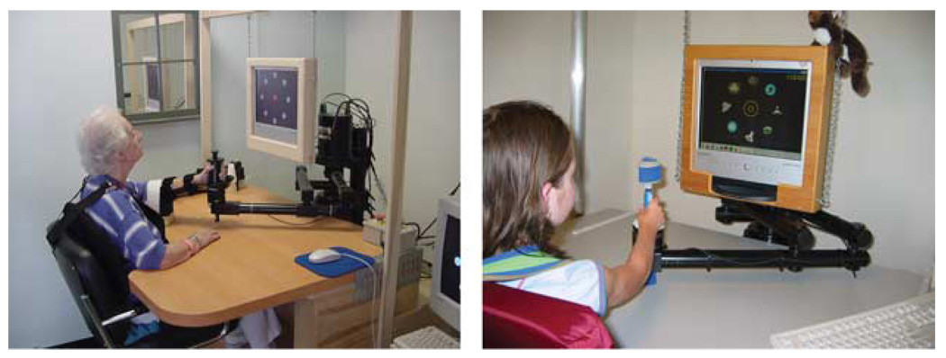
Our approach was to tackle a difficult and common clinical problem, stroke rehabilitation for the upper limb, using the best available technology. In this case, we had to invent the technology, since in 1989 there were no available alternatives. The centerpiece of this effort became known as MIT-MANUS, from MIT Motto’s Mens et Manus (Mind and Hand). MIT-MANUS is a robot designed for clinical neurological applications [27]–[29]. Unlike most industrial robots, MIT-MANUS was configured for safe, stable, and highly compliant operation in close physical contact with humans. This was achieved using impedance control, a key feature of the robot control system. Its computer control system modulates the way the robot reacts to mechanical perturbation from a patient or clinician and ensures a gentle compliant behavior. The machine was designed to have a low intrinsic end-point impedance (i.e., be back-drivable) to allow weak patients to express movements without constraint and offer minimal resistance at speeds up 2 m/s (the approximate upper limit of unimpaired human performance, hence, the target of therapy, and the maximum speed observed in some pathologies, e.g., the shock-like movements of myoclonus). The design achieved a low and nearly isotropic inertia (1 ± 0.33 kg, maximum anisotropy 2 : 1) and friction (0.84 ± 0.28 N, maximum anisotropy 2 : 1). It is also capable of producing a predetermined range of forces (0–45 N) and impedances (stiffness 0–5 N/mm and damping 0–220 Ns/m). Furthermore, weight, visibility, and accessibility to the patient’s hand were key requirements. MIT-MANUS has two active degrees of freedom (DOFs) and weighs 390 N. It consists of a semidirect-drive five-bar-linkage selective compliance assembly robot arm (SCARA) mechanism driven by brushless motors rated to 7.86 Nm of continuous stall torque (Considerably higher torques can be produced but only for limited periods of time) with 16-bit resolvers for position and velocity measurements. Redundant velocity sensing is provided by dc-tachometers with a sensitivity of 1.75 V/rad/s. Torque sensors on the motor shafts are rated to 22.6 Nm with a torsional stiffness of 2302 Nm/rad. To minimize noncollocation effects, actuators and sensors are aligned on the same axis. The resolvers and torque sensors are mounted on the brushless motor shaft outputs, while the tachometers are mounted on the motor shaft tips. The mechanism is mounted on a tubular base that allows its vertical position to be adjusted. Since then several variants have been manufactured with basically the same configuration, but incorporating more powerful motors (9.65 N.m continuous stall torque), eliminating the 16-bit resolvers in favor of 16-bit optical encoders for position measurement, eliminating the torque sensors and adding a 6-DOF force transducer at the robot tip. Fig. 1 shows the robot during therapy.
Fig. 1.
Planar shoulder-and-elbow stroke therapy. The left photo shows a person with acute stroke (inpatient), while the photo on the right shows a child with cerebral palsy during robot training at Spaulding Rehabilitation Hospital.
IV. ANTIGRAVITY SHOULDER-AND-ELBOW ROBOT
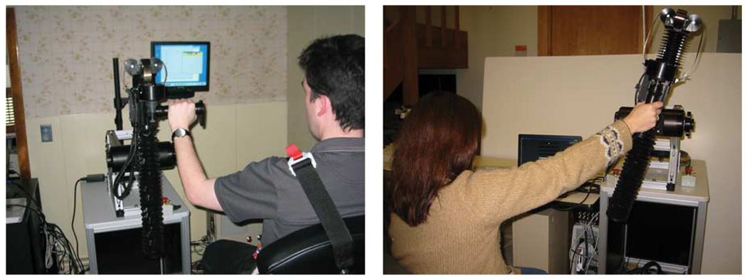
Following successful clinical trials with MIT-MANUS, a 1-DOF module was conceived to extend the benefits of planar robotic therapy to spatial arm movements, including movements against gravity. Incorporated into the design are therapists’ suggestions that functional reaching movements often occur in a range of motion close to shoulder scaption. That is, this robotic module was designed for therapy to focus on movements within the 45° to 65° range of shoulder abduction and from 30° to 90° of shoulder elevation or flexion [30]. The module allows 0.49 m of linear motion and it is capable of exerting a maximum endpoint force of 65 N upward and 45 N downward. The module can be used independently or mounted at the tip of MIT-MANUS for movement in a limited spatial workspace of 0.38 m × 0.46 m × 0.49 m. It incorporates recent changes in linear induction motor technology, which completely enclose the magnets within the motor forcer. Without increasing magnetic field strength, this dramatically increases its line integral. We employ a Copley Control ThrustTube TB2504 brushless dc linear motor (see Fig. 2). The module can permit free motion of the patient’s arm, or can provide partial or full assistance or resistance as the patient moves against gravity. In comparison to MIT-MANUS, the antigravity module has a greater effective endpoint friction (2.48 ± 0.65 N) and comparable inertia, though the resulting system is still highly backdrivable. The antigravity module’s impedance range is a stiffness from 0 to 40 N/mm and a damping from 0 to 800 Ns/m.
Fig. 2.
Vertical 1-DOF module using linear electromechanical technology. This commercial version of MIT’s prototype module can be operated in standalone fashion or integrated with the planar module to allow spatial movements. Note that in the standalone mode it can be operated at any angle relative to the horizontal and vertical planes with adjustable handle positions. The left photo demonstrates an example of subject positioning during therapy aimed at neurological recovery. The right photo demonstrates an example of subject positioning during therapy aimed at orthopedic recovery following rotator cuff surgery (Courtesy Baltimore Veterans Administration Medical Center).
V. WRIST ROBOT
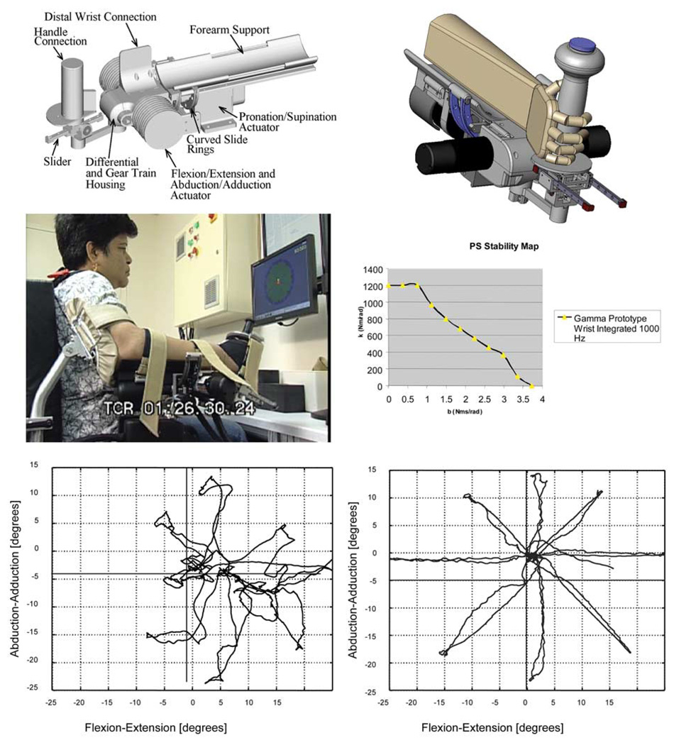
To extend the treatment envelope beyond the shoulder and elbow, we designed and built a wrist module for robotic therapy [31]. Fig. 3 shows a solid model, the completed device during therapy, and its achievable impedance in pronation/supination. A curved rail sits between four guide wheels, which allow it to rotate. Torque transmission from the abduction/adduction and flexion/extension actuators to the patient’s arm is accomplished by a differential mechanism with a total speed reduction of 8 : 1. This speed-reduction ratio in a compact, low-friction transmission is a key aspect of the design as it permits the use of smaller and lighter actuators than a direct-drive design of comparable performance, while maintaining a low robot output impedance (i.e., the device is highly “backdrivable”). Ideally, a patient attempting to move the robot at speeds from 0 to 38 rad/s should encounter no significant friction, inertia or stiffness. In this design, the apparent stiffness is zero, the maximum apparent inertia for each wrist DOF is estimated to be 30 to 45 × 10−4 kg-m2 and the maximum apparent static friction torque is 0.29 N.m for pronation/supination, 0.075 N.m for flexion/extension, and 0.075 N.m for abduction/adduction. The device accommodates the range of motion of a normal wrist in everyday tasks, i.e., flexion/extension 60°/60°, abduction/adduction 30°/45°, pronation/supination 70°/70°. The torque output from the device is capable of lifting the patient’s hand against gravity, accelerating the inertia, and appears to be able to overcome most forms of hypertonicity. The present device can produce a range of continuous stall torques with no cogging (1.85 N.m for pronation/supination, 1.43 N.m of flexion/extension, and 1.43 N.m of abduction/adduction), impedances (0 to 60 N.m/rad for pronation/supination, 0 to 40 N.m/rad of flexion/extension, and 0 to 40 N.m/rad of abduction/adduction), and damping (0 to 1 N.m.s/rad for pronation/supination, 0 to 0.45 N.m.s/rad for flexion/extension, and 0 to 0.45 N.m.s/rad for abduction/adduction). A torque sensor is mounted at each axis with an end-of-scale equivalent to the continuous stall torque of each axis.
Fig. 3.
Wrist robot. The top row shows solid models of the prototype design. The middle row shows the device during therapy at Burke Rehabilitation Hospital (left) and the achievable impedance range of the device (right) in pronation and supination. The bottom row shows measurement of a person with chronic left hemiparesis due to stroke prior to (left) and after (right) training with the wrist robot.
The commonest form of unimpaired upper-extremity movement appears to be a combination of translating the hand (with the shoulder and elbow) to a location in space and orienting the hand (with the wrist) to facilitate object manipulation. Furthermore, it is plausible that different neural structures may subserve the control of translation and rotation, which prompts the questions: what is the optimum sequence of treatment for upper-extremity movement disorders? Should proximal joints (e.g., shoulder an elbow) be treated before distal joints (e.g., the wrist) or vice versa? Recovering the ability to orient the hand without the ability to bring it to an object would seem to have limited functional value but the converse could also be argued. Or must all relevant joints be treated simultaneously to obtain maximum benefit? At the time of writing, these important questions about movement therapy remain unanswered. However, the wrist module can be operated in isolation or mounted at the tip of the shoulder-and-elbow modules thereby affording, a rigorous, quantitative empirical approach (with a minimum of uncontrolled and confounding variables) to test whether the ability to transport the hand and preposition it to manipulate an object are better recovered simultaneously or sequentially.
VI. HAND ROBOT
The next module required to afford whole-arm therapy is a hand robot. Moving a patient’s hand is not a simple task, since the human hand has 15 joints with a total of 22 DOFs so it was prudent to determine how many DOFs are necessary for a patient to perform the majority of everyday functional tasks. Here our clinical experience with over 300 stroke patients was invaluable in that it allowed us to identify what was most likely to work in the clinic (and what probably would not). Though individual digit opposition (e.g., thumb to pinkie) may be important for the unimpaired human hand, it is clearly beyond the realistic expectations of most of our patients whose impairment level falls between severe and moderate; a device to manipulate 22 DOFs is unnecessary (or at least premature). However, the exquisite sensitivity of the skin and its sensors, i.e., Meissner corpuscles, Merkell disks, Ruffini organs, and Pacinian corpuscles, indicates that the requirement of back-drivability will be especially important, requiring minimal cogging and force/torque ripple to afford an experience ranging from “feather-touch” unconstrained movement to simulated hard-surface contact.
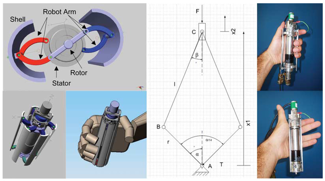
The hand therapy module is a novel design that converts rotary into linear movement using a single brushless dc electrical motor as a free-base mechanism with what is traditionally called the stator being allowed to rotate freely [32]. The stator (strictly, the “second rotor”) is connected to a set of arms, while the rotor is connected to another set of arms: when commanded to rotate, the rotor and stator work like a double crank & slider mechanism, in opposing configuration, where the crank is represented by a single arm and the slider is the shell or panel which interacts with the hand of the patient (see Fig. 4). The hand robot is used to simulate grasp and release with its impedance determined by the torque between stator and rotor. A torsional spring (connected in geometric parallel) is available to compensate for a patient’s hypertonicity (inability to relax) and, in addition, the device is capable of delivering a net torque to grasp and release of 800 mNm. The hand robot is capable of providing continuous passive motion, strength, sensory, and sensorimotor training for grasp and release and it can be employed in stand-alone operation or mounted at the tip of the planar, wrist, or antigravity modules or the combination of all the above to afford a whole-arm functional training.
Fig. 4.
Hand robot. The left columns show solid models of the device. The middle column sketches the principle of operation, while the right column shows the device during operation (some shells were removed for clarity). Note the black line indicating the rotation of both the rotor and stator.
VII. RECONFIGURABLE ROBOTS FOR WHOLE-ARM THERAPY
To increase the effectiveness of robotic therapy we are investigating whole-arm therapy approaches that integrate robotic therapy with clinical practice to enhance the carry-over of robot trained movements to functional tasks. We expect that a treatment protocol, properly targeted to emphasize the sequence and timing of sensory and motor stimuli similar to those naturally occurring in daily life tasks, will facilitate carry-over of observed gains in motor abilities, to confer greater improvements in functional recovery. From one perspective, a whole-arm therapy approach is a departure from our previous research into therapeutic robotics. Our initial emphasis on the use of standalone robotic modules is a “bottom-up” approach, which assumes that improvements in underlying capacities will enhance motor function during daily life activities and tasks. Whole-arm therapy may be considered an alternative “top-down” approach to rehabilitation in which a person’s identified goals for task performance are used in conjunction with robot-derived evaluation data to establish a treatment plan. However, these alternatives are not mutually exclusive and both approaches may be needed to maximize recovery.
Robotic technology may not only provide remediation for impairments at the capacity levels (e.g. strength, isolated movement), but also provide task-specific, intensive therapy for impaired abilities (e.g. speed or coordination of limb movement) that underlie task performance and ultimately assisted practice of tasks in their normal functional context. One goal of our present research is to identify the best blend of “top down” and “bottom up” approaches and to begin building a scientific basis for the “best” rehabilitation practice. The modularity of our suite of therapeutic robots, which is a cornerstone of our design philosophy, is uniquely suited to this investigation. It allows us to employ them in standalone mode to deliver therapy for particular impairments or to integrate the modules to deliver whole-arm functional robotic therapy (see Fig. 5). To the best of our knowledge, no other research group can presently pursue this objective, because there are no other reconfigurable rehabilitation robots.
Fig. 5.
Reconfigurable robotic modules. The robotic therapy modules can operate in standalone mode or be integrated into a coordinated functional unit. The left panel shows a solid model and the right panel shows a hardware implementation of the planar (elbow & shoulder) and wrist modules integrated into a single unit.
VIII. ANKLEBOT (ANKLE ROBOT)
The devices described above were intended to treat the upper extremity, and as we completed the enabling technology for reconfigurable whole-arm therapy, we initiated the development of lower-extremity devices [33]. Again, we are aiming for maximum reconfigurability and have taken a modular approach. The first lower extremity robot module deployed to the clinic has been dubbed the “anklebot.” The ankle joint is of particular importance in stroke rehabilitation due to the prevalence of “foot drop,” which is a simple name for a complex problem. The foot needs to clear the ground during the swing phase of gait and it needs to have a controlled landing during heel strike. Lack of proper control during these two phases increases the likelihood of trips and falls. Foot drop can be defined as weakness of ankle and toe dorsiflexors which include the tibialis anterior, extensor hallucis longus, and extensor digitorum longus. Patients typically compensate by walking with an exaggerated hip hike (increased abduction) to prevent the toes from catching on the ground during swing phase and the foot slapping the ground following heel strike but this yields an unsightly and tiring gait. At present, foot drop is typically addressed in the clinic via an ankle foot orthosis which restricts the ankle’s range of motion but that approach has limitations and offers little hope of reducing the impairment.
To address impairment reduction, we recently introduced to the clinic (at the Center on Task-Oriented Exercise and Robotics in Neurological Disease at the Baltimore Veterans Administration Medical Center) a novel ankle robot, which allows normal range of motion in all three DOFs of the foot relative to the shank during walking overground or on a treadmill. Specifically, it allows 25° dorsiflexion, 45° plantar flexion, 25° inversion, 15° eversion, and 15° of internal or external rotation. These are near the limits of the range of comfortable motion for normal subjects and beyond what is required for typical gait. The anklebot provides independent, active assistance in two of these three degrees-of-freedom, namely, dorsi/plantar flexion and in-version/eversion, and a passive degree-of-freedom for internal/external rotation. Internal/external rotation is limited at the ankle with the orientation of the foot in the transverse plane being controlled primarily by rotation of the leg. Furthermore, internal/external rotation plays a marginal role in foot drop and is coupled to inversion/eversion.
The device was designed primarily to position the foot during the swing phase of the impaired leg while providing some assistance during the stance phase. Ankle moments in stance can be one to two orders of magnitude larger than those during the swing phase; the motors cannot supply ankle moments big enough to lift the weight of the patient. The device was designed to deliver a target torque of approximately 17 N-m in either actuated DOF. This value is larger than that required to position the foot of unimpaired subjects during swing phase and pilot trials suggest that it is adequate to drive the foot of patients with significant hypertonicity.
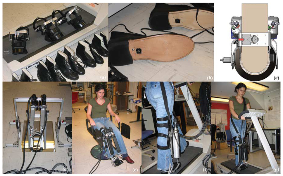
The clinically deployed prototype weighs exactly 34.5 N mounted at the knee and it follows our philosophy of aiming for minimum friction and inertia and maximizing back-drivability. Furthermore, as with all our devices, we purposely under-actuated the ankle robot with fewer DOFs than are anatomically present. Not only does this simplify the mechanical design, it allows the device to be installed quickly without problems of misalignment with the patient’s joint axes, in this case the ankle and subtalar joints, causing excessive forces or torques. Ease of use is another critical consideration in all our designs as we consider it a major determinant of success or failure of the device in rehabilitation. The ankle robot must be attached to or removed from the patient within 2 min. To afford such speedy deployment, patient first dons a pair of shoes retrofitted with a quick connector placed unobtrusively in the shank and a knee-brace that can be mounted from the front of the leg (without requiring the foot to be threaded through it, see Fig. 6). While still seated, patient steps into the ankle robot foot piece or is helped by the therapist. The patient’s foot (with shoe on) is secured to this piece with the quick connector and a single strap over the hind foot for safety and to minimize spastic reaction. The robot’s foot piece does not extend the entire length of the patient’s shoe but is designed to end near the midtarsals, to allow forefoot mobility. The robot is then mounted onto the knee brace with a bicycle-type quick lock/release mechanism (see Fig. 6).
Fig. 6.
Anklebot Beta-prototype for foot drop. (a) Shows shoes and knee-braces of different sizes. (b) Shows quick connect/release cleat, while (c) shows a solidworks representation of the shoe stepping into the locking mechanism, (d) shows the actual prototype, (e) shows an unimpaired subject using the device in a sitting position during driving-simulator training, and (f) and (g) show the same subject during treadmill walking.
In this clinically deployed beta-prototype, we used two dc brushless Kollmorgen RBE(H) 00714 motors with a maximum continuous torque of 0.249 N-m each. Two of these motors produce a maximum continuous torque of 0.50 N-m (0.25 N-m each), which is amplified and transmitted to the foot piece via a pair of parallel linear traction drives. The net torque is approximately 23 N-m in dorsi/plantar flexion and 15 N-m in inversion/eversion. Position (and velocity) information is provided by Gurley R19 encoders mounted coaxial with the motors and by a pair of Renishaw encoders with 1-µm resolution mounted on the traction drive. Furthermore, to avoid interference with the unimpaired leg, we kept the inside of the leg (the medial border) free of any obstructions.
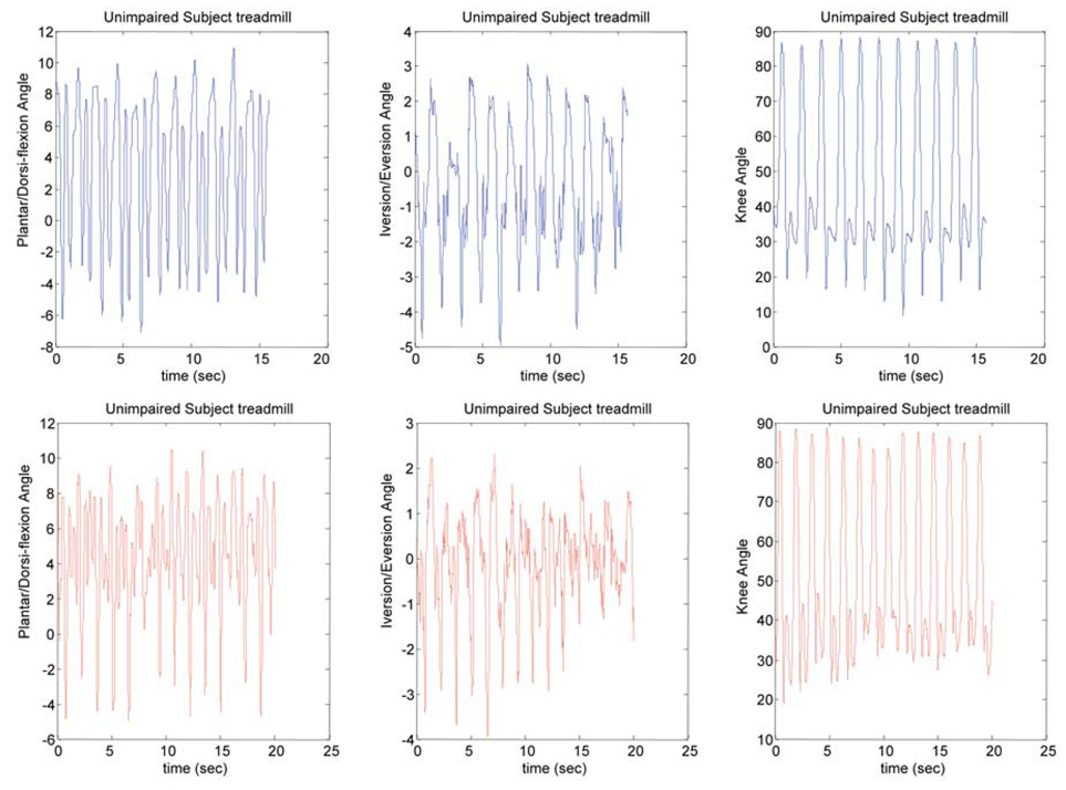
Fig. 7 shows the kinematics of the initial prototype during trials with unimpaired subjects comparing three different training conditions: subject in a sitting position, with the anklebot serving as the “accelerator pedal” of a driving simulator [Fig. 7(a)], subject walking with asymmetric loading over a treadmill (ankle module on one leg) [Fig. 7(b)], and subject walking overground with asymmetric loading (ankle module on one leg) [Fig. 7(c)].
Fig. 7.
Anklebot kinematic comparison during treadmill training with asymmetric loading due to the ankle prototype with unimpaired subject. The left column shows plantar-dorsiflexion (deg), the middle columns shows inversion-eversion (deg), and the right column shows knee angle (deg). The top row shows an unimpaired subject walking in the device (teach mode) and the bottom row shows data from another unimpaired subject during training (playback mode). The teach mode reference gait is wrapped-around, which allows to extend the duration of training as needed.
IX. CONCLUSION
We presented a retrospective and chronological review of our efforts to revolutionize the way physical medicine is practiced by introducing robotics. We showed a sample of our results (now with well over 300 stroke patients, both inpatients and outpatients) which proved first, that movement therapy has a significant and measurable impact on neurorecovery following injury, and second, that effective movement therapy can be delivered by a robot. These results have since been replicated by many different groups elsewhere and the focus of recent research has shifted to far more complex questions: A multitude of variables might influence recovery, but which ones significantly impact the outcome of therapy? Are they independent or do they interact? If the latter, can the interaction among variables be used to further improve the clinical outcome? This exciting area of clinical research is in its infancy. Due to the complexity of human movement and its recovery after injury, an empirical, evidence-based approach is mandatory. A “gym” of robots such as we have developed (planar, antigravity, wrist, hand, and ankle modules, with more in progress) provides essential tools to help clinicians unravel this knot. These reconfigurable modules facilitate tightly controlled experiments on the relative importance of different DOFs and the best sequence for their treatment.
It is probably obvious that robots can deliver reproducible therapy and measure behavior, thereby further accelerating the pace of research. It is perhaps less obvious that to design robots to provide data relevant to a study of recovery is a difficult challenge. Ideally, the robot should not encumber the patient, but allow the expression of whatever movement may be possible, however weak or partial it may be. Measuring forces or torques exerted on an unyielding machine may provide valuable information but it cannot be used to infer movement behavior; to study movement the patient must be allowed to move. Minimal encumbrance (i.e. maximal “backdrivability”) continues to be the guiding principle behind all of our designs.
Another important feature of successful therapeutic robots is their “interactivity,” which seems to be an essential component of effective robotic therapy. Our designs achieve this by controlling the robot’s mechanical impedance. Shaping the robot’s mechanical impedance enables therapy to become “directed mobility” rather than imposed motion. Of course, our designs to date achieve minimal and controllable intrinsic mechanical impedance with varying degrees of success, and there is ample room for improvement.
We claimed above that clinical research on the best form of motor rehabilitation therapy is in its infancy. A thoughtful reader might equally wonder if it is simply our engineering arrogance to suggest that the first compelling evidence-based results are being published only now. Why have the previous 75 years of compassionate rehabilitation practice not provided answers? Of course, they have to some degree, but the pace of progress has been sluggish at best. The lack of means to deliver therapy reproducibly and the lack of measurement instruments to evaluate outcomes objectively made it difficult to conduct definitive experiments and made the data that was obtained open to multiple interpretations. Robotics has “broken open” this problem. Interactive robots that can participate effectively in the practice of rehabilitation therapy will enable an explosive growth in definitive, evidence-based research in the hands of compassionate clinicians.
Acknowledgment
Dr. H. I. Krebs and Dr. N. Hogan are coinventors in the MIT-held patent for the robotic device used to treat patients in this work. They hold equity positions in Interactive Motion Technologies, Inc., the company that manufactures this type of technology under license to MIT.
This work was supported in part by the National Institutes of Health (NIH) under Grant R01-HD-36827, Grant R01-HD-37397, and Grant R01-HD-045343, in part by the VA Veterans Affairs under Grant V512(P)P-407-01, Grant V512(P)P-521-02, Grant B3688R, and Grant B3607R, in part by the NYSCORE, and in part by a grant from the Burke Medical Research Institute.
Biographies

Hermano Igo Krebs received the electrician degree in 1976 from Escola Tecnica Federal de Sao Paulo, Brazil, the B.S. and M.S. degrees in naval engineering (option electrical) from University of Sao Paulo, in 1980 and 1987, respectively. He received the M.S. degree in ocean engineering from Yokohama National University, Yokohama, Japan, in 1989, and the Ph.D. degree in ocean engineering from the Massachusetts Institute of Technology (MIT), Cambridge, in 1997, with the thesis: “Robot-Aided Neuro-Rehabilitation and Functional Imaging.”
From 1977 to 1978, he taught electrical design at Escola Tecnica Federal de Sao Paulo. From 1978 to 1979, he worked at the University of Sao Paulo in a project aiming at the identification of hydrodynamic coefficients during ship maneuvers. From 1980 to 1986, he was a surveyor of ships, offshore platforms, and containercranes at the American Bureau of Shipping—Sao Paulo office. In 1989, he was a Visiting Researcher at Sumitomo Heavy Industries—Hiratsuka Laboratories—Japan. From 1993 to 1996, he worked at Casper, Phillips & Associates in containercranes and control systems. He joined the Mechanical Engineering Department, MIT, in 1997 where he is a Principal Research Scientist and Lecturer—Newman Laboratory for Biomechanics and Human Rehabilitation. He also holds an affiliate position as an Adjunct Research Professor of Neuroscience a Weill Medical College of Cornell University, Ithaca, NY. He is one of the founders of Interactive Motion Technologies, a Cambridge-based start-up company commercializing robot technology for rehabilitation. His present goal is to revolutionize the way rehabilitation medicine is practiced today by applying robotics and information technology to assist, enhance, and quantify rehabilitation; particularly neurorehabilitation. This goal translates into research interests in neurorehabilitation, functional imaging, human-machine interactions, robotics, and dynamic systems modeling and control.

Neville Hogan was born in Dublin, Ireland. He received the Dip.Eng. (with distinction) from Dublin College of Technology and the M.S., M.E., and Ph.D. degrees from Massachusetts Institute of Technology (MIT), Cambridge.
He is Professor of Mechanical Engineering, Professor of Brain and Cognitive Sciences, and Director of the Newman Laboratory for Biome-chanics and Human Rehabilitation at MIT. He is a cofounder of Interactive Motion Technologies, Inc., and a board member of Advanced Mechanical Technologies, Inc. Following industrial experience in engineering design, he joined MIT's School of Engineering faculty in 1979 and has served as Head and Associate Head of the Mechanical Engineering department's System Dynamics and Control division. His research is broad and multidisciplinary, extending from biology to engineering—it has made significant contributions to motor neuroscience, rehabilitation engineering, and robotics—but its focus converges on an emerging class of machines designed to cooperate physically with humans. His recent work pioneered the creation of robots sufficiently gentle to provide physiotherapy to frail and elderly patients recovering from neurological injury such as stroke, a novel therapy that has already proven its clinical significance.
Dr. Hogan received the Honorary Doctorate from the Delft University of Technology, Delft, The Netherlands; the Silver Medal of the Royal Academy of Medicine in Ireland; and an Honorary Doctorate from the Dublin Institute of Technology.
Footnotes
Reprinted from Proceedings of IEEE.
This material is posted here with permission of the IEEE. Such permission of the IEEE does not in any way imply IEEE endorsement of any of this web site's products or services. Internal or personal use of this material is permitted. However, permission to reprint/republish this material for advertising or promotional purposes or for creating new collective works for resale or redistribution must be obtained from the IEEE by writing to pubs-permissions@ieee.org.
By choosing to view this document, you agree to all provisions of the copyright laws protecting it.
Contributor Information
Hermano Igo Krebs, Mechanical Engineering Department, Massachusetts Institute of Technology, Cambridge, MA 02139 USA. He is also with the Department Neurology and Neuroscience, Burke Medical Research Institute, Weill Medical College of Cornell University, White Plains, NY 10605 USA (e-mail: hikrebs@mit.edu)..
Neville Hogan, Mechanical Engineering Department, and the Department of Brain and Cognitive Sciences, Massachusetts Institute of Technology, Cambridge, MA 02139 USA (e-mail: neville@mit.edu)..
REFERENCES
- 1.American Heart Association. 2003. Heart disease and stroke statistics. update. [Google Scholar]
- 2.Japan’s National Institute of Population and Social Security Research. [Online]. Available: http://www.ipss.go.jp/index-e.html.
- 3.Volpe BT, Krebs HI, Hogan N. Is robot-aided sensorimotor training in stroke rehabilitation a realistic option? Curr. Opin. Neurol. 2001;vol. 14:745–752. doi: 10.1097/00019052-200112000-00011. [DOI] [PubMed] [Google Scholar]
- 4.Hogan N. Impedance control: An approach to manipulation. J. Dyn. Syst. Meas. Control. 1985;vol. 107:1–24. [Google Scholar]
- 5.Emken JL, Bobrow JE, Reinkensmeyer DJ. Robotic movement training as an optimization problem: Designing a controller that assists only as needed. Proc. ICORR 2005—9th Int. Conf. Rehabilitation Robotics; 2005. pp. 307–312. [Google Scholar]
- 6.Riener R, Lunenburger L, Jezernik S, Anderschitz M, Colombo G, Dietz V. Patient-cooperative strategies for robot-aided treadmill training: First experimental results. IEEE Trans Neural Syst. Rehabil. Eng. 2005 Sep.vol. 13(no 3):380–394. doi: 10.1109/TNSRE.2005.848628. [DOI] [PubMed] [Google Scholar]
- 7.Aisen ML, Krebs HI, Hogan N, McDowell F, Volpe BT. The effect of robot-assisted therapy and rehabilitative training on motor recovery following stroke. Arch. Neurol. 1997;vol. 54:443–446. doi: 10.1001/archneur.1997.00550160075019. [DOI] [PubMed] [Google Scholar]
- 8.Krebs HI, Volpe BT, Aisen ML, Hogan N. Increasing productivity and quality of care: Robot-aided neurorehabilitation. VA J. Rehabil. Res. Develop. 2000;vol. 37(no 6):639–652. [PubMed] [Google Scholar]
- 9.Volpe BT, Krebs HI, Hogan N, Edelstein OL, Diels C, Aisen M. A novel approach to stroke rehabilitation: Robot-aided sensorimotor stimulation. Neurology. 2000;vol. 54:1938–1944. doi: 10.1212/wnl.54.10.1938. [DOI] [PubMed] [Google Scholar]
- 10.Volpe BT, Krebs HI, Hogan N, Edelstein L, Diels CM, Aisen ML. Robot training enhanced motor outcome in patients with stroke maintained over 3 years. Neurology. 1999;vol. 53:1874–1876. doi: 10.1212/wnl.53.8.1874. [DOI] [PubMed] [Google Scholar]
- 11.Ferraro M, Demaio JH, Krol J, Trudell C, Edelstein L, Christos P, England J, Fasoli S, Aisen ML, Krebs HI, Hogan N, Volpe BT. Assessing the motor status score: A scale for the evaluation of upper limb motor outcomes in patients after stroke. Neurorehabil. Neural Repair. 2002;vol. 16(no 3):301–307. doi: 10.1177/154596830201600306. [DOI] [PubMed] [Google Scholar]
- 12.Burgar CG, Lum PS, Shor PC, Van der Loos HFM. Development of robots for rehabilitation therapy: The Palo Alto VA/Stanford experience. VA J. Rehabil. Res. Develop. 2000;vol. 37:663–673. [PubMed] [Google Scholar]
- 13.Reinkensmeyer DJ, Kahn LE, Averbuch M, McKenna-Cole A, Schmit BD, Rymer WZ. Understanding and treating arm movement impairment after chronic brain injury: Progress with the ARM guide. VA J. Rehabil. Res. Develop. 2000;vol. 37:653–562. [PubMed] [Google Scholar]
- 14.Lum PS, Burgar CG, Shor PC, Majmundar M, Van der Loos M. Robot-assisted movement training compared with conventional therapy techniques for the rehabilitation of upper-limb motor function after stroke. Arch. Phys. Med. Rehabil. 2002;vol. 83:952–959. doi: 10.1053/apmr.2001.33101. [DOI] [PubMed] [Google Scholar]
- 15.Lum PS, Lehman SL, Reinkensmeyer DJ. The bimanual lifting rehabilitator: An adaptive machine for therapy of stroke patients. IEEE Trans. Rehabil. Eng. 1995 Jun.vol. 3(no 2):166–174. [Google Scholar]
- 16.Lum PS, Reinkensmeyer DJ, Lehman S. Robotic assist devices for bimanual physical therapy: Preliminary experiments. IEEE Trans. Rehabil. Eng. 1993 Sep.vol. 1(no 3):185–191. [Google Scholar]
- 17.Shor PC, Lum PS, Burgar CG, Van der Loos HFM, Majmundar M, Yap R. Mokhtari M. Integration of Assistive Technology in the Information Age. Amsterdam, The Netherlands: IOS; 2001. The effect of robot aided therapy on upper extremity joint passive range of motion and pain; pp. 79–83. [Google Scholar]
- 18.Fasoli SD, Krebs HI, Stein J, Frontera WR, Hogan N. Effects of robotic therapy on motor impairment and recovery in chronic stroke. Arch. Phys. Med. Rehabil. 2003;vol. 84:477–482. doi: 10.1053/apmr.2003.50110. [DOI] [PubMed] [Google Scholar]
- 19.Fasoli SD, Krebs HI, Stein J, Frontera WR, Hughes R, Hogan N. Robotic therapy for chronic motor impairments after stroke: Follow-up results. Arch. Phys. Med. Rehabil. 2004;vol. 85:1106–1111. doi: 10.1016/j.apmr.2003.11.028. [DOI] [PubMed] [Google Scholar]
- 20.Fasoli SD, Krebs HI, Ferraro M, Hogan N, Volpe BT. Does shorter rehabilitation limit potential recovery poststroke? Neurorehabil. Neural Repair. 2004;vol. 18(no 2):88–94. doi: 10.1177/0888439004267434. [DOI] [PubMed] [Google Scholar]
- 21.Stein J, Krebs HI, Frontera WR, Fasoli SE, Hughes R, Hogan N. Comparison of two techniques of robot-aided upper limb exercise training after stroke. Amer. J. Phys. Med. Rehabil. 2004;vol. 83(no 9):720–728. doi: 10.1097/01.phm.0000137313.14480.ce. [DOI] [PubMed] [Google Scholar]
- 22.Ferraro M, Palazzolo JJ, Krol J, Krebs HI, Hogan N, Volpe BT. Robot aided sensorimotor arm training improves outcome in patients with chronic stroke. Neurology. 2003;vol. 61:1604–1607. doi: 10.1212/01.wnl.0000095963.00970.68. [DOI] [PubMed] [Google Scholar]
- 23.Finley MA, Fasoli SE, Dipietro L, Ohlhoff J, MacClellan L, Meister C, Whitall J, Macko R, Bever CT, Krebs HI, Hogan N. Short duration upper extremity robotic therapy in stroke patients with severe upper extremity motor impairment. VA J. Rehabil. Res. Develop. 2005;vol. 42(no 5):683–692. doi: 10.1682/jrrd.2004.12.0153. [DOI] [PubMed] [Google Scholar]
- 24.Daly J, Hogan N, Perepezko E, Krebs HI, Rogers J, Goyal K, Dohring M, Fredrickson E, Nethery J, Ruff R. Response to upper limb robotics and functional neuromuscular stimulation following stroke. VA J. Rehabil. Res. Develop. 2005;vol. 42(no 6):723–736. doi: 10.1682/jrrd.2005.02.0048. [DOI] [PubMed] [Google Scholar]
- 25.MacClellan LR, Bradham DD, Whitall J, Wilson PD, Ohlhoff J, Meister C, Hogan N, Krebs HI, Bever CT. Robotic upper extremity neuro-rehabilitation in chronic stroke patients. VA J. Rehabil. Res. Develop. 2005;vol. 42(no 6):717–722. doi: 10.1682/jrrd.2004.06.0068. [DOI] [PubMed] [Google Scholar]
- 26.Dipietro L, Ferraro M, Palazzolo JJ, Krebs HI, Volpe BT, Hogan N. Customized interactive robotic treatment for stroke: EMG-triggered therapy. IEEE Trans. Neural Syst. Rehabil. Eng. 2005 Sep.vol. 13(no 3):325–334. doi: 10.1109/TNSRE.2005.850423. [DOI] [PMC free article] [PubMed] [Google Scholar]
- 27.Hogan N, Krebs HI, Charnnarong J, Srikrishna P, Sharon A. MIT-MANUS: A workstation for manual therapy and training I. Proc. IEEE Int. Workshop Robot and Human Communication RO-Man92; 1992. pp. 161–165. [Google Scholar]
- 28.Hogan N, Krebs HI, Sharon A, Charnnarong J, inventors. Interactive robot therapist. 5 466 213. U.S. Patent. 1995 Nov. 14
- 29.Krebs HI, Hogan N, Aisen ML, Volpe BT. Robot-aided neurorehabilitation. I.EEE Trans. Rehabil.Eng. 1998 Mar.vol. 6(no 1):75–87. doi: 10.1109/86.662623. [DOI] [PMC free article] [PubMed] [Google Scholar]
- 30.Krebs HI, Ferraro M, Buerger SP, Newbery MJ, Makiyama A, Sandmann M, Lynch D, Volpe BT, Hogan N. Rehabilitation robotics: Pilot trial of a spatial extension for MIT-MANUS. J. Neuroeng. Rehabil., Biomedcentral. 2004 doi: 10.1186/1743-0003-1-5. [Online]. 1. Available: http://www.jneuroengrehab.com/content/1/1/5. [DOI] [PMC free article] [PubMed] [Google Scholar]
- 31.Williams DJ, Krebs HI, Hogan N. A robot for wrist rehabilitation. presented at the 23rd IEEE- EMBS Conf.; Istanbul, Turkey. 2001. [Google Scholar]
- 32.Masia L, Krebs HI, Cappa P, Hogan N. Whole-arm rehabilitation following stroke: Hand module. presented at the BioRob; Pisa, Italy. 2006. [DOI] [PMC free article] [PubMed] [Google Scholar]
- 33.Wheeler JW, Krebs HI, Hogan N. An ankle robot for a modular gait rehabilitation system. presented at the IROS; Sendai, Japan. 2004. [Google Scholar]


