BACKGROUND
Templating is the method where a surgeon calculates the correct-sized prosthesis from a pre-operative radiograph.1–3 Digital radiography and PACS (Picture Archiving and Communications System) create images of variable magnification. This has led to expensive software to allow templating. We present a simple method to allow acetate templates to be used on PACS images.
TECHNIQUE
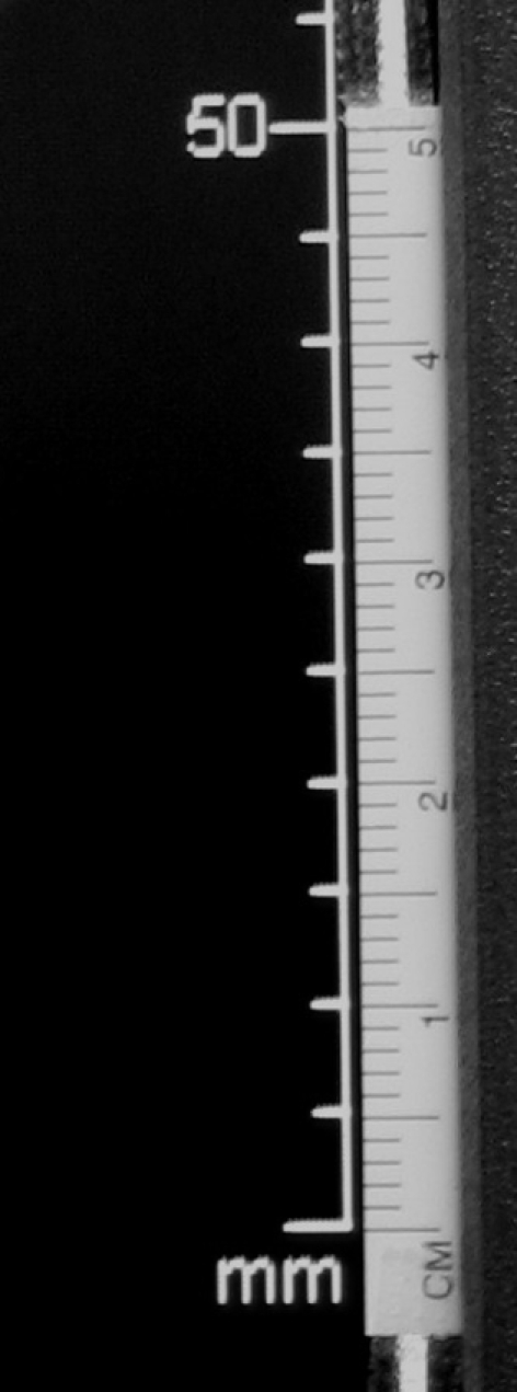
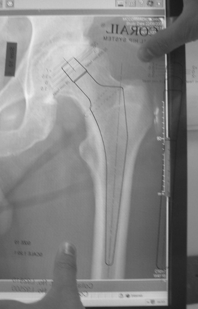
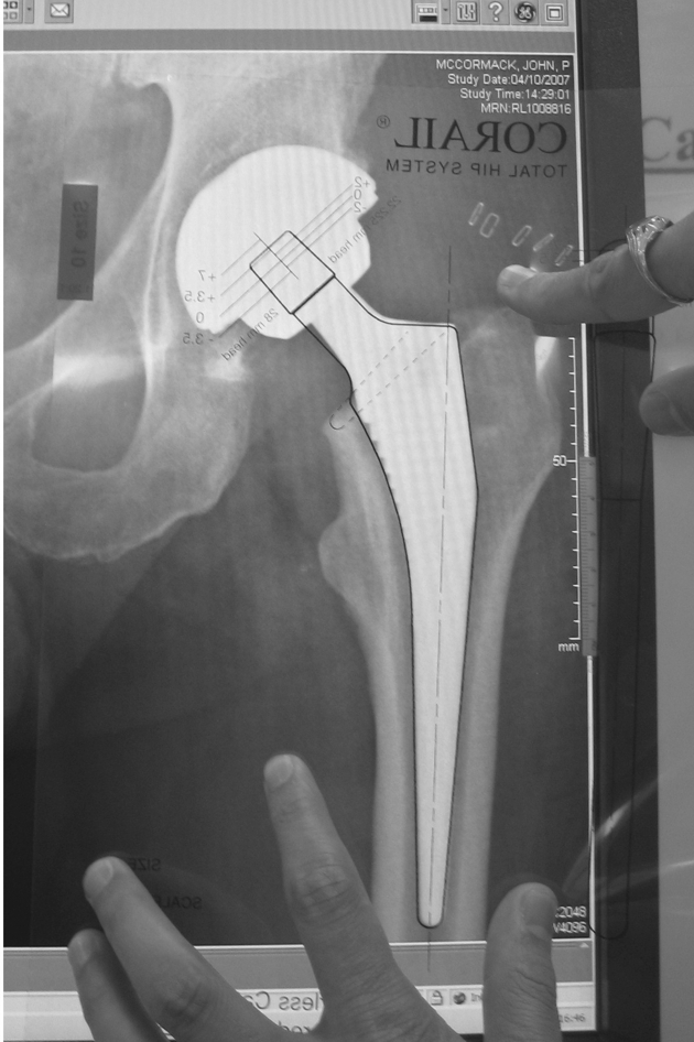
An anteroposterior pelvis X-ray is 20% (± 6%, 2 SDs) magnified as the X-ray plate is separated from the hip by buttock muscles and fat.4 Acetate templates are, therefore, 20% oversized (e.g. Corail hip). A digital anteroposterior pelvis image is taken by the same method but uses a ‘digital’ plate. The size of the displayed image depends on the screen size and resolution of the screen. The image can be further magnified or shrunk. This variation in size makes it difficult to template. PACS systems have a calibration tool, displayed as an unmagnified measure. A ruler can be fixed to the screen (we used the plastic ruler packaged with a skin marker). The display is magnified or shrunk until 5 cm on the screen equals 5 cm on the ruler (Fig. 1). The digital image is now 20% oversized and equates to a hard copy X-ray (Fig. 2). To prove this, Figure 3 shows the postoperative film with the chosen template perfectly superimposed, confirming the digital image is 20% oversize.
Figure 1.

Calibration of image magnification using plastic ruler.
Figure 2.

Acetate templates being used on PACS image.
Figure 3.

Confirmation of 20% oversized image.
DISCUSSION
With the change to digital radiography, many hospitals no longer print hard-copy films. This method provides an alternative to expensive digital software that is as effective as hard-copy templating.
References
- 1.Capello WN. Preoperative planning of total hip arthroplasty. Am Acad Orthop Surg Instr Course Lect. 1986;35:249–57. [PubMed] [Google Scholar]
- 2.D'antonio JA. Preoperative templating and choosing the implant for primary THR in the young patient. Am Acad Orthop Surg Instr Course Lect. 1994;43:339–46. [PubMed] [Google Scholar]
- 3.Dore DD, Rubash HE. Primary total hip arthroplasty in the older patient: optimising the results. Am Acad Orthop Surg Instr Course Lect. 1994;43:47–57. [PubMed] [Google Scholar]
- 4.Clark KC, Naylor E, Roebuck EJ, editors. Clark's Positioning in Radiography. 12th edn. London: Hodder Arnold; 2005. [Google Scholar]


