Abstract
We present a case of gallstone obstruction of the duodenum in a post total gastrectomy patient without a cholecystoenteric fistula. The patient presented with epigastric pain. On abdominal computed tomography and percutaneous transhepatic choangiography imaging, the patient was found to have duodenal obstruction. At operation, the cause of obstruction was found to be a large gallstone in the third part of the duodenum, but there was no associated cholecystoenteric fistula. This report is the first to describe duodenal obstruction by a gallstone formed within the duodenum, in a patient post total gastrectomy with Roux-en-Y reconstruction, and highlights what can be a difficult diagnosis in such patients.
Keywords: Gallstone, Duodenal obstruction, Total gastrectomy complications
Gallstone ileus is not uncommon accounting for 1–3% of non-resolving small bowel obstructions.1,2 The level of obstruction may correlate with the size of the stone with larger stones resulting in more proximal obstruction. The terminal ileum is the most common level of obstruction; however, Bouveret's syndrome (gastric or duodenal obstruction by gallstone in the presence of cholecystoenteric fistula) is described.3,4 We present the first report of duodenal gallstone obstruction in a post-gastrectomy patient without cholecystoenteric fistula.
Case report
A72-year-old man was admitted with a 2-week history of worsening epigastric pain. The pain was colicky in nature, exacerbated by eating and relieved by vomiting. His bowel habit was normal.
Six years prior to this admission, he had undergone a total gastrectomy with Roux-en-Y reconstruction for gastric necrosis secondary to gastric volvulus in a hiatus hernia. For 2 years prior to admission, he had complained of epigastric discomfort and nausea after eating. Barium swallow and upper gastrointestinal endoscopy investigations at that time had proved inconclusive. He had no other significant past medical history.
On examination, the patient was apyrexial and haemodynamically stable with epigastric tenderness but no peritonitis. Blood test revealed a serum amylase level of 647 IU/l, serum bilirubin level of 12 mg/l, a white cell count of 15.3, with otherwise normal renal and haematological results. The patient was treated for acute pancreatitis with intravenous fluid resuscitation, supplementary oxygen and analgesia, and his pain lessened but did not completely resolve.
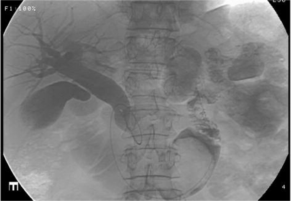
Ultrasound examination of the upper abdomen was reported as showing a dilated common bile duct of 2-cm diameter, a dilated intrahepatic biliary tree, and a 2.8-cm ill-defined mass in the region of the gallbladder which did not cast an acoustic shadow. A subsequent abdominal computed tomography examination (CT) revealed a distended proximal duodenum of 5-cm diameter with an abrupt transition to normal diameter at the third part of the duodenum. Just proximal to the transition point, a collection of high-density material was seen in the duodenal lumen, but no mass lesion was identified. Intraand extrahepatic biliary dilatation was confirmed. A percutaneous transhepatic cholagiogram (PTC) was performed to investigate the biliary dilatation, which demonstrated free flow of contrast through the biliary tree into the grossly dilated duodenal loop. Immediately proximal to the duodenojejunal junction, a 4-cm filling defect was seen, which was thought to represent a polyp, lipoma or collection of inspissated bile (Fig. 1).
Figure 1.

Percutaneous transhepatic cholagiogram showing dilated biliary tree and duodenum with filling defect in third part of duodenum.
A laparotomy was performed which revealed a grossly distended proximal and mid duodenum, with a clearly palpable intraluminal stone at the junction of the third and fourth parts of the duodenum. The gallbladder was distended, but free of adhesions or cholecystoenteric fistula. The jejuno-jejunal anastomosis was widely patent.
A longitudinal duodenotomy was made, and a 5–6 cm obstructing vivid yellow homogeneous gallstone and smaller fragments were milked back and removed from the lumen. The enterotomy was closed transversely. The patient subsequently made an uncomplicated recovery and, at 6 months postoperatively, remains asymptomatic and well, with resolution of all his symptoms particularly epigastric pain and post-prandial nausea.
Discussion
The classic ‘gallstone ileus’ small bowel obstruction is caused by a large gallstone or faceted gallstones in the shape of a cast of the gallbladder of origin. This case differs from all other reports of gallstone-related small bowel obstruction in that there was no associated cholecystoenteric fistula through which a stone originating in the gallbladder could pass into the duodenum or small bowel.
It, therefore, follows that this gallstone must have formed in the duodenum from inspissated bile. The previous gastrectomy and Roux-en-Y reconstruction may be causative, as the duodenum was excluded from the passage of large volumes of gastric contents, resulting in possible stasis of bile and/or bacterial overgrowth within its lumen promoting bile inspissation. Additionally, there is a jejuno-jejunal anastomosis just distal to the fourth part of the duodenum which may have lead to functional delay in duodenal emptying. The pathophysiology for his pancreatitis, which was his presenting diagnosis, may have been back pressure in the duodenum due the gallstone.
This previously undescribed complication should be considered in the investigation of atypical epigastric pain in post total gastrectomy patients. Dilatation of the biliary tree is a possible indicator of the diagnosis. Diagnosis is difficult as the obstruction is only to the free passage of bile through an otherwise defunctioned duodenumgiving a confusing clinical picture. Investigation is also difficult as endoscopic access to the duodenum is usually not possible, but CT scanning and PTC are useful investigations.
References
- 1.Shalowitz JL. Gallstone emesis. Am J Gastroenterol. 1989;84:334–6. [PubMed] [Google Scholar]
- 2.Clavien PA, Richon J, Burgan S, Rohner A. Gallstone ileus. Br J Surg. 1990;77:737–42. doi: 10.1002/bjs.1800770707. [DOI] [PubMed] [Google Scholar]
- 3.Lowe AS, Stevenson S, Kay CL, May J. Duodenal obstruction by gallstones (Bouveret's syndrome): a review of the literature. Endoscopy. 2005;37:82–7. doi: 10.1055/s-2004-826100. [DOI] [PubMed] [Google Scholar]
- 4.Harthun NL, Long SM, Wilson W, Choudhury A. An unusual case of Bouveret's syndrome. J Laparoendosc Adv Surg Tech. 2002;12:69–72. doi: 10.1089/109264202753486975. [DOI] [PubMed] [Google Scholar]


