Abstract
Background
Repetitive deformation enhances intestinal epithelial migration across tissue fibronectin (tFN) via Src but inhibits migration across collagen. Since cell spreading generally precedes motility, we compared the effects of cyclic strain on Caco-2 spreading and migration on tFN, collagen-I, and plasma fibronectin (pFN) and investigated the role of Src in deformation-influenced spreading and migration.
Materials and Methods
Human Caco-2 intestinal epithelial cells on tFN, collagen-I or pFN were subjected to an average 10% strain at 10 cycles/min for 2 hours. Src was inhibited with 10μM PP2 or Src was reduced with siRNA. Parallel studies assessed deformation effects on monolayer wound closure.
Results
Deformation, Src-inhibition or reduction each inhibited spreading on tFN but Src-inhibition or reduction prevented further inhibition of spreading by deformation without preventing further inhibition of motility. Deformation did not alter spreading on collagen-I or pFN, but inhibited wound closure.
Conclusions
Although cell spreading generally precedes and parallels motility, repetitive deformation regulates motility independently of spreading. Since deformation activates Src, the ability of Src blockade to mimic strain-associated inhibition of spreading on tFN suggests that this effect occurs by a separate mechanism that may also require basal Src activity. Further delineation of the mechanisms by which strain disparately modulates spreading and motility may permit acceleration of mucosal healing by targeted interventions to separately promote spreading and epithelial motility.
Keywords: collagen, deformation, enterocyte, fibronectin, motility, Src, spreading
Introduction
The intestinal mucosa experiences repetitive deformation due to peristalsis, villous motility, and physical interactions with relatively non-compressible luminal chime (1-4). In vitro, such repetitive deformation promotes the migration of human Caco-2 intestinal epithelial cells and non-transformed rat IEC-6 intestinal epithelial cells across substrates of tissue fibronectin (5). However, on fibronectin-poor substrates such as type I collagen, type IV collagen, or laminin, deformation stimulates intestinal epithelial proliferation and differentiation but inhibits motility (6, 7). The deposition of tissue fibronectin into inflamed or injured tissues (8) may therefore trigger an important switch in the manner in which intestinal epithelial cells respond to deformation, turning off a normal trophic response that stimulates proliferation and differentiation, and turning on a migratory phenotype adapted to wound healing and the maintenance of the mucosal barrier.
Epithelial sheet migration is an ordered process that requires lamellipodial extension, traction at the leading edge of the cells at the migrating front, detachment of cell-matrix interaction sites at the trailing edge, and the application of traction force along sites of cell-cell contact to pull along cells behind the wound margin (9). Lamellipodial extension and cell spreading across a wound defect represents a key first step in this process that is itself governed by cell-matrix interaction. It therefore becomes an important question whether the regulation of epithelial sheet migration by repetitive deformation reflects effects on cell spreading or whether deformation may affect motility at a different step, such as the generation of traction force by the myosin motor. We therefore sought to determine whether the matrix-dependent effects of repetitive deformation upon intestinal epithelial cell motility were initiated by parallel regulation of intestinal epithelial cell spreading. Adding plasma fibronectin to cell culture media in vitro inhibits the mitogenic effects of repetitive deformation on collagen-cultured intestinal epithelial cells (7), but the effects of using plasma fibronectin as a substrate for wound closure have not yet been studied, so we also evaluated intestinal epithelial cell motility and spreading on membranes precoated with plasma fibronectin and collagen type I. Finally, since Src activation is required for both the stimulation by repetitive deformation of intestinal epithelial proliferation on collagen substrates (10) and of intestinal epithelial migration on fibronectin substrates (5, 11). we also sought to determine the effects of Src blockade on intestinal epithelial cell spreading and motility in the presence of repetitive deformation of the matrix substrate.
We used human Caco-2BBE intestinal epithelial cells as a differentiated model for intestinal epithelial cell biology (12), and applied repetitive deformation to cells cultured on matrix-precoated flexible membranes at an average 10% strain for 10 cycles per minute, parameters resembling the deformation caused in vivo by peristalsis or villous motility (6).
Materials and Methods
Materials
We obtained Dulbecco’s modified Eagle’s medium (DMEM) Oligofectamine, and Plus Reagent were obtained from Invitrogen (Carlsbad, CA) and human transferrin from Roche Applied Science (Indianapolis, IN). Plasma fibronectin and tissue fibronectin were obtained from Calbiochem (Gibbstown, NJ). Trypsin and collagen type I was obtained from Sigma (St. Louis, MO). Six-well Flex I Collagen I precoated (P1001C) and Flex I amino uncoated (P1001A) plates were obtained from Flexcell International Corporation (Hillsborough, NC). Double-stranded short interfering RNAs (siRNAs) targeting human forms of Src and control nontargeting siRNA 1 (NT1 siRNA) were purchased from Dharmacon (Lafayette, CO). The sequences targeted by siRNA were selected using Dharmacon Smartdesign® as follows: human Src1, NNCUCGGCUCAUUGAAGACAA; and human Src2, NNUGGCCUACUACUCCAAACA. We have previously used these siRNAs and observed Src-reduction 60-80% at protein level (10)
Cell culture
We studied Caco-2BBE intestinal epithelial cells, a subclone of the original Caco-2 cell line selected for its ability to differentiate in culture as indicated by the formation of an apical brush border and the expression of brush-border enzymes (12). We maintained the cells at 37°C with 8% CO2 in DMEM with 4,500 mg/l D-glucose, 4 mM glutamine, 1 mM sodium pyruvate, 100 U/ml penicillin, 100 μg/ml streptomycin, 10 μg/ml transferrin, 10 mM HEPES (pH 7.4), and 3.7 g/l NaHCO3 supplemented with 10% heat-inactivated FBS. All studies were performed on cells within 10-15 passages.
Matrix precoating and medium treatments
Uncoated six-well flex I amino plates were precoated with 12.5 μg/ml of tissue fibronectin or plasma fibronectin at saturating concentrations in precoating buffer (15 mM Na2CO3, 35 mM NaHCO3, pH 9.4) as previously described (5). PP2 (a potent and selective inhibitor of Src family tyrosine kinases) was obtained from Calbiochem (Gibbstown, NJ). The PP2 was dissolved in DMSO, aliquoted and stored at -20°C, and diluted immediately before use in culture medium. For studies of Src inhibition, we pretreated Caco-2 cells with PP2 (10 μM) or equivalent amounts of DMSO as a vehicle control for 60 minutes before exposing the cells to cyclic strain to assess cell spreading during Src blockade.
Application of mechanical strain
We plated Caco-2 cells on elastomer membranes coated with tissue fibronectin, plasma fibronectin, or and collagen I and exposed them to a continuous cycles of strain/relaxation generated by a cyclic vacuum produced by a computer-driven system (Flexcell 3000, Flexcell) as previously described (10). A 20-kPa vacuum at 10 cycles/min deforms the membranes from below to an average 10% strain with a stretch-to-relaxation ratio of 1:1 (3.0-second deformation alternating with 3.0 second in neutral conformation). The strain is transmitted to adherent cells cultured on the upper surface of the membrane, which experience similar elongation (6, 13). The six-well plates were incubated at 37°C with 5% CO2 during the repetitive strain, and similarly treated cells on similar plates were maintained in the same incubator without strain as controls.
Spreading Assay
Caco-2 intestinal epithelial cells were trypsinized, resuspended in complete-DMEM and plated at 100, 000 cells/well on flexible membranes precoated with tissue fibronectin, plasma fibronectin, or collagen I for one hour at 37°C. The non-adherent cells were gently washed away and the adherent cells were subjected to an average 10% repetitive deformation at 10 cycles/min for 2 hours. Cells were then formalin-fixed and stained with crystal violet. Cells were photographed for experimental conditions using a QCOLOR3 digital camera (Olympus America Inc) attached to a Nikon Eclipse TE 300 microscope (Japan), and cell areas were calculated using Kodak 1D software (version 3.6) on a Kodak Image Station (Perkin-Elmer, Boston, MA). We measured cell area for at least 100 cells for each condition in each experiment, and pooled at least 4 similar studies for each set of observations.
Transfection with siRNA
Caco-2 cells were plated on flexible membranes precoated with tissue fibronectin at 30–40% confluence 1 day before transfection with siRNAs as described previously (10). Briefly, oligofectamine in Opti-MEM was used for transfection at 10 μg/ml according to the manufacturer’s protocol. The final siRNA concentration was 100 nM unless otherwise indicated. After 6–8 hours of transfection, 0.5 volume of DMEM containing 20% heat-inactivated FBS was added to the cells, and transfection was continued for 72 hours. For spreading experiments, cells were trypsinized, replated onto flexible membranes precoated with tissue fibronectin at 100, 000 cells per well for 1 hour, and then incubated for an additional 2 hours under static conditions or conditions of repetitive deformation as in the description of the spreading assay above.
Migration experiments
We assessed cell migration using a previously described wound healing model (5, 11).. Briefly, Caco-2 monolayers on deformable membranes precoated with tissue fibronectin, plasma fibronectin, or collagen I were subjected to 10% deformation at 10 cycles/min for 0–24 hour after the induction of a small uniform circular wound. As previously described by other investigators (14), we created wounds in confluent cell monolayers using a 1.0-ml pipette tip (1.5 mm diameter) attached to a vacuum source. We applied the tip gently to the monolayer and applied suction for approximately 1 second. We photographed each hole at 0 and 24 hour after the initiation of strain using a QCOLOR3 digital camera (Olympus America Inc) attached to a Nikon Eclipse TE 300 microscope (Japan), and calculated wound areas using Kodak 1D software (version 3.6) on a Kodak Image Station (Perkin-Elmer, Boston, MA). To account for variability in the size of the initial holes, we expressed the remaining wound area in each wound at 24 hour as a ratio to the initial size of each individual wound. Migration was then expressed as the percentage of wound closure in each wound and calculated as follows: 100 × (Initial wound — Final wound)/Initial wound. Similar procedures were used to calculate migration in control monolayers wounded simultaneously but not subjected to repetitive strain
Statistical analysis
We performed all experiments independently at least three times unless otherwise indicated. We expressed all data as means ± SE and analyzed our results using paired or unpaired t-tests using Bonferroni corrections as appropriate and seeking 95% confidence.
Results
Effect of repetitive deformation on cell motility on tissue fibronectin, collagen I and plasma fibronectin
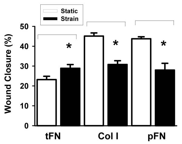
Repetitive deformation accelerated wound closure across tissue fibronectin (Figure 1A, n=30, p<0.05), but inhibited wound closure by 19±2% across type I collagen (Figure 1A, n=30, p<0.005), consistent with previous observations (5, 11). Caco-2 cells cultured on flexible membranes precoated with plasma fibronectin responded to repetitive deformation differently than cells on tissue fibronectin substrates. Caco-2 cells subjected to repetitive deformation on plasma fibronectin exhibited 30±2% decreased wound closure compared with control cells on plasma fibronectin substrates not subjected to deformation. (Figure 1A, n=25, p<0.005).
Figure 1. Effect of repetitive deformation on cell migration and cell spreading across tissue fibronectin, collagen I and plasma fibronectin substrates.
(A) After Caco-2 cells had been cultured to confluence on tissue fibronectin, collagen I or plasma fibronectin substrates, circular wounds were created and photographed in the monolayers and the monolayers were maintained under static conditions (open bars) or conditions of repetitive deformation (shaded bars) for 24 hours before reimaging and quantitation of wound closures. Repetitive deformation accelerated wound closure on tissue fibronectin in comparison to undeformed static control monolayers (n=30, p<0.05). In contrast, repetitive deformation inhibited wound closure on collagen I or plasma fibronectin substrates in comparison to unstretched static control cells (n>25, p<0.05).
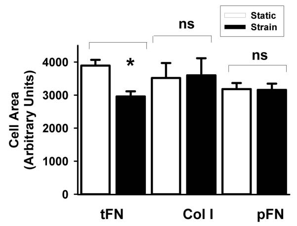
(B) Caco-2 cells were allowed to adhere to tissue fibronectin, collagen I or plasma fibronectin for one hour. Then, unattached cells were washed away and the attached cells were maintained under static conditions (open bars) or subjected to repetitive deformation (shaded bars) for 2 hours to allow cell spreading prior to image quantitation of cell areas. Repetitive deformation reduced cell areas compared to static control cells on tissue fibronectin (n=400, P<0.005). Repetitive deformation did not change cell size compared to unstretched control cells on either collagen I or plasma fibronectin substrates (n=400, P=n.s.).
Effect of repetitive deformation on cell spreading on tissue fibronectin, collagen I and plasma fibronectin
Repetitive deformation significantly decreased Caco-2 cell spreading on tissue fibronectin substrates compared with control cells on the same matrix not subject to repetitive deformation. (Figure 1B, n=400, p<0.005). In contrast, repetitive deformation did not significantly alter cell spreading on collagen I (Figure 1B, n=400, p=n.s.).We further studied the effect of repetitive deformation on cell spreading across a plasma fibronectin substrate. Repetitive deformation did not significantly alter cell spreading across plasma fibronectin in comparison to unstretched control cells (Figure 1C, n=400 p=n.s.).
Effect of Src inhibition on cell spreading on collagen type I
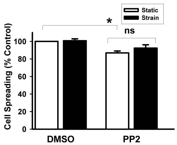
We have previously observed that repetitive deformation activates Src in intestinal epithelial cells cultured on type I collagen substrates and that Src-inhibition blocks the stimulation of cell proliferation on collagen I by repetitive deformation (11). We further assessed the effect of Src blockade by PP2 on cell spreading on type I collagen in the setting of repetitive deformation. Src-inhibition by PP2 reduced basal cell spreading on collagen I in the absence of deformation by 14±3% (Figure 2A, n=400, p<0.005), and no further decrease in cell spreading was observed in response to deformation in Src-blocked cells.
Figure 2. Effect of Src inhibition by PP2 on cell spreading and motility across collagen I.
(A) Caco-2 cells were pretreated with a DMSO vehicle control or 10μM PP2 for 60 minutes prior to incubation without (open bars) or with (shaded bars) repetitive deformation for 2 hours. PP2 and repetitive deformation each reduced cell area in comparison to unstretched static control cells treated with the DMSO vehicle control (n=400, P<0.005). No further decrease was observed in cells subjected to both PP2 and repetitive deformation.
(B) Circular wounds were created in Caco-2 monolayers on collagen I that had been pretreated with a DMSO vehicle control or 10μM PP2 for 60 minutes and cells were subjected to repetitive deformation (shaded bars) for 24 hours or maintained under control conditions without deformation (open bars). PP2 and repetitive deformation each reduced migration compared to to unstretched cells treated with the DMSO vehicle control (n=15, p<0.05). A further decrease in cell migration was observed in cells subjected to both PP2 and repetitive deformation (n=15, p<0.05).
Effect of Src inhibition on cell motility on collagen type I
We further assessed the effect of Src blockade by PP2 on cell motility across type I collagen in the setting of repetitive deformation. Src-inhibition by PP2 reduced basal cell migration on collagen I in the absence of deformation (Figure 2B, n=15, p<0.05). However, in contrast to our observations on cell spreading on collagen I, repetitive deformation further inhibited cell migration even in Src-blocked cells (Figure 2B, n=15, p<0.05).
Effect of Src inhibition and Src reduction on cell spreading on tissue fibronectin
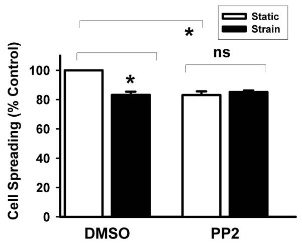
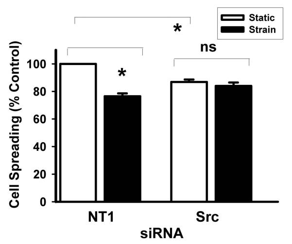
We have previously observed repetitive deformation activates Src in intestinal epithelial cells cultured on fibronectin substrates and that Src-inhibition blocks the stimulation of motility across fibronectin by repetitive deformation (11). We further assessed the effect of Src blockade by PP2 or Src reduction by specific siRNAs targeted to Src on cell spreading across tissue fibronectin in the setting of repetitive deformation. Src-inhibition by PP2 reduced basal cell spreading on tissue fibronectin in the absence of deformation by 17±2% (Figure 3A, n=400, p<0.005), and no further decrease in cell spreading was observed in response to deformation in Src-blocked cells. We performed further studies with siRNA targeted to Src confirm these results. Transfection with siRNA targeted to Src reduced Src protein levels by 70% (not shown). In the absence of repetitive deformation, Src reduction by specific Src-targeted siRNAs reduced cell spreading on tissue fibronectin (13±2%, Figure 3B, n=400, p<0.005) similarly to the reduction induced by PP2 in the absence of deformation. Moreover, as in the case of Src blockade by PP2, repetitive deformation did not induce any further significant reduction in cell spreading in Src-reduced cells.
Figure 3. Effect of Src inhibition with PP2 and Src reduction with siRNA targeted to Src on cell spreading across tissue fibronectin.
(A) Cao-2 cells were pretreated with 10μM PP2 or vehicle control (DMSO) for 60 minutes and then cells were subjected to repetitive deformation (shaded bars) for 2 hours or maintained under control conditions without deformation (open bars). PP2 and repetitive deformation each resulted in reduced cell areas in comparison to unstretched static control cells treated with the DMSO vehicle control (n=400, P<0.005). No further decrease was observed in cells subjected to both PP2 and repetitive deformation.
(B) Caco-2 cells transfected with non-targeting (NT1) human siRNA or siRNA targeted to human Src for 72 hours on tissue fibronectin. Cells were then trypsinized and replated onto tissue fibronectin for 60 minutes, nonadherent cells were washed away, the remaining adherent cells were then subjected to repetitive deformation (shaded bars) for 2 hours or maintained under control conditions without deformation (open bars). Repetitive deformation reduced cell areas in NT1-transfected cells, as did Src reduction by siRNA in the absence of repetitive deformation in comparison to unstretched static control cells transfected with NT1 siRNA (n=400, P<0.005). However, no further decrease was observed in Src-reduced cells that were then subjected to repetitive deformation.
Discussion
The intestine is subject to constant injuries even during normal gut function which must be closed by intestinal epithelial migration, sometimes followed by proliferation, in order to maintain the mucosal barrier (15). We have previously demonstrated that repetitive deformation can stimulate or inhibit intestinal epithelial migration in vitro, depending upon the matrix substrate across which the migration occurs (5). Our present results demonstrate that the effects of deformation on cell motility occur independently of cell spreading, that Src activation by deformation is essential for the modulation of cell spreading by deformation independently of matrix, and that migration across tissue fibronectin is affected differently than migration across a substrate of plasma fibronectin.
After wounding, cells at the edge of a defect spread in the direction of migration, extending lamellipodia which provide an anchoring point for the traction that will move the epithelial sheet forward. Cell spreading generally precedes and parallels cell motility in response to many stimuli. We observed that the effects of repetitive deformation on cell spreading do not correlate with effects on motility. Epithelial sheet migration involves several different processes, including initial lamellipodial extension and spreading in the direction of the defect, the generation of traction force by the cellular myosin motor, release of the trailing edge of the cell from the underlying substrate, the transmittal of forces between the cells at the migrating edge and cells behind the migrating front via cell-cell contacts. Indeed, these cells behind the migrating front must themselves coordinately regulate the force of their substrate-binding interactions to permit further movement. This complex series of events might be influenced at any step along the way. However, cell spreading and cell motility often correlate, and it seemed an attractive hypothesis that strain and fibronectin regulate intestinal epithelial wound closure by influencing cell motility. In fact, our present results suggest that this does not occur, as the effects of deformation and matrix on spreading and motility do not parallel. We have previously described the stimulation of myosin light chain kinase phosphorylation in intestinal epithelial cells subjected to repetitive deformation (5), so the regulation of the myosin motor may be an attractive target for further mechanistic study beyond the scope of the current study, although these other alternatives also await investigation. Thus, the dissociation of motility and spreading observed here suggests that deformation influences motility primarily via this later step in migration rather than at the level of cell spreading. Others have also observed dissociation of cell spreading and motility. For instance, Griffith and colleagues reported that the overexpression of a tumor suppressor gene increases cell spreading on fibronectin and decreased cell motility (16). Recent evidence suggests that the myosin II isoforms IIA and IIB may differentially affect spreading and motility (17).
Interestingly, Src may contribute to each in a complex fashion. Although we have previously reported that Src is required for deformation to promote migration across tissue fibronectin (11), our present results extend this finding to demonstrate that Src is not required for the inhibition of migration by deformation across a collagen substrate. In contrast, Src blockade did inhibit the effects of deformation on cell spreading independently of matrix. In this manuscript, we inhibited Src with 10μM PP2. Although PP2 has previously been reported to inhibit Src at concentrations as low as 100 nM (18), this was in a cell free in vitro system of purified proteins. In contrast, the 10μM concentration used here was consistent with that used by our group (10, 11, 19-22) and others (23-27) in intact cells. Moreover, we observed similar effects on cell spreading when Src was specifically reduced by siRNA and have previously reported that siRNA to Src yields similar results to PP2 at this concentration with regard to other endpoints like proliferation, adhesion, migration and differentitations (10, 11, 19, 24-27). Thus, the effects reported here of pharmacological Src blockade seem less likely to reflect non-specific off-target effects than specific inhibition of Src. The disparities between the matrix-dependence of the requirement for Src in the modulation of migration by deformation and the matrix-independence of the regulation of cell spreading by deformation further emphasize that the effects of deformation on cell motility are not simply effects on cell spreading.
We have previously reported that both plasma and tissue fibronectin can inhibit the mitogenic effect of strain on intestinal epithelial cells (7), while strain stimulates intestinal epithelial migration across a tissue fibronectin substrate (5). Our present results further demonstrate that plasma fibronectin does not support the motogenic effect of strain. Since we have previously reported that deformation affects cells on collagen I, collagen IV, and laminin similarly (7), this suggests that tissue fibronectin may uniquely facilitate the promotion of intestinal epithelial wound closure by repetitive deformation. Tissue fibronectin is highly homologous to plasma fibronectin, but includes an extra 20 kD domain at the C-terminal end of the molecule, containing Arg-Gly-Asp (RGD) like sequences that represent consensus sites for integrin-binding (28). The promotion of motility and inhibition of spreading on tissue fibronectin by repetitive deformation may therefore involve adhesion to this extra 20 kD domain, absent in pFN.
Tissue fibronectin is deposited into the gut mucosa in settings of chronic inflammation and injury such as Crohn’s disease, ulcerative colitis, or other mucosal ulcerations (29-33). Plasma fibronectin is elevated systemically in the setting of acute illness (34, 35). Thus, while elevated plasma fibronectin generalized stress suppresses the mitogenic actions of strain, only localized deposition of tissue fibronectin at the site of injury can invoke the motogenic effect required to promote mucosal healing. This would seem teleologically appropriate, since there would be no reason to further promote intestinal epithelial migration in the absence of or at sites distant from mucosal injury. Although it is conceivable that tissue fibronectin levels might in the future be specifically manipulated at the site of an injury by some localized molecular technique to invoke this response, tissue fibronectin levels are already substantially increased at sites of chronic injury. Thus, it may be more important to understand the downstream mechanisms by which deformation and fibronectin together promote mucosal healing so that these can be further supplemented pharmacologically or by molecular techniques in the future. This will require a more detailed understanding of these downstream signaling mechanisms, a major target of ongoing (36) and future investigations beyond the scope of the current manuscript. However, the current study certainly suggests that such mechanistic studies will be more profitably directed at the regulation of intestinal epithelial cell motility itself by strain and fibronectin than at the regulation of cell spreading.
Clearly these in vitro studies await in vivo validation. We have previously reported that repetitive deformation activates tyrosine kinase signaling within the gut mucosa (37) and that the chronic pathological pressure of a partial small bowel obstruction can inhibit mucosal healing (38). Others have shown that chronic constant strain can accelerate intestinal epithelial proliferation (39). However, although these studies certainly suggest that physical forces can modulate intestinal epithelial cell biology, it is more difficult to specifically examine cell spreading and movement in the intact gut in vivo during repetitive deformation. Nevertheless, these studies raise the possibility that further delineation of the mechanisms by which strain disparately modulates spreading and motility may permit acceleration of mucosal healing by targeted interventions to separately promote spreading and subsequent epithelial cell movement.
Acknowledgments
Author LS Chaturvedi and SA Saad have equal contributions for the first authorship (*). This research work was supported in part by a Veterans Affairs Merit Research Award and by National Institute of Diabetes and Digestive and Kidney Diseases Grant RO1-DK-067257 to MDB.
Supported in part by a VA MERIT Award and NIH RO1 DK067257 (MDB).
Footnotes
Publisher's Disclaimer: This is a PDF file of an unedited manuscript that has been accepted for publication. As a service to our customers we are providing this early version of the manuscript. The manuscript will undergo copyediting, typesetting, and review of the resulting proof before it is published in its final citable form. Please note that during the production process errors may be discovered which could affect the content, and all legal disclaimers that apply to the journal pertain.
Reference
- 1.Hennig GW, Costa M, Chen BN, Brookes SJ. Quantitative analysis of peristalsis in the guinea-pig small intestine using spatio-temporal maps. J Physiol. 1999;517(Pt 2):575–590. doi: 10.1111/j.1469-7793.1999.0575t.x. [DOI] [PMC free article] [PubMed] [Google Scholar]
- 2.Storkholm JH, Zhao J, Villadsen GE, Hager H, Jensen SL, Gregersen H. Biomechanical remodeling of the chronically obstructed Guinea pig small intestine. Dig Dis Sci. 2007;52:336–346. doi: 10.1007/s10620-006-9431-7. [DOI] [PubMed] [Google Scholar]
- 3.Friedman HI, Cardell RR., Jr. Alterations in the endoplasmic reticulum and Golgi complex of intestinal epithelial cells during fat absorption and after termination of this process: a morphological and morphometric study. Anat Rec. 1977;188:77–101. doi: 10.1002/ar.1091880109. [DOI] [PubMed] [Google Scholar]
- 4.Womack WA, Barrowman JA, Graham WH, Benoit JN, Kvietys PR, Granger DN. Quantitative assessment of villous motility. Am J Physiol. 1987;252:G250–256. doi: 10.1152/ajpgi.1987.252.2.G250. [DOI] [PubMed] [Google Scholar]
- 5.Zhang J, Owen CR, Sanders MA, Turner JR, Basson MD. The motogenic effects of cyclic mechanical strain on intestinal epithelial monolayer wound closure are matrix dependent. Gastroenterology. 2006;131:1179–1189. doi: 10.1053/j.gastro.2006.08.007. [DOI] [PubMed] [Google Scholar]
- 6.Basson MD, Li GD, Hong F, Han O, Sumpio BE. Amplitude-dependent modulation of brush border enzymes and proliferation by cyclic strain in human intestinal Caco-2 monolayers. J Cell Physiol. 1996;168:476–488. doi: 10.1002/(SICI)1097-4652(199608)168:2<476::AID-JCP26>3.0.CO;2-#. [DOI] [PubMed] [Google Scholar]
- 7.Zhang J, Li W, Sanders MA, Sumpio BE, Panja A, Basson MD. Regulation of the intestinal epithelial response to cyclic strain by extracellular matrix proteins. FASEB J. 2003;17:926–928. doi: 10.1096/fj.02-0663fje. [DOI] [PubMed] [Google Scholar]
- 8.Wilson AJ, Gibson PR. Epithelial migration in the colon: filling in the gaps. Clin Sci (Lond) 1997;93:97–108. doi: 10.1042/cs0930097. [DOI] [PubMed] [Google Scholar]
- 9.Casanova JE. Epithelial cell cytoskeleton and intracellular trafficking V. Confluence of membrane trafficking and motility in epithelial cell models. Am J Physiol Gastrointest Liver Physiol. 2002;283:G1015–1019. doi: 10.1152/ajpgi.00255.2002. [DOI] [PubMed] [Google Scholar]
- 10.Chaturvedi LS, Marsh HM, Shang X, Zheng Y, Basson MD. Repetitive deformation activates focal adhesion kinase and ERK mitogenic signals in human Caco-2 intestinal epithelial cells through Src and Rac1. J Biol Chem. 2007;282:14–28. doi: 10.1074/jbc.M605817200. [DOI] [PubMed] [Google Scholar]
- 11.Chaturvedi LS, Gayer CP, Marsh HM, Basson MD. Repetitive deformation activates Src-independent FAK-dependent ERK motogenic signals in human Caco-2 intestinal epithelial cells. Am J Physiol Cell Physiol. 2008;294:C1350–1361. doi: 10.1152/ajpcell.00027.2008. [DOI] [PMC free article] [PubMed] [Google Scholar]
- 12.Peterson MD, Mooseker MS. Characterization of the enterocyte-like brush border cytoskeleton of the C2BBe clones of the human intestinal cell line, Caco-2. J Cell Sci. 1992;102(Pt 3):581–600. doi: 10.1242/jcs.102.3.581. [DOI] [PubMed] [Google Scholar]
- 13.Basson MD, Turowski G, Emenaker NJ. Regulation of human (Caco-2) intestinal epithelial cell differentiation by extracellular matrix proteins. Exp Cell Res. 1996;225:301–305. doi: 10.1006/excr.1996.0180. [DOI] [PubMed] [Google Scholar]
- 14.Lotz MM, Rabinovitz I, Mercurio AM. Intestinal restitution: progression of actin cytoskeleton rearrangements and integrin function in a model of epithelial wound healing. Am J Pathol. 2000;156:985–996. doi: 10.1016/S0002-9440(10)64966-8. [DOI] [PMC free article] [PubMed] [Google Scholar]
- 15.McNeil PL, Ito S. Gastrointestinal cell plasma membrane wounding and resealing in vivo. Gastroenterology. 1989;96:1238–1248. doi: 10.1016/s0016-5085(89)80010-1. [DOI] [PubMed] [Google Scholar]
- 16.Griffith E, Coutts AS, Black DM. Characterisation of chicken TES and its role in cell spreading and motility. Cell Motil Cytoskeleton. 2004;57:133–142. doi: 10.1002/cm.10162. [DOI] [PubMed] [Google Scholar]
- 17.Betapudi V, Licate LS, Egelhoff TT. Distinct roles of nonmuscle myosin II isoforms in the regulation of MDA-MB-231 breast cancer cell spreading and migration. Cancer Res. 2006;66:4725–4733. doi: 10.1158/0008-5472.CAN-05-4236. [DOI] [PubMed] [Google Scholar]
- 18.Karni R, Mizrachi S, Reiss-Sklan E, Gazit A, Livnah O, Levitzki A. The pp60c-Src inhibitor PP1 is non-competitive against ATP. FEBS Lett. 2003;537:47–52. doi: 10.1016/s0014-5793(03)00069-3. [DOI] [PubMed] [Google Scholar]
- 19.Chaturvedi LS, Marsh HM, Basson MD. Src and focal adhesion kinase mediate mechanical strain-induced proliferation and ERK1/2 phosphorylation in human H441 pulmonary epithelial cells. Am J Physiol Cell Physiol. 2007;292:C1701–1713. doi: 10.1152/ajpcell.00529.2006. [DOI] [PubMed] [Google Scholar]
- 20.Thamilselvan V, Craig DH, Basson MD. FAK association with multiple signal proteins mediates pressure-induced colon cancer cell adhesion via a Src-dependent PI3K/Akt pathway. FASEB J. 2007;21:1730–1741. doi: 10.1096/fj.06-6545com. [DOI] [PubMed] [Google Scholar]
- 21.Sanders MA, Basson MD. Collagen IV regulates Caco-2 migration and ERK activation via alpha1beta1- and alpha2beta1-integrin-dependent Src kinase activation. Am J Physiol Gastrointest Liver Physiol. 2004;286:G547–557. doi: 10.1152/ajpgi.00262.2003. [DOI] [PubMed] [Google Scholar]
- 22.Thamilselvan V, Basson MD. Pressure activates colon cancer cell adhesion by inside-out focal adhesion complex and actin cytoskeletal signaling. Gastroenterology. 2004;126:8–18. doi: 10.1053/j.gastro.2003.10.078. [DOI] [PubMed] [Google Scholar]
- 23.Salazar EP, Rozengurt E. Bombesin and platelet-derived growth factor induce association of endogenous focal adhesion kinase with Src in intact Swiss 3T3 cells. J Biol Chem. 1999;274:28371–28378. doi: 10.1074/jbc.274.40.28371. [DOI] [PubMed] [Google Scholar]
- 24.Salsmann A, Schaffner-Reckinger E, Kabile F, Plancon S, Kieffer N. A new functional role of the fibrinogen RGD motif as the molecular switch that selectively triggers integrin alphaIIbbeta3-dependent RhoA activation during cell spreading. J Biol Chem. 2005;280:33610–33619. doi: 10.1074/jbc.M500146200. [DOI] [PubMed] [Google Scholar]
- 25.Hopkins AM, Bruewer M, Brown GT, Pineda AA, Ha JJ, Winfree LM, Walsh SV, Babbin BA, Nusrat A. Epithelial cell spreading induced by hepatocyte growth factor influences paxillin protein synthesis and posttranslational modification. Am J Physiol Gastrointest Liver Physiol. 2004;287:G886–898. doi: 10.1152/ajpgi.00065.2004. [DOI] [PubMed] [Google Scholar]
- 26.Di Florio A, Capurso G, Milione M, Panzuto F, Geremia R, Delle Fave G, Sette C. Src family kinase activity regulates adhesion, spreading and migration of pancreatic endocrine tumour cells. Endocr Relat Cancer. 2007;14:111–124. doi: 10.1677/erc.1.01318. [DOI] [PubMed] [Google Scholar]
- 27.Daoud G, Le bellego F, Lafond J. PP2 regulates human trophoblast cells differentiation by activating p38 and ERK1/2 and inhibiting FAK activation. Placenta. 2008;29:862–870. doi: 10.1016/j.placenta.2008.07.011. [DOI] [PubMed] [Google Scholar]
- 28.Kornblihtt AR, Vibe-Pedersen K, Baralle FE. Human fibronectin: molecular cloning evidence for two mRNA species differing by an internal segment coding for a structural domain. EMBO J. 1984;3:221–226. doi: 10.1002/j.1460-2075.1984.tb01787.x. [DOI] [PMC free article] [PubMed] [Google Scholar]
- 29.Yagi K. The immunohistochemical study on the recurrent mechanism of acetic acid induced gastric ulcer in rats—extracellular matrix and cellular kinetics. Nippon Shokakibyo Gakkai Zasshi. 1992;89:1155–1164. [PubMed] [Google Scholar]
- 30.Mori N, Doi Y, Hara K, Yoshizuka M, Ohsato K, Fujimoto S. Role of multipotent fibroblasts in the healing colonic mucosa of rabbits. Ultrastructural and immunocytochemical study. Histol Histopathol. 1992;7:583–590. [PubMed] [Google Scholar]
- 31.Tominaga K, Higuchi K, Watanabe T, Fujiwara Y, Kim S, Arakawa T, Iwao H, Kuroki T. Expression of gene for EIIIA- and EIIIB- fibronectin, fetal types of fibronectin, during gastric ulcer healing in rats. Dig Dis Sci. 2001;46:311–317. doi: 10.1023/a:1005541305255. [DOI] [PubMed] [Google Scholar]
- 32.Lacy ER. Rapid epithelial restitution in the stomach: an updated perspective. Scand J Gastroenterol Suppl. 1995;210:6–8. doi: 10.3109/00365529509090260. [DOI] [PubMed] [Google Scholar]
- 33.Ernst H, Grunert S, Schneider HT, Beck WS, Brune K, Hahn EG. Distribution of extracellular matrix proteins in indomethacin-induced lesions in the rat stomach. Scand J Gastroenterol. 1995;30:847–853. doi: 10.3109/00365529509101590. [DOI] [PubMed] [Google Scholar]
- 34.Allan A, Wyke J, Allan RN, Morel P, Robinson M, Scott DL, Alexander-Williams J. Plasma fibronectin in Crohn’s disease. Gut. 1989;30:627–633. doi: 10.1136/gut.30.5.627. [DOI] [PMC free article] [PubMed] [Google Scholar]
- 35.Verspaget HW, Biemond I, Allaart CF, van Weede H, Weterman IT, Gooszen HG, Pena AS, Lamers CB. Assessment of plasma fibronectin in Crohn’s disease. Hepatogastroenterology. 1991;38:231–234. [PubMed] [Google Scholar]
- 36.Gayer CP, Basson MD. The effects of mechanical forces on intestinal physiology and pathology. Cell Signal. 2009 doi: 10.1016/j.cellsig.2009.02.011. [DOI] [PMC free article] [PubMed] [Google Scholar]
- 37.Basson MD, Coppola CP. Repetitive deformation and pressure activate small bowel and colonic mucosal tyrosine kinase activity in vivo. Metabolism. 2002;51:1525–1527. doi: 10.1053/meta.2002.36303. [DOI] [PubMed] [Google Scholar]
- 38.Flanigan TL, Owen CR, Gayer C, Basson MD. Supraphysiologic extracellular pressure inhibits intestinal epithelial wound healing independently of luminal nutrient flow. Am J Surg. 2008;196:683–689. doi: 10.1016/j.amjsurg.2008.07.016. [DOI] [PMC free article] [PubMed] [Google Scholar]
- 39.Spencer AU, Sun X, El-Sawaf M, Haxhija EQ, Brei D, Luntz J, Yang H, Teitelbaum DH. Enterogenesis in a clinically feasible model of mechanical small-bowel lengthening. Surgery. 2006;140:212–220. doi: 10.1016/j.surg.2006.03.005. [DOI] [PMC free article] [PubMed] [Google Scholar]


