Abstract
Human lymphatic filariasis is a mosquito-vectored disease caused by the nematode parasites Wuchereria bancrofti, Brugia malayi and Brugia timori. These are relatively large roundworms that can cause considerable damage in compatible mosquito vectors. In order to assess how mosquitoes respond to infection in compatible mosquito-filarial worm associations, microarray analysis was used to evaluate transcriptome changes in Aedes aegypti at various times during B. malayi development. Changes in transcript abundance in response to the different stages of B. malayi infection were diverse. At the early stages of midgut and thoracic muscle cell penetration, a greater number of genes were repressed compared to those that were induced (20 vs. 8). The non-feeding, intracellular first-stage larvae elicited few differences, with 4 transcripts showing an increased and 9 a decreased abundance relative to controls. Several cecropin transcripts increased in abundance after parasites molted to second-stage larvae. However, the greatest number of transcripts changed in abundance after larvae molted to third-stage larvae and migrated to the head and proboscis (120 induced, 38 repressed), including a large number of putative, immunity-related genes (∼13% of genes with predicted functions). To test whether the innate immune system of mosquitoes was capable of modulating permissiveness to the parasite, we activated the Toll and Imd pathway controlled rel family transcription factors Rel1 and Rel2 (by RNA interference knockdown of the pathway's negative regulators Cactus and Caspar) during the early stages of infection with B. malayi. The activation of either of these immune signaling pathways, or knockdown of the Toll pathway, did not affect B. malayi in Ae. aegypti. The possibility of LF parasites evading mosquito immune responses during successful development is discussed.
Author Summary
Filarial worms that cause human lymphatic filariasis (LF) are transmitted by many species of mosquitoes. Within susceptible mosquitoes, Brugia malayi develop from microfilariae (mf) to infective-stage larvae (L3s), in approximately eight days. These nematodes develop as intracellular parasites within mosquito flight muscle cells, in which they ingest cellular material and eventually cause cell death when L3s migrate to the mosquito's proboscis. We examined the effects of B. malayi parasitism on Aedes aegypti by analyzing changes in mosquito gene expression at different stages of parasite development. We found that a few genes were differentially expressed at the RNA level relative to non-infected controls. The majority of changes occurred at two time periods, when the filarial worms began feeding and when the L3s were in the head and proboscis. Many transcriptional changes in the later group concur with documented descriptions of tissue damage, clean-up and repair that occurs in mosquitoes infected with filarial worms. In addition, we activated two innate immunity signaling pathways and observed the effects on filarial worm development. B. malayi seems to be capable of evading these immune responses, because its development was not impeded by the activation of either the Toll or Imd signal pathways in Ae. aegypti.
Introduction
It is estimated that 120 million people are infected with Wuchereria bancrofti, Brugia malayi, or B. timori, the mosquito-transmitted, parasitic nematodes that cause human lymphatic filariasis (LF). In approximately 40% of cases, the disease is manifested by lymphedema of the extremities or hydrocoele. Although human LF does not increase mortality in endemic areas, morbidity causes major economic losses and often leads to psychosocial and psychosexual conditions in infected individuals [1]. Recent efforts by the Global Program for the Elimination of Lymphatic Filariasis (GPELF) have decreased the numbers of individuals infected with, and at risk for, this parasitic disease [2].
Several different mosquito species within the genera Culex, Anopheles, Aedes and Mansonia can serve as primary vectors of LF parasites. The geographical location and habitat type influence which mosquito species function as vectors in any particular endemic area. Biological transmission of filarial worms is termed cyclodevelopmental, i.e., the parasite undergoes development within the vector to become infective to the vertebrate host, but does not multiply. In competent vectors, microfilariae (mf), produced by adult female worms and found circulating in the peripheral blood, are ingested with a blood meal and will quickly (within 2 hr) penetrate the midgut epithelium to access the hemocoel [3]. Mf migrate in the mosquito's hemolymph to reach the thoracic musculature and from there penetrate into the indirect flight muscles. This tissue is the site of development, where mf undergo two molts and emerge as infective-stage larvae (L3s). Approximately eight days after exposure, L3s migrate to the head and proboscis from where they escape by penetrating the labellum of the proboscis when the mosquito takes a blood meal. Within the human host, the parasites undergo two additional molts and grow as they migrate to lymphatic vessels where adult male and female worms mate and females give birth to mf. Mf then make their way into the circulating blood from where they can be ingested by another blood feeding mosquito.
LF parasites grow nearly seven times in length (B. malayi grow from ∼200 to ∼1,350 µm in length, from mf to L3s respectively) during the extrinsic developmental period within the mosquito [4]. As parasites develop, the mosquito must tolerate a series of insults due to parasite activities, e.g., migrating mf damage both midgut [5] and muscle cells as they penetrate through or into them; second-stage larvae (L2s) actively ingest mosquito cellular components; L3s are very large and migrate out of the thoracic muscles and through the body cavity to reach the head and proboscis. Ultrastructural studies of Aedes mosquitoes infected with Brugia parasites have revealed that nuclear enlargement (a sign of a putative repair response) occurs in both Brugia-infected and neighboring non-infected muscle cells, and that complete degeneration of infected muscle cells occurs once L3s exit the flight muscles [6]. Other studies have shown that mosquito flight muscle cells become devoid of glycogen granules following infection with Brugia parasites [7],[8]. Considering the amount of tissue damage observed in muscle cells, it is not surprising that Brugia-infected mosquitoes are known to have decreased flight activity and longevity [9],[10]. But the successful development (and subsequent transmission) of LF parasites depends on the ability of competent mosquito vectors to survive infection.
Some mosquitoes are able to limit or prevent filarial worm infections with various refractory or resistance mechanisms. For example, mf can be damaged during ingestion by an armed pharyngeal and/or cibarial pump (often found in Anopheles spp.), inhibiting them from penetrating the midgut wall [11],[12]. Mf that successfully penetrate the midgut and enter the hemocoel come in contact with hemolymph components, including circulating blood cells called hemocytes. Melanotic encapsulation is a hemocyte-mediated, innate immune response that can be very specific and robust, and can limit or prevent parasite development in some mosquito species [13]. In contrast, the involvement of a humoral immune response is not well understood in many compatible filarial worm-mosquito systems. Some parasites are able to evade or suppress a host's immune system in order to survive, but it is unknown if such interactions occur between LF parasites and mosquitoes [14].
Previous studies have investigated the effect of an activated mosquito immune response on filarial worm development, but the results remain inconclusive. When bacteria are inoculated into the mosquito hemocoel, to induce the expression of infection responsive immune factors prior to filarial worm exposure, a reduced B. malayi prevalence and mean intensity in Ae. aegypti was observed as compared to non-inoculated controls [15]. However, when the same bacterial strains and inoculation procedures were used to pre-activate the immune response of Culex pipiens prior to W. bancrofti exposure, there was no difference in prevalence or mean intensity between bacteria-inoculated and control mosquitoes [16]. Further investigation is needed to assess the effects of an activated mosquito immune response on a LF parasite infection.
The role of Toll and Imd signaling pathways in the immune recognition, modulation, and response of mosquitoes to LF parasites has yet to be examined. Recently, Xi et al. [17] developed methods to manipulate these immune signaling pathways in Ae. aegypti by (1) gene silencing of Cactus, a negative regulator of the Toll pathway, (2) gene silencing of MyD88, an adaptor required for endogenous Toll pathway signal transduction, and (3) gene silencing of Caspar, a negative regulator of the Imd pathway. Using these tools, the Toll and Imd pathways are transiently relieved from endogenous suppression (1 and 3 above) or made unresponsive to detected stimuli (2 above).
In this study we assess transcriptome changes associated with the development of B. malayi in Ae. aegypti and investigate the effect of the immune signaling pathways, Toll and Imd, on parasite development.
Materials and Methods
All animals were handled in strict accordance with good animal practice as defined by the University of Wisconsin-Madison School of Veterinary Medicine/University of Wisconsin Research Animal Resource Center, and all animal work was approved by these entities.
Mosquito Maintenance
Aedes aegypti black-eyed, Liverpool (LVP) strain used in this study were maintained at the University of Wisconsin-Madison as previously described [18]. Briefly, mosquitoes were maintained on 0.3 M sucrose in an environmental chamber at 26.5±1°C, 75±10% RH, and with a 16 hr light and 8 hr dark photoperiod with a 90 minute crepuscular period at the beginning and end of each light period. Ae. aegypti LVP was originally selected for susceptibility to Brugia malayi by Macdonald in 1962. This strain supports the development of mf to L3s. Four- to five-day-old mosquitoes were sucrose starved for 14 to 16 hours prior to blood feeding.
Brugia malayi Infections
Mosquitoes were exposed to B. malayi (originally obtained from the University of Georgia NIH/NIAD Filariasis Research Reagent Repository Center) by feeding on ketamine/xylazine anesthetized, dark-clawed Mongolian gerbils, Meriones unguiculatus. The same animals were used for all three biological replicates. Microfilaremias were determined, using blood from orbital punctures, immediately before each feeding and ranged from 50–150 mf per 20 µl of blood. Control mosquitoes were exposed to anesthetized, uninfected gerbils. Mosquitoes that fed to repletion were separated into cartons and maintained on 0.3 M sucrose in the laboratory.
Mosquito Dissections
In early stages of development (1 h to 3 d post-infection [PI]), individual mosquitoes were separated into midgut, thorax and abdomen (with midgut removed) and dissected in Aedes saline [19]. Tissue dissections were cover-slipped and parasites were observed with a compound microscope using phase-contrast optics. The same procedure was used for dissections at 5–6 d PI, except only thoraces were dissected and examined. At 8–9 d PI, the thorax was processed as described above, but the abdomen and head & proboscis were dissected in separate drops of Aedes saline to observe L3s using a dissection microscope. Individual mosquitoes were dissected in a drop of Aedes saline for the recovery of L3s at 13–14 d PI. Images of B. malayi developmental stages were captured and processed as previously described, with the addition of Nomarski optics [20].
Mosquito Collection
Five sample groups were created to study the transcriptional response of mosquitoes to B. malayi development. In each group, 20 mosquitoes were collected for RNA extraction. These sample groups are defined by the time after the blood meal and represent significantly different stages of parasite development. Briefly, Group 1 consisted of mosquitoes collected at 1, 6, 12 and 24 h PI. At these early time points, mf are penetrating the mosquito midgut, migrating through the hemocoel and penetrating thoracic muscle cells. Group 2 was collected at 2–3 d PI, a time when mf have differentiated into intracellular first-stage larvae (L1s). At 5–6 d PI, B. malayi complete the molt to second-stage larvae (L2s) and actively feed on mosquito muscle tissue (Group 3). In Group 4, at 8–9 d PI, parasite development is complete with a second molt to the L3s. Tissue damage continues as L3s break out of the thoracic muscles and migrate to the mosquito's head and proboscis. The final collection (Group 5), made at 13–14 d PI, occurs when the majority of L3s are located in the head and proboscis (see [4],[21]). Five mosquitoes, from both B. malayi-infected and uninfected blood meals, were collected at 1, 6, 12, and 24 h PI and ten mosquitoes at 2, 3, 5, 6, 8, 9, 13 and 14 d PI for transcriptional analysis. Mosquitoes were pooled (5 mosquitoes/tube), flash frozen in liquid nitrogen and stored at −80°C prior to total RNA extraction using RNeasy (QIAGEN). In addition, five B. malayi-infected mosquitoes were dissected to verify filarial worm infection and to determine the stage of parasite development at each time point. Three biological replications were completed.
Microarray Procedures and Analysis
Microarray assays were conducted and analyzed as reported previously [17],[22]. A full genome microarray platform (Agilent; 4×44k) was used with the probe sequences identical to the previous version (1×22k) [22]. In brief, 2–3 µg total RNA was used for probe synthesis of Cy3- and Cy5-labeled dCTP. Hybridizations were conducted with an Agilent Technologies In Situ Hybridization kit at 60°C according to the manufacturer's instructions. Three independent biological replicate assays were performed. Hybridization intensities were determined with an Axon GenePix 4200AL scanner, and images were analyzed with Gene Pix software. To produce the expression data, the background-subtracted median fluorescent values were normalized according to a LOWESS normalization method to reduce dye-specific biases, and Cy5/Cy3 ratios from replicate assays were subjected to t-tests at a significance level of p<0.05 using TIGR MIDAS and MeV software [23]. Expression data from all replicate assays were averaged with the GEPAS microarray preprocessing software prior to logarithm (base 2) transformation. Self-self hybridizations have been used to determine the cut-off value for the significance of gene abundance on these microarrays to 0.8 in log2 scale, which corresponds to 1.74- fold regulation [22]. For genes with P<0.05, the average ratio was used as the final fold change; for genes with P>0.05, the inconsistent probes (with distance to the median of replicate probe ratios larger than 0.8 log2) were removed, and only the value from a gene with at least two replicates was further averaged. The robustness of these microarray gene expression assays were validated through qPCR (Text S1).
Real-Time PCR Assays
Real time PCR assays were conducted as previously described to validate gene silencing efficiency and microarray expression data for selected genes [24]. Briefly, RNA samples were treated with Turbo DNAse (Ambion, Austin, Texas, United States) and reverse-transcribed using Superscript III (Invitrogen, Carlsbad, California, United States) with random hexamers. Transcript relative quantification was performed using the QuantiTect SYBR Green PCR kit (Qiagen) and ABI Detection System ABI Prism 7300 (Applied Biosystems, Foster City, California, United States). qRT-PCR reactions were conducted using a 10 minute step at 94°C and 40 cycles of 15 seconds at 94°C, 15 seconds at 55°C and 15 seconds at 72°C. Three independent biological replicates were conducted and all PCR reactions were performed in triplicate. Transcript abundance was normalized against the ribosomal protein S7 gene. All primers used for qPCR assay are presented in Table S1. The primer sequences used to verify gene knockdown efficiency included: S7 (AAEL009496-RA) forward: 5′-GGGACAAATCGGCCAGGCTATC-3′, reverse: 5′- TCGTGGACGCTTCTGCTTGTTG-3′; Caspar (AAEL003579-RA) forward: 5′-GAATCCGAGCGAGCCGATGC-3′, reverse: 5′-CGTAGTCCAGCGTTGTGAGGTC-3′; Cactus (AAEL000709-RA) forward: 5′-AGACAGCCGCACCTTCGATTCC-3′, reverse: 5′-CGCTTCGGTAGCCTCGTGGATC-3′; MyD88 (AAEL007768) forward: 5′-CATCCCATTCAGTTTCTCAGC-3′, reverse: 5′-ACCGGTTGGAAGTTCTGATG-3′. A complete list of PCR primer sequences is presented in Table S1.
Gene Silencing Assays
RNAi was conducted by intrathoracic injection of dsRNA using described methodology [24],[25]. Mosquitoes were three to four days old at the time of blood feeding and dsRNA was injected either 48 h before or after parasite exposure, i.e., dsRNA injections were performed on non-blood fed one- to two-day-old mosquitoes and on blood fed, five- to six-day-old mosquitoes. Approximately 0.5 µl of dsRNAs (1.0 or 0.5 µg/µl) were injected into the thorax of cold-anesthetized mosquitoes. The primers used to synthesize Cactus, Caspar and MyD88 dsRNA have been published previously [17]. To synthesize GFP dsRNA, methods described by Bartholomay et al. [25] were used with minor changes. The following sequences were annealed by heating at 95°C for 5 min and slow cooling: GFP_F 5′-TAGTACAACTACAACAGCCACAACGTCTATATCATGGCCGACAAGCAGA AGAACGGCATCAAGGTGAACTTCAAGATCCGCCACAACA-3′ and GFP_R: 5′-TCGATGTTGTGGCGGATCTTGAAGTTCACCTTGATGCCGTTCTTCTGCTTGTCGGCCATGATATAGACGTTGTGGCTGTTGTAGTTGTA-3′ (Integrated DNA Technologies, Inc., Coralville, IA). This dsDNA was ligated into pBlueScript KS+ (Stratagene) at XbaI (T7) and SalI (T3) sites. A complete list of PCR primer sequences is presented in Table S1.
Infection
B. malayi exposures were performed as described above and microfilaremias ranged from 35–162 mf per 20 µl of blood. Mosquitoes injected with GFP dsRNA were blood fed on each infected gerbil and used as a control for parasite infections. Mosquito mortality was observed every 24 h and mosquitoes were dissected at 6 d or 12–13 d PI to observe parasite development. To verify gene knockdown, five mosquitoes were collected 48 h post dsRNA injection, placed in a microcentrifuge tube, flash frozen and stored at −80°C until RNA extraction. Real-time PCR was used to quantify gene silencing efficiency. Silencing of Cactus, Caspar and MyD88 resulted in a reduction of mRNA levels by 60%, 84% and 27%, respectively. For each exposure, the prevalence and mean intensity of infection was calculated. Comparisons of mean intensities and mosquito mortality curves were done with the Mann-Whitney and Log-Rank tests, respectively, using GraphPad Prism 5 (GraphPad Software, Inc., La Jolla, CA). Results were considered significant at P≤0.05.
Results
Brugia malayi Development in Aedes aegypti
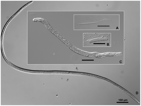
The development of B. malayi was observed at 1 h to 14 d PI for each biological replicate and is summarized in Table S2. Worms were recovered from 166 of the 180 mosquitoes examined for an overall infection prevalence of 92%. Mf were recovered from 1 h to 3 d PI, but after 24 h PI mf were no longer the most abundant developmental stage recovered. At 2 d PI, mf recovery began to decrease and almost all worms had differentiated into intracellular L1s by 3 d PI. Parasites molted to L2s in the thoracic musculature by 5 d PI, and were the only developmental stage identified in transcriptional group 3. The molt from L2 to L3 occurred at 8–9 d PI. At 8 d PI, L2s and L3s were recovered in the thorax, and only 4% of the worms were located in the head and proboscis. In contrast, at 9 d PI, both L2s and L3s were observed in the thorax region, but the majority were L3s and 47% of all recovered worms were located in the head and proboscis. By 13–14 d PI, all parasites had developed into L3s. Images of B. malayi development, from mf to L3s, are presented in Figure 1. The prevalence of L3s (for all three biological replicates at 13–14 d PI) was 80% (n = 30) and the mean intensity was 6.9±5.7.
Figure 1. Relative sizes of Brugia malayi developmental stages that occur within compatible mosquito hosts: An example of cyclo-developmental transmission.
A. microfilariae are ingested during blood feeding; B. parasites differentiate into non-feeding, first-stage larvae within mosquito indirect flight muscle cells; C. following the first molt, second-stage larvae remain intracellular parasites which ingest cellular material into their newly developed digestive tract. D. third-stage larvae leave the muscle cells and migrate to the mosquito's head and proboscis where they will exit through the mosquito cuticle during blood feeding.
Global Response of Ae. aegypti to B. malayi Infection
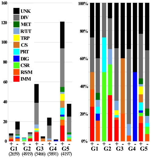
The Ae. aegypti global transcript responses to the successful development of B. malayi were determined using a genome microarray expression approach. These transcriptome infection-response patterns differed significantly, in both the number of regulated genes and their direction of regulation, at the different parasite development stages (Fig. 2A). A general suppression of transcription was evident during the early stages of infection (group 1); 20 genes were down-regulated while only 8 genes were up-regulated. However, this transcriptional suppression was reduced when parasites developed into L1s (group 2), and reversed when they became L2s (group 3); 59 genes were up-regulated and only 5 genes were down-regulated at this stage. Parasite infection at later stages of infection mainly caused transcriptional up-regulation (groups 4 and 5). Strikingly, 120 genes were up-regulated in group 5 that represented mosquitoes in which L3s had migrated to the head and proboscis from where they can be transmitted to a host upon blood feeding. As many as 158 mosquito genes were differentially expressed at this late stage. The second largest number of infection-responsive genes was observed when L2s were present (group 3).
Figure 2. Global transcriptional analysis of the response of a compatible mosquito infected with successfully developing Brugia malayi.
A. Regulated (differentially expressed above a 1.7-fold threshold) genes for each experimental group. (+) indicates up-regulated in infected mosquitoes and (−) indicates down-regulated. Assayable genes (those that gave signal intensities above a standard cutoff threshold) are indicated below the graph. Functional group distributions are the same to those used in Nene et al., 2007 and Xi et al., 2008: IMM: immunity, R/S/M: redox, stress, mitochondrial, CSR: chemosensory reception, DIG: blood digestive, PRT: proteolysis, C/S: cytoskeletal, structural, TRP: transport, R/T/T: replication, transcription, translation, MET: metabolism, DIV: diverse, UKN: unknown. B. Percent distribution of functional group for each up- and down-regulated experimental group. Transcription data are presented in Table S5.
Interestingly, the transcripts that were over represented in the groups that displayed the most prominent transcriptional regulation (3 and 5), were highly enriched with putative immune genes (Fig. 2). Specifically, several antimicrobial peptide effector genes were strongly induced in group 3 and a number of putative pattern recognition receptor and signal modulator genes were up-regulated in group 5 (Table 1). Group 5 also contained several serine protease cascade components and putative melanization –related factor transcripts in increased abundance (Tables 1 and 2).
Table 1. Cluster analysis of Aedes aegypti immunity genes transcribed by Brugia malayi-infected mosquitoes.
| Cluster | Vectorbase ID | Gene Name | Relative Transcript Abundancea | ||||
| Group 1 (Infection) | Group 2 (Non-feeding L1s) | Group 3 (Feeding L2s) | Group 4 (Molt to L3s) | Group 5 (Persistent L3s) | |||
| 1 | AAEL005748-RA | elastase, putative | 1.70 | ||||
| 1 | AAEL003857-RA | DEF D | 1.94 | ||||
| 2 | AAEL000598-RA | CEC D | 1.85 | ||||
| 2 | AAEL000611-RA | CEC E | 3.04 | ||||
| 2 | AAEL000625-RA | CEC F | −1.97 | 2.12 | |||
| 2 | AAEL000627-RA | CEC A | −1.89 | 2.43 | |||
| 2 | AAEL015515-RA | CEC G | 1.99 | ||||
| 2 | AAEL000621-RA | CEC N | 2.01 | ||||
| 2 | AAEL001929-RA | SPZ 5 | 2.11 | ||||
| 2 | AAEL012958-RA | conserved hypothetical protein | 1.89 | ||||
| 2 | AAEL010606-RA | down syndrome cell adhesion molecule | 1.77 | ||||
| 3 | AAEL003294-RA | FREP | 1.73 | ||||
| 3 | AAEL009178-RA | GNBPB4 | 1.68 | ||||
| 3 | AAEL014989-RA | peptidoglycan recognition protein-1, putative | 2.02 | ||||
| 3 | AAEL007037-RA | PGRPS4 | 1.86 | ||||
| 3 | AAEL005374-RA | SCRB1 | 1.96 | ||||
| 3 | AAEL014349-RA | CLIPSP | 1.72 | ||||
| 3 | AAEL005060-RA | CLIPSP | 2.24 | ||||
| 3 | AAEL005644-RA | CLIPSPH | 1.82 | ||||
| 3 | AAEL010267-RA | serine protease | 1.72 | ||||
| 3 | AAEL009558-RA | serine protease, putative | 1.80 | ||||
| 3 | AAEL010769-RA | SRPN6 | 1.74 | ||||
| 3 | AAEL004000-RA | TOLL10 | 1.75 | ||||
| 3 | AAEL002583-RA | TOLL7 | 1.70 | ||||
| 3 | AAEL013499-RA | PPO2 | 1.74 | ||||
Fold change, by Mosquito Transcriptional Group (Stage of Filarial Worm Development).
Table 2. Few Aedes aegypti genes are differentially transcribed in multiple stages of the infection response to Brugia malayi.
| Vectorbase ID | Description | Function | Relative Transcript Abundancea | ||||
| Group 1 (Infection) | Group 2 (Non-feeding L1s) | Group 3 (Feeding L2s) | Group 4 (Molt to L3s) | Group 5 (Persistent L3s) | |||
| AAEL002621-RA | hypothetical protein | DIV | 2.02 | 1.83 | |||
| AAEL003014-RA | hypothetical protein | DIV | 1.82 | 1.72 | |||
| AAEL003015-RA | protein phosphatase 2a, regulatory subunit | DIV | 1.87 | 1.76 | |||
| AAEL004706-RA | conserved hypothetical protein | DIV | −1.92 | 2.56 | |||
| AAEL006167-RA | runt | DIV | 2.10 | 1.73 | |||
| AAEL006567-RA | max binding protein, mnt | DIV | 1.75 | 1.71 | |||
| AAEL008194-RA | protein phosphatase 2a, regulatory subunit | DIV | 1.69 | 1.71 | |||
| AAEL009136-RA | hypothetical protein | DIV | 1.87 | 1.92 | |||
| AAEL009417-RA | hypothetical protein | DIV | −1.75 | 2.42 | 1.87 | ||
| AAEL013349-RA | lethal(2)essential for life protein, l2efl | DIV | 1.70 | 2.45 | |||
| AAEL000625-RA | CECF | IMM | −1.97 | 2.12 | |||
| AAEL000627-RA | CECA | IMM | −1.89 | 2.43 | |||
| AAEL015257-RA | hypothetical protein | PROT | 1.86 | 1.73 | |||
| AAEL013350-RA | heat shock protein 26 kD, putative | RSM | 2.00 | 2.44 | |||
| AAEL001912-RA | forkhead protein/forkhead protein domain | RTT | 1.96 | 1.82 | |||
| AAEL006944-RA | hypothetical protein | RTT | 1.71 | 1.70 | |||
| AAEL007166-RA | hypothetical protein | RTT | −2.95 | −2.04 | 2.30 | ||
| AAEL001392-RA | hypothetical protein | UNK | −3.06 | 1.79 | 1.98 | ||
| AAEL004149-RA | hypothetical protein | UNK | 2.03 | 1.70 | |||
| AAEL005233-RA | hypothetical protein | UNK | 2.28 | 1.69 | |||
| AAEL005265-RA | conserved hypothetical protein | UNK | 1.91 | 1.73 | |||
| AAEL007108-RA | hypothetical protein | UNK | 2.34 | 2.02 | |||
| AAEL009473-RA | conserved hypothetical protein | UNK | 2.22 | 1.68 | |||
| AAEL011351-RA | hypothetical protein | UNK | −1.89 | −2.02 | |||
| AAEL013178-RA | hypothetical protein | UNK | 1.80 | 1.78 | |||
| AAEL015259-RA | hypothetical protein | UNK | −1.82 | 1.75 | |||
Fold change, by Mosquito Transcriptional Group (Stage of Filarial Worm Development).
Effect of the Toll and Imd Pathways on B. malayi Development
To investigate whether the mosquito's two major immune signaling pathways, Toll and Imd, had any effect on B. malayi infection we used an established gene silencing approach to either simulate the activation of the Toll and Imd pathway, by depleting their negative regulators Cactus and Caspar, respectively, or inhibit the Toll pathway through the depletion of the MyD88 factor [17],[26].
Mosquito survival
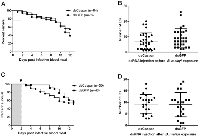
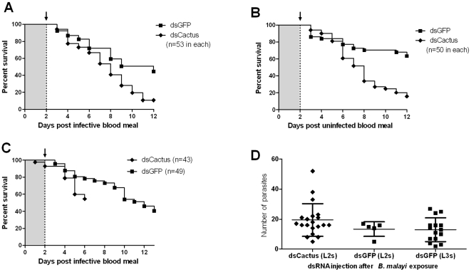
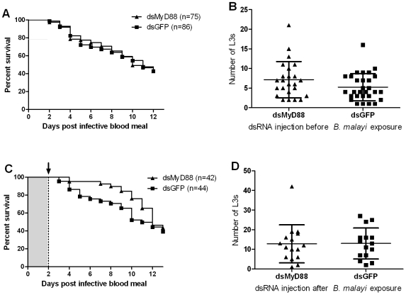
Caspar and MyD88 gene knockdown did not change the mortality rate in either pre- or post-bloodfed dsRNA-injected mosquito groups compared to their respective GFP dsRNA controls (Log-rank tests, P-values ranging from 0.23 to 0.41; see Fig. 3 and 4). In contrast, Cactus gene knockdown, that results in the activation of the Rel1 factor, resulted in a significantly increased mosquito mortality in both pre- and post-blood feeding Cactus dsRNA injected groups (Log-rank test, P<0.001; data not shown). This increased mortality was not related to infection status (Fig. 5A and B).
Figure 3. Gene knockdown of Ae. aegypti Caspar does not affect B. malayi development.
A, B. Gene knockdown of Ae. aegypti Caspar at the time of parasite ingestion did not affect mosquito mortality (A; P = 0.23) or B. malayi development (B; P = 0.10), in comparison to control mosquitoes. C, D. Gene knockdown of Ae. aegypti Caspar at the time of the parasite's first molt (L1 to L2) did not affect mosquito mortality (C; P = 0.32) or B. malayi development (D; P = 0.72). Bars indicate the mean intensity (total number of L3s recovered per infected individual) and standard deviation (B and D). Log-rank and Mann-Whitney tests were used to compare mosquito mortality curves and parasite mean intensities, respectively. Arrow: dsRNA was injected intrathoracically two days following infective blood meal.
Figure 4. Gene knockdown of Ae. aegypti MyD88 does not affect B. malayi development.
A, B. Gene knockdown of Ae. aegypti MyD88 during parasite ingestion did not affect mosquito mortality (A; P = 0.94) or B. malayi development (B; P = 0.14), compared to control mosquitoes. C, D. Knockdown of Ae. aegypti MyD88 when parasites first molt (L1 to L2) did not affect mosquito mortality (C; P = 0.30) or B. malayi development (D; P = 0.77). Bars indicate the mean intensity (total number of L3s recovered per infected individual) and standard deviation (B and D). Log-rank and Mann-Whitney tests were used to compare mosquito mortality curves and parasite mean intensities, respectively. Arrow: dsRNA was injected intrathoracically two days following infective blood meal.
Figure 5. Gene knockdown of Ae. aegypti Cactus decreases mosquito survival but does not affect B. malayi development (until death of the host).
A. Cactus-silenced mosquitoes showed a greater mortality compared to the GFP dsRNA treated controls (P<0.001). B. Mortality of Cactus-silenced mosquitoes fed on uninfected blood displayed a similar mortality rate to those that ingested parasite infected blood (4A). C. Cactus-silenced mosquitoes were dissected at 6 d post blood meal to observe parasite development. D. Gene silencing of Ae. aegypti Cactus did not affect the development of B. malayi (no difference in L2 mean intensities, P = 0.16). Bars indicate the mean intensity (total number of L3s recovered per infected individual) and standard deviation (D). Log-rank and Mann-Whitney tests were used to compare mosquito mortality curves and parasite mean intensities, respectively. Arrows indicate dsRNA was injected intrathoracically two days following the infective blood meal.
Parasite development
There was no significant difference in B. malayi mean intensities between Caspar, Cactus or MyD88 silenced mosquitoes compared to the GFP dsRNA injected controls that had fed on the same microfilaremic gerbil. Likewise, there was no difference in prevalence or mean intensity of L3s between Caspar and MyD88 depleted mosquitoes as compared to their respective GFP dsRNA injected controls before (Fig. 3B and 4B) or after (Fig. 3D and 4D) blood feeding. Although knockdown of Cactus increased the mortality rate of Ae. aegypti, parasites that were recovered from live mosquitoes at six and 12 d PI had developed normally and there was no difference in the infection prevalence or mean intensity compared to the controls (Figure 5).
Discussion
In this study, we provide insights into the interactions between filarial worms that cause human LF and their compatible mosquito vectors. As Brugia and Wuchereria parasites develop, the mosquito experiences a series of insults that include: (1) the penetration of cells and tissues by mf, (2) the consumption of cellular material by developing larvae, and (3) the migration of L3s through the body cavity.
The infection response of mosquitoes is surprisingly diverse during the course of nematode development, as different gene transcripts and regulatory trends were observed in each of the five different developmental time points examined. By infection response we refer to the overall transcriptional and physiological change that occurs in the mosquito as a result of parasite infection, and it includes a vast array of distinct types of responses (e.g., repair, immune, metabolic, reproductive, behavioral, etc.). Overall, the response to filarial worm infection in this compatible system is mainly comprised of molecules involved in cellular signaling, proteolysis, stress response, transcriptional regulation, and repair (see Table S3). And these include several genes that have traditionally been classified as immunity related (Table 1). Very few transcriptional changes were observed until L2s were present, and the most profound transcriptional changes were observed in mosquitoes that harbored infective-stage parasites for 4–5 days. A large proportion of the regulated transcripts represented genes of unknown function (32.9%), and genes that have multiple or diverse functions (40%).
As expected, the transcriptomic profiles of B. malayi-infected Ae. aegypti are very different than those previously described in B. malayi-infected Armigeres subalbatus (see Table S4) [27]. In this non-compatible relationship, B. malayi development does not occur due to the rapid recognition and melanization of mf in the hemocoel of Ar. subalbatus [28]. The different transcriptional changes following infection of mosquitoes that support parasite development and those that do not can provide clues to the molecular mechanisms that determine compatible versus incompatible mosquito-filarial worm associations. Such comparisons have been made between Cx. pipiens and Ae. aegypti infected with W. bancrofti [29], but transcriptional responses that occur in these mosquitoes may not represent genes that are used to deter filarial worm infection in an incompatible system, i.e., it is quite possible that differences in gene transcription of mosquitoes in different genera could represent unique strategies for overcoming damage caused by filarial worms and therefore do not represent anti-filarial worm responses. Similarly, identification of immune-responsive genes activated in response to filarial worm infection does not indicate that the mosquito is/has mounted an immune response against the parasites itself [30]. It is possible that the observed response could be an indirect effect caused by the infection, i.e., mf midgut penetration or muscle cell damage that occurs later in development in compatible mosquitoes. The current study provides transcriptional data from a strain of Ae. aegypti that is highly compatible with B. malayi, and can help guide the planning of future studies measuring transcriptional changes in a strain of Ae. aegypti that does not support the development of B. malayi.
The differences in parasite size (Fig. 1) and behavior among developmental stages provide a foundation for discussing the response of Ae. aegypti to a successful B. malayi infection. In mosquitoes sampled from 1–24 h PI, mf are in the process of penetrating the midgut epithelium and migrating through the hemocoel to penetrate the indirect flight muscle cells. Differentiation to L1s begins as soon as mf become intracellular parasites. At this early stage of infection, transcriptional profiles suggest the presence of B. malayi may alter blood digestion/proteolysis (four serine proteases; sterol trafficking), chitin-related interactions (two transcripts contain the chitin-binding Peritrophin-A domain; IPR002557), and immune function (DEF D; AAEL003857).
B. malayi-infected mosquitoes sampled at 2–3 d PI harbor L1s, a stage when parasites have a markedly decreased mobility within the indirect flight muscle cells and are in the process of developing a digestive track. The few infection-responsive transcripts (13 genes) during infection with L1s include three down-regulated immunity-related genes: CEC F (AAEL000625), CEC A (AAEL000627), and a hypothetical protein (AAEL003843) which is a putative knottin with an interesting genomic location; just upstream and on the opposite strand from DEF A (AAEL003841). The regulation of cecropin transcripts in response to pathogens is complex [31], and the interpretation of their decreased transcriptional abundance therefore remains speculative.
Mosquitoes sampled at 5–6 d PI contain worms that have molted to L2s. At this stage in development, the parasites are actively ingesting cellular material, have developed a digestive system (with an open mouth but an anus still closed) and have grown four times in length compared to mf. Even though filarial worms remain intracellular until the molt to L3s, these internally damaged cells are likely to provide the necessary stimuli for the mosquito's infection response. Genes involved in cellular signaling (e.g., G-protein coupled receptors, Spaetzle 5, DSCAM) and transcriptional regulation (i.e., changing patterns in transcription factors) were identified as components of the infection response at this time interval (Table S3). Another component of the infection response to L2s is the increased abundance of six cecropin transcripts (Table 1).
At 8–9 d PI, worms have molted to L3s and begun migrating from the thoracic musculature to the mosquito head and proboscis. In Ae. aegypti, the onset of severe muscle damage occurs when L3s have exited the infected muscle cells [6]. It has been noted that this muscle damage, and the subsequent damage repair response, also occurs in mosquitoes that are mechanically damaged by external thoracic punctures [32]. Considering these ultrastructural observations by Beckett et al. [6], [32]–[34], it is interesting to find that the infection response in this group involved only modest changes in transcript abundance. Of the 26 infection responsive transcripts identified during L3 migration, the majority are induced and putatively involved in stress response (n = 4) and transcriptional regulation (n = 4; see Table S3).
The most profound transcriptomic changes, in response to infection, occurred at 13–14 d PI, an infection stage when mosquitoes have harbored L3s for approximately 4–5 days. Many components of intra- and extracellular signaling pathways (n = 12; see Table S3) were differentially transcribed in these infected mosquitoes, again lending support to the fact that detection and communication of stimuli is key to mounting a response and repairing tissues. L3-infected mosquitoes seem to be responding to tissue damage with 15 genes identified with possible functions in proteolysis and seven insect cuticle protein transcripts in increased abundance. There are multiple sources of tissue damage at this point in infection: (1) the degrading muscle cells that supported the development of the parasites [6], (2) L3 migration throughout the mosquito body cavity [35], and (3) the ability of L3s to penetrate through the cuticular surface [36]. The data also show that infected mosquitoes are responding to the stressful conditions of harboring L3s (9 stress response-related transcripts).
Studies have shown that mosquito behavior can be modified by filarial worm infection, and occurs in an intensity-dependent manner [9]. Comparisons between an earlier study of spontaneous flight behavior changes in Brugia-infected Ae. aegypti [9] and transcriptional changes seen in the present study could be made for three of our five groups. The estimated mean intensities of Brugia infections in mosquitoes collected for transcriptional analysis fit within the categories of low (1–10 parasites) to moderate (11–20 parasites) intensities created by Berry et al. [9]. Changes in the transcriptome of Brugia-infected mosquitoes are associated with an increase in flight behavior during the time L2s are feeding (Group 3; 5–6 d PI), a marked decrease followed by recovery of flight when L3s emerge and migrate from infected muscle cells (Group 4; 8–9 d PI), and up to a 60% decrease in spontaneous flight activity when mosquitoes harbor Brugia L3s (Group 5; 13 d PI). The data from ultrastructural [6], [32]–[34], behavioral [9], and transcriptional observations (presented herein) all support the fact that filarial worm development is not a benign infection to mosquitoes.
Certain mosquito-borne pathogens are known to be controlled by vector immune responses, which are regulated by intracellular signaling pathways, such as Toll and Imd. For example, the Toll and Imd pathways in An. gambiae regulates infection with malaria parasites (Plasmodium berghei and P. falciparum) and is required for antibacterial defenses [26],[37]. Innate immune response in tsetse flies has also been implicated in regulating the intensity of trypanosome infection [38]. An immune response is considered a mechanism by which a host attempts to eliminate or reduce an infection. A host's immune response to parasitism may however not always lead to an elimination of parasites because of the latter's capacity to evade the immune defense mechanisms [39]. Previous studies in our laboratory suggest that LF parasites either elicit an immune response, e.g., melanotic encapsulation, or go undetected and therefore unmolested by an immune response in certain mosquitoes [28],[40]. Although the interactions between these nematodes and the mosquito immune system are mechanistically undescribed, there is potential for LF parasites to evade and/or suppress the mosquito immune system [41]–[44]. In this study, we manipulated the mosquito immune system in an effort to activate immune response pathways to determine what effects, if any, they might have on parasite infection and development.
We used a RNAi–mediated gene knockdown approach to transiently activate the two major immune signaling pathways, Toll and Imd, by targeting their negative regulators, Cactus and Caspar, respectively. Post-transcriptional silencing of these pathway regulators leads to pathway-specific immune responses. Previous studies on the effect of mosquito immune responses on filarial worm development utilized bacterial challenges to activate the immune system [15],[16]. We selected time points for the activation of immune pathways that would specifically target the parasites early in development, i.e., when they might be most vulnerable to the mosquito immune system; when microfilariae migrate to the thoracic musculature and when parasites undergo the first molt (first- to second-stage larvae). The activation of these immune pathways had no detectable effect on B. malayi development in Ae. aegypti (Fig. 3– 5). The lack of an anti-parasite effect as a result of activating Toll and Imd pathways suggest that the parasite limiting mechanism, that was observed in bacterial-challenged Ae. aegypti, was not attributed to a Rel1 or Rel2 nuclear translocation. This may imply that the bacteria challenge induced some other defense system, independently of the Toll or Imd pathways, or that the bacteria exerted a direct anti-parasitic effect on the filarial worms.
Infection of Ae. aegypti with a compatible filarial parasite, B. malayi, resulted in fairly few changes in the mosquito transcriptome; however, these infection responses were diverse and differed vastly between the different infection stages. The majority of these transcriptional infection responses are most likely a reflection of the mosquito's attempt to repair tissue damage resulting from nematode development. We have also shown that removing the inhibitors of Rel1 and Rel2 activation did not affect the permissiveness of this mosquito to B. malayi infection. This observation may indicate a resistance, immune evasion and/or suppression stategy(ies) by the parasite, whereby it remains inert to destruction by the mosquito's immune system.
Not all mosquitoes respond the same to LF parasite infection, and differences between natural and artificial systems should be carefully considered. Aedes aegypti is a common laboratory vector that has been genetically selected for susceptibility to many pathogens, including B. malayi [45], but is not a natural vector of LF parasites [46]. Investigations of flight muscle cell damage caused by developing B. malayi in natural and artificial vectors have concluded that tissue damage is more severe in Ae. aegypti compared to Mansonia uniformis, which is a natural vector [6],[34]. This increased pathology occurs when L3s migrate out of the flight muscle cells, and is reflected by a spike in Ae. aegypti mortality [47]. It is apparent that Ae. aegypti may utilize different mechanism(s) for surviving infection, and future studies comparing the infection response of natural mosquito-LF parasite systems would allow a better assessment of these differences.
As advancements are made within the field of lymphatic filariasis parasite-host interactions, it will be interesting to compare the infection responses of both the vertebrate and invertebrate hosts. Mf and L3s are the developmental stages transmitted between hosts, and are known to elicit a vertebrate immune response [48]–[50]. The short time interval between L3s escaping the mosquito and infecting the vertebrate host exemplifies the link between the two host environments. For example, the unknown mechanism(s) employed by L3s to suppress the infection response of vertebrates [51] might be functional before L3 escape and may therefore also act on the innate immune response of the mosquito.
Supporting Information
PCR primer sequences.
(0.11 MB RTF)
The development of Brugia malayi in Aedes aegypti (black-eyed, Liverpool) was recorded each time mosquitoes were collected for transcriptional analysis.
(0.07 MB RTF)
Aedes aegypti transcriptome data. The Aedes aegypti response to filarial worm infection is comprised of molecules involved in cellular signaling, proteolysis, stress response, transcriptional regulation, and repair.
(0.08 MB XLS)
Comparative analysis to filarial infection responses of a non-compatible mosquito (Armigeres subalbatus).
(0.05 MB XLS)
Aedes aegypti transcriptome data. Log2 transformed and non-transformed expression ratio (infected/non-infected) data of the five experimental groups.
(1.38 MB XLS)
Validation of microarray gene expression with real-time RT-PCR (RT-qPCR). The expression values (log2 ratios) for four genes in three separate time points are plotted against the RT-qPCR expression values. The Pearson's correlation coefficient of 0.889 and the goodness of the fit (R2 = 0.790) indicates a high degree of correlation.
(1.19 MB RTF)
Acknowledgments
We thank Jeremy Fuchs, Heather Free, Young-Jun Choi and Erin Reddy for assisting with mosquito and parasite procedures and the Johns Hopkins Malaria Research Institute Genome Core Facility for their assistance.
Footnotes
The authors have declared that no competing interests exist.
This study was supported by US National Institutes of Health grants AI19769 (to BMC), 1 R01 AI061576-01A1 and 1 R01 AI059492-01A1 and Johns Hopkins Malaria Research Institute (to GD and ZX). The funders had no role in study design, data collection and analysis, decision to publish, or preparation of the manuscript.
References
- 1.WHO. Global Programme to Eliminate Lymphatic Filariasis: Annual Report on Lymphatic Filariasis 2001 . Geneva: World Health Organization.; 2002. 76p [Google Scholar]
- 2.WHO. Global programme to eliminate lymphatic filariasis. Wkly Epidemiol Rec. 2007;82:361–380. [PubMed] [Google Scholar]
- 3.Christensen BM, Sutherland DR. Brugia pahangi: Exsheathment and midgut penetration in Aedes aegypti. Transactions of the American Microscopical Society. 1984;103:423–433. [Google Scholar]
- 4.Murthy PK, Sen AB. Sequential development changes in microfilariae of subperiodic Brugia malayi to infective larvae in susceptible strain of Aedes aegypti (Macdonald). J Commun Dis. 1981;13:102–109. [PubMed] [Google Scholar]
- 5.Perrone JB, Spielman A. Microfilarial perforation of the midgut of a mosquito. J Parasitol. 1986;72:723–727. [PubMed] [Google Scholar]
- 6.Beckett EB. Histological changes in mosquito flight muscle fibres associated with parasitization by filarial larvae. Parasitology. 1971;63:365–372. doi: 10.1017/s0031182000079907. [DOI] [PubMed] [Google Scholar]
- 7.Kan SP, Ho BC. Development of Brugia pahangi in the flight muscles of Aedes togoi: Ultrastructural changes in the infected muscle fibers and the infecting filarial larvae. Am J Trop Med Hyg. 1973;22:179–188. doi: 10.4269/ajtmh.1973.22.179. [DOI] [PubMed] [Google Scholar]
- 8.Lehane MJ, Laurence BR. Flight muscle ultrastructure of susceptible and refractory mosquitoes parasitized by larval Brugia pahangi. Parasitology. 1977;74:87–92. doi: 10.1017/s0031182000047557. [DOI] [PubMed] [Google Scholar]
- 9.Berry WJ, Rowley WA, Christensen BM. Influence of developing Brugia pahangi on spontaneous flight activity of Aedes aegypti (Diptera: Culicidae). J Med Entomol. 1986;23:441–445. doi: 10.1093/jmedent/23.4.441. [DOI] [PubMed] [Google Scholar]
- 10.Hockmeyer WT, Schiefer BA, Redington BC, Eldridge BF. Brugia pahangi: effects upon the flight capability of Aedes aegypti. Exp Parasitol. 1975;38:1–5. doi: 10.1016/0014-4894(75)90031-4. [DOI] [PubMed] [Google Scholar]
- 11.McGreevy PB, Bryan JH, Oothuman P, Kolstrup N. The lethal effects of the cibarial and pharyngeal armatures of mosquitoes on microfilariae. Trans R Soc Trop Med Hyg. 1978;72:361–368. doi: 10.1016/0035-9203(78)90128-1. [DOI] [PubMed] [Google Scholar]
- 12.Shoukry A, Soliman BA. The ultrastructure of the foregut and its influence on bancroftian microfilariae ingestion in three Egyptian mosquito species. J Egypt Soc Parasitol. 1995;25:367–375. [PubMed] [Google Scholar]
- 13.Christensen BM, Li J, Chen CC, Nappi AJ. Melanization immune responses in mosquito vectors. Trends Parasitol. 2005;21:192–199. doi: 10.1016/j.pt.2005.02.007. [DOI] [PubMed] [Google Scholar]
- 14.Schmid-Hempel P. Immune defence, parasite evasion strategies and their relevance for ‘macroscopic phenomena’ such as virulence. Philos Trans R Soc Lond B Biol Sci. 2009;364:85–98. doi: 10.1098/rstb.2008.0157. [DOI] [PMC free article] [PubMed] [Google Scholar]
- 15.Lowenberger CA, Ferdig MT, Bulet P, Khalili S, Hoffmann JA, et al. Aedes aegypti: induced antibacterial proteins reduce the establishment and development of Brugia malayi. Exp Parasitol. 1996;83:191–201. doi: 10.1006/expr.1996.0066. [DOI] [PubMed] [Google Scholar]
- 16.Bartholomay LC, Farid HA, Ramzy RM, Christensen BM. Culex pipiens pipiens: characterization of immune peptides and the influence of immune activation on development of Wuchereria bancrofti. Mol Biochem Parasitol. 2003;130:43–50. doi: 10.1016/s0166-6851(03)00143-9. [DOI] [PubMed] [Google Scholar]
- 17.Xi Z, Ramirez JL, Dimopoulos G. The Aedes aegypti toll pathway controls dengue virus infection. PLoS Pathog. 2008;4:e1000098. doi: 10.1371/journal.ppat.1000098. [DOI] [PMC free article] [PubMed] [Google Scholar]
- 18.Hillyer JF, Schmidt SL, Fuchs JF, Boyle J, Christensen BM. Age-associated mortality in the mosquito Aedes aegypti after Escherichia coli immune challenge correlates with a decrease in hemocytes. Cellular Microbiology. 2004;7:39–51. doi: 10.1111/j.1462-5822.2004.00430.x. [DOI] [PubMed] [Google Scholar]
- 19.Hayes RO. Determination of a physiological saline solution for Aedes aegypti (L.). Journal of Economic Entomology. 1953;46:624–627. [Google Scholar]
- 20.Kato N, Mueller CR, Fuchs JF, Wessely V, Lan Q, et al. Regulatory mechanisms of chitin biosynthesis and roles of chitin in peritrophic matrix formation in the midgut of adult Aedes aegypti. Insect Biochem Mol Biol. 2006;36:1–9. doi: 10.1016/j.ibmb.2005.09.003. [DOI] [PubMed] [Google Scholar]
- 21.Beckett EB, Macdonald WW. The survival and development of subperiodic Brugia malayi and B. pahangi larvae in a selected strain of Aedes aegypti. Trans R Soc Trop Med Hyg. 1971;65:339–346. doi: 10.1016/0035-9203(71)90011-3. [DOI] [PubMed] [Google Scholar]
- 22.Nene V, Wortman JR, Lawson D, Haas B, Kodira C, et al. Genome sequence of Aedes aegypti, a major arbovirus vector. Science. 2007;316:1718–1723. doi: 10.1126/science.1138878. [DOI] [PMC free article] [PubMed] [Google Scholar]
- 23.Saeed AI, Sharov V, White J, Li J, Liang W, et al. TM4: a free, open-source system for microarray data management and analysis. Biotechniques. 2003;34:374–378. doi: 10.2144/03342mt01. [DOI] [PubMed] [Google Scholar]
- 24.Dong Y, Aguilar R, Xi Z, Warr E, Mongin E, et al. Anopheles gambiae immune responses to human and rodent Plasmodium parasite species. PLoS Pathog. 2006;2:e52. doi: 10.1371/journal.ppat.0020052. [DOI] [PMC free article] [PubMed] [Google Scholar]
- 25.Bartholomay LC, Fuchs JF, Vizioli J, Lowenberger C, Christensen BM. Reassessing the role of defensin in the innate immune response of the mosquito, Aedes aegypti. Insect Molecular Biology. 2004;13:125–132. doi: 10.1111/j.0962-1075.2004.00467.x. [DOI] [PubMed] [Google Scholar]
- 26.Garver LS, Dong Y, Dimopoulos G. Caspar controls resistance to Plasmodium falciparum in diverse anopheline species. PLoS Pathog. 2009;5:e1000335. doi: 10.1371/journal.ppat.1000335. [DOI] [PMC free article] [PubMed] [Google Scholar]
- 27.Aliota MT, Fuchs JF, Mayhew GF, Chen CC, Christensen BM. Mosquito transcriptome changes and filarial worm resistance in Armigeres subalbatus. BMC Genomics. 2007;8:463. doi: 10.1186/1471-2164-8-463. [DOI] [PMC free article] [PubMed] [Google Scholar]
- 28.Beerntsen BT, Luckhart S, Christensen BM. Brugia malayi and Brugia pahangi: inherent difference in immune activation in the mosquitoes Armigeres subalbatus and Aedes aegypti. J Parasitol. 1989;75:76–81. [PubMed] [Google Scholar]
- 29.Magalhaes T, Oliveira IF, Melo-Santos MA, Oliveira CM, Lima CA, et al. Expression of defensin, cecropin, and transferrin in Aedes aegypti (Diptera: Culicidae) infected with Wuchereria bancrofti (Spirurida: Onchocercidae), and the abnormal development of nematodes in the mosquito. Exp Parasitol. 2008;120:364–371. doi: 10.1016/j.exppara.2008.09.003. [DOI] [PubMed] [Google Scholar]
- 30.Kumar BA, Paily KP. Identification of immune-responsive genes in the mosquito Culex quinquefasciatus infected with the filarial parasite Wuchereria bancrofti. Med Vet Entomol. 2008;22:394–398. doi: 10.1111/j.1365-2915.2008.00757.x. [DOI] [PubMed] [Google Scholar]
- 31.Bartholomay LC, Mayhew GF, Fuchs JF, Rocheleau TA, Erickson SM, et al. Profiling infection responses in the haemocytes of the mosquito, Aedes aegypti. Insect Mol Biol. 2007;16:761–776. doi: 10.1111/j.1365-2583.2007.00773.x. [DOI] [PubMed] [Google Scholar]
- 32.Beckett EB. The non-specific nature of the response of mosquito flight muscle to filarial parasitization. Parasitol Res. 1990;76:360–366. doi: 10.1007/BF00928193. [DOI] [PubMed] [Google Scholar]
- 33.Beckett EB. The pattern of flight muscle damage in relation to the distribution of developing filarial larvae in Aedes aegypti and Mansonia uniformis. Ann Trop Med Parasitol. 1974;68:353–357. doi: 10.1080/00034983.1974.11686958. [DOI] [PubMed] [Google Scholar]
- 34.Beckett EB. Species variation in mosquito flight-muscle damage resulting from a single filarial infection and its repercussions on a second infection. Parasitol Res. 1990;76:606–609. doi: 10.1007/BF00932571. [DOI] [PubMed] [Google Scholar]
- 35.Paily KP, Hoti SL, Manonmani AM, Balaraman K. Longevity and migration of Wuchereria bancrofti infective larvae and their distribution pattern in relation to the resting and feeding behaviour of the vector mosquito, Culex quinquefasciatus. Ann Trop Med Parasitol. 1995;89:39–47. doi: 10.1080/00034983.1995.11812927. [DOI] [PubMed] [Google Scholar]
- 36.Zielke E. On the escape of infective filarial larvae from the mosquitoes. Tropenmed Parasitol. 1977;28:461–466. [PubMed] [Google Scholar]
- 37.Meister S, Kanzok SM, Zheng XL, Luna C, Li TR, et al. Immune signaling pathways regulating bacterial and malaria parasite infection of the mosquito Anopheles gambiae. Proc Natl Acad Sci U S A. 2005;102:11420–11425. doi: 10.1073/pnas.0504950102. [DOI] [PMC free article] [PubMed] [Google Scholar]
- 38.Hu C, Aksoy S. Innate immune responses regulate trypanosome parasite infection of the tsetse fly Glossina morsitans morsitans. Mol Microbiol. 2006;60:1194–1204. doi: 10.1111/j.1365-2958.2006.05180.x. [DOI] [PubMed] [Google Scholar]
- 39.Boete C, Paul RE, Koella JC. Direct and indirect immunosuppression by a malaria parasite in its mosquito vector. Proc Biol Sci. 2004;271:1611–1615. doi: 10.1098/rspb.2004.2762. [DOI] [PMC free article] [PubMed] [Google Scholar]
- 40.Christensen BM, Sutherland DR, Gleason LN. Defense reactions of mosquitoes to filarial worms: comparative studies on the response of three different mosquitoes to inoculated Brugia pahangi and Dirofilaria immitis microfilariae. J Invertebr Pathol. 1984;44:267–274. doi: 10.1016/0022-2011(84)90024-7. [DOI] [PubMed] [Google Scholar]
- 41.Christensen BM, LaFond MM. Parasite-induced suppression of the immune response in Aedes aegypti by Brugia pahangi. J Parasitol. 1986;72:216–219. [PubMed] [Google Scholar]
- 42.Christensen BM, Forton KF, Lafond MM, Grieve RB. Surface changes on Brugia pahangi microfilariae and their association with immune evasion in Aedes aegypti. J Invertebr Pathol. 1987;49:14–18. doi: 10.1016/0022-2011(87)90120-0. [DOI] [PubMed] [Google Scholar]
- 43.Beerntsen BT, James AA, Christensen BM. Genetics of mosquito vector competence. Microbiol Mol Biol Rev. 2000;64:115–137. doi: 10.1128/mmbr.64.1.115-137.2000. [DOI] [PMC free article] [PubMed] [Google Scholar]
- 44.Lafond MM, Christensen BM, Lasee BA. Defense reactions of mosquitoes to filarial worms: potential mechanism for avoidance of the response by Brugia pahangi microfilariae. J Invertebr Pathol. 1985;46:26–30. doi: 10.1016/0022-2011(85)90126-0. [DOI] [PubMed] [Google Scholar]
- 45.Macdonald WW, Ramachandran CP. The Influence of the Gene Fm (Filarial Susceptibility, Brugia malayi) on the Susceptibility of Aedes aegypti to Seven Strains of Brugia, Wuchereria and Dirofilaria. Ann Trop Med Parasitol. 1965;59:64–73. doi: 10.1080/00034983.1965.11686284. [DOI] [PubMed] [Google Scholar]
- 46.Ramachandran CP, Edeson JF, Kershaw WE. The development of Brugia malayi in Aedes aegypti. Ann Soc Belg Med Trop. 1961;41:283–284. [PubMed] [Google Scholar]
- 47.Lindsay SW, Denham DA. The ability of Aedes aegypti mosquitoes to survive and transmit infective larvae of Brugia pahangi over successive blood meals. J Helminthol. 1986;60:159–168. doi: 10.1017/s0022149x00026031. [DOI] [PubMed] [Google Scholar]
- 48.Babu S, Blauvelt CP, Kumaraswami V, Nutman TB. Regulatory networks induced by live parasites impair both Th1 and Th2 pathways in patent lymphatic filariasis: implications for parasite persistence. J Immunol. 2006;176:3248–3256. doi: 10.4049/jimmunol.176.5.3248. [DOI] [PubMed] [Google Scholar]
- 49.Babu S, Blauvelt CP, Nutman TB. Filarial parasites induce NK cell activation, type 1 and type 2 cytokine secretion, and subsequent apoptotic cell death. J Immunol. 2007;179:2445–2456. doi: 10.4049/jimmunol.179.4.2445. [DOI] [PubMed] [Google Scholar]
- 50.Babu S, Nutman TB. Proinflammatory cytokines dominate the early immune response to filarial parasites. J Immunol. 2003;171:6723–6732. doi: 10.4049/jimmunol.171.12.6723. [DOI] [PubMed] [Google Scholar]
- 51.Semnani RT, Law M, Kubofcik J, Nutman TB. Filaria-induced immune evasion: suppression by the infective stage of Brugia malayi at the earliest host-parasite interface. J Immunol. 2004;172:6229–6238. doi: 10.4049/jimmunol.172.10.6229. [DOI] [PubMed] [Google Scholar]
Associated Data
This section collects any data citations, data availability statements, or supplementary materials included in this article.
Supplementary Materials
PCR primer sequences.
(0.11 MB RTF)
The development of Brugia malayi in Aedes aegypti (black-eyed, Liverpool) was recorded each time mosquitoes were collected for transcriptional analysis.
(0.07 MB RTF)
Aedes aegypti transcriptome data. The Aedes aegypti response to filarial worm infection is comprised of molecules involved in cellular signaling, proteolysis, stress response, transcriptional regulation, and repair.
(0.08 MB XLS)
Comparative analysis to filarial infection responses of a non-compatible mosquito (Armigeres subalbatus).
(0.05 MB XLS)
Aedes aegypti transcriptome data. Log2 transformed and non-transformed expression ratio (infected/non-infected) data of the five experimental groups.
(1.38 MB XLS)
Validation of microarray gene expression with real-time RT-PCR (RT-qPCR). The expression values (log2 ratios) for four genes in three separate time points are plotted against the RT-qPCR expression values. The Pearson's correlation coefficient of 0.889 and the goodness of the fit (R2 = 0.790) indicates a high degree of correlation.
(1.19 MB RTF)


