Abstract
Microglial activation and adaptive immunity have been implicated in the neurodegenerative processes in Parkinson disease. It has been proposed that these responses may be triggered by modified forms of α-synuclein (αSYN), particularly nitrated species, which are released as a consequence of dopaminergic neurodegeneration. To examine the relationship between αSYN, microglial activation and adaptive immunity, we used a mouse model of Parkinson disease in which human αSYN is overexpressed by a recombinant adeno-associated virus vector (AAV2-SYN); this overexpression leads to slow degeneration of dopaminergic neurons. Microglial activation and components of the adaptive immune response were assessed using immunohistochemistry; quantitative PCR was used to examine cytokine expression. Four weeks after injection, there was a marked increase in CD68-positive microglia and greater infiltration of B and T lymphocytes in the SN of the AAV2-SYN group than in controls. At 12 weeks, CD68 staining declined but B and T cell infiltration persisted. Expression of proinflammatory cytokines was enhanced whereas markers of alternative activation, (i.e. Arginase I and interleukins-4 and -13), were not altered. Increased immunoreactivity for mouse immunoglobulin was detected at all time points in the AAV2-SYN animals. These data show that overexpression of α-SYN alone, in the absence of overt neurodegeneration, is sufficient to trigger neuroinflammation with both microglial activation and stimulation of adaptive immunity.
Keywords: Adeno-associated virus, Dopamine, Glia, Immunoglobulin, Lymphocytes, Neurodegeneration, Neuroinflammation
INTRODUCTION
Parkinson disease (PD) is characterized by a progressive loss of dopamine neurons from the substantia nigra pars compacta (SN); recent studies have revealed an important role for neuro-inflammatory processes in its pathogenesis and progression. Microglial activation is consistently observed in the SN of PD patients and in animal models of PD (1–3). Activated microglia release a wide array of proinflammatory molecules that ultimately may lead to neuronal injury. In a mouse model of PD produced using the neurotoxin 1-methyl 4-phenyl 1,2,3,6-tetrahydropyridine (MPTP), there is lymphocytic infiltration of CD4- and CD8-positive T cells in the substantia nigra and striatum and elevated MHC class I and II antigens expression on microglia (4). Toxicity in this model can be attenuated by the adoptive transfer of T cells from copolymer-1-immunized mice, an effect that involves CD4-positive CD25-positive T regulatory cells (5). There is also evidence of activation of adaptive immune responses in PD. For example, postmortem samples of PD brains show binding of immunoglobulin (IgG) to dopaminergic neurons of the SN and the presence of IgG-binding receptors (FcγRI) on microglia (6); patients with PD also have been found to have an increase in peripheral CD3-positive CD4 bright-positive CD8 dull-positive lymphocytes (7); neurotoxicity resulting from a microglial response to IgG from PD patients can be demonstrated in vivo and in vitro (8, 9); injection of IgG purified from the sera of PD patients into the SN of rats results in a specific loss of dopamine neurons; and cerebrospinal fluid from PD patients is cytotoxic to dopaminergic cell lines (10, 11).
An important question is the nature of the antigens responsible for the activation of the microglial and adaptive immune response in PD. A prominent neuropathological finding in PD is the presence of aggregates of the protein α-synuclein (αSYN) both in Lewy bodies within dopamine neurons, and in degenerating Lewy neurites (12). Point mutations in the αSYN gene and duplication and triplication of the αSYN locus cause familial forms of PD that have earlier onset and a more aggressive disease phenotype compared to idiopathic PD. Promoter polymorphisms αSYN are also risk factors for PD (13–15) and in sporadic PD, there is evidence for impaired clearance of αSYN (16). The extent of microglial activation in the SN and the degree of αSYN accumulation have been correlated in PD (17), and it has been suggested that modified forms of αSYN, particularly nitrated species, may be released as a consequence of dopaminergic neurodegeneration and that they trigger subsequent immune responses (18). In support of this hypothesis are the observations that after administration of MPTP to mice, nitrated αSYN can be detected in cervical lymph nodes and antibodies to both normal and nitrated αSYN are observed. Moreover, the transfer of T cells from mice immunized with nitrated αSYN leads to a neuroinflammatory response and dopaminergic cell loss (19). Nitrated αSYN can also activate microglia in vitro (18).
We have recently described a model of PD in mice that is generated by targeted overexpression of normal human αSYN in the SN using an AAV2 viral vector. This model differs in several important respects from the MPTP model. After treatment with MPTP, neuronal injury is induced by inhibition of Complex I of the electron transport chain leading to rapid necrosis of the majority of the dopaminergic neurons. MPTP does induce extensive oxidative modification of αSYN but it also leads to oxidative damage to DNA, lipids, and other proteins (20, 21). In the AAV2 viral model, cell injury is gradual; there is no detectable dopamine cell loss at three months after administration of the vector, and at six months there is about a 30% reduction in the total number of dopaminergic neurons in the SN (22). In addition, no superimposed oxidative injury is required. This model may, therefore, more closely mimic the gradual nature of neurodegeneration that occurs in human PD. The vector itself is associated with little or no toxicity, and it encodes no viral proteins. Therefore, it is likely that the neurodegeneration results directly from overexpression of αSYN.
We sought to use this AAV2 viral model to determine whether overexpression of human αSYN alone, in the absence of overt neurodegeneration or the oxidative injury associated with MPTP, was sufficient to induce microglial activation and an adaptive immune response. We found that there is striking microglial activation and adaptive immune activation in this model. These features appear early (i.e. prior to the onset of neuronal loss), suggesting that overexpression of αSYN alone in the absence of overt neurodegeneration is sufficient to trigger neuroinflammation similar to that associated with Parkinson disease.
MATERIALS AND METHODS
Animals and Treatment
Male C57BL/6 mice (8- to 12-weeks old) were used for the study. The mice were housed under 12 hour light/12 hour dark cycle and were provided standard mouse diet. Construction of the rAAV vectors rAAV-CBA-IRES-EGFP-WPRE (CIGW) and rAAV-CBA-SYNUCLEIN-IRES-EGFP-WPRE (CSIGW) is described elsewhere (22). The rAAV2 plasmids CSIGW and CIGW and the helper plasmid pDG-1 (a kind gift from Dr. Ponnazhagan, University of Alabama at Birmingham) were purified using a cesium chloride density gradient ultracentrifugation. Recombinant adeno-associated virus, serotype 2 (rAAV2), containing the gene for human αSYN (AAV2-SYN) or green fluorescent protein (AAV2-GFP) was packaged by co-transfecting CSIGW/CIGW and pDG-1 into HEK-293 cells using a previously published protocol (23). AAV2-SYN and AAV2-GFP were purified by a single step column purification protocol using heparin agarose columns (24). The viral titer was 3.5×10¹² viral genome/ml for AAV2-SYN and 5.3×10¹² viral genome/mlfor AAV2-GFP as estimated by quantitative PCR. Under isoflurane anesthesia, the mice were injected stereotaxically with 2 μl of AAV2-SYN or AAV2-GFP into the right SN; coordinates were anterior-posterior, −3.2 mm from bregma, medio-lateral, −1.2 from midline and dorso-ventral, −4.6 from the dura.
Immunohistochemistry for CD68 and IgG
At 2, 4 and 12 weeks post-treatment, the animals were deeply anesthetized with pentobarbital sodium and transcardially perfused with 4% paraformaldehyde in phosphate buffered saline. Following post-fixing in the same fixative overnight and impregnation with 30% sucrose, 40-μm sections of midbrain were cut using a sliding microtome. Free-floating tissue sections were first blocked with 10% normal goat serum for 30 minutes followed by incubation with a rabbit anti-human-αSYN (1:1000, Biosource, Camarillo, CA), rabbit anti-green fluorescent protein (1:1000, Abcam, Cambridge, MA) or 1:1000 dilution of rat anti-CD68 (AbD Serotec, Oxford, UK) antibodies followed by a 1: 500 dilution of alexa-488 conjugated goat anti-rabbit (Molecular Probes, Carlsbad, CA) or CY3-conjugated goat anti-rat (Jackson Immunoresearch, West Grove, PA) secondary antibodies. The presence of IgG was detected using a CY3-conjugated goat antibody against mouse IgG (Jackson Immunoresearch). The slides were then rinsed and coverslipped with Vectashield mounting medium (Vector Labs, Burlingame, CA).
Imaging and Quantification of IgG and CD68
Confocal images were captured using a Leica TCS-SP5 laser scanning confocal microscope. The images were processed using the Leica software and exported as Tiff files and processed using Adobe Photoshop CS2. For quantitation of IgG and CD68 staining, the slides were observed using a Nikon Eclipse E800M fluorescent microscope. Coded slides for IgG and CD68 stained sections were scored using a numerical scale from 0 (no staining) to 4 (most intense) by an observer blinded to the treatment paradigm. Only IgG or CD68 staining in close proximity to SN neurons was considered for scoring; staining along the needle tract was ignored. Scores obtained from six mice per group were analyzed using the Mann-Whitney U test.
Estimation of Markers of Neuroinflammation in the SN Using Quantitative-PCR
Male C57BL/6 mice were injected stereotaxically with AAV2-SYN or AAV2-GFP into the right SN. Two or 4 weeks later, the animals were killed and the SN from the injected side was dissected out and stored at −80°C until assayed by quantitative PCR. Lipopolysaccharide (LPS; Sigma, St. Louis, MO) was injected into the right SN at a volume of 2 μl at 2.5 μg/μl concentration to a separate group of mice as a positive control. The SN from naive mice was used as negative control. Total RNA from the injected SN was isolated using the TRI reagent (Sigma) and purified using the RNeasy mini kit (Qiagen, Valencia, CA). The RNA was then reverse transcribed into cDNA using a superscript III kit (Invitrogen, Carlsbad, CA) and cDNA was measured spectrophotometrically and stored at −20°C. Primers for the neuroinflammatory markers were designed using the Primer3 program (http://frodo.wi.mit.edu/). Quantitative PCR was performed using a Bio-Rad IQ5 multicolor real time PCR system. 100 ng/μl of cDNA was used for the reaction. Serial dilutions of cDNA from LPS injected mice served as positive control and also as the source of standard curve from which the values for proinflammatory molecules were extrapolated. The levels of expression of examined markers were normalized against glyceraldehyde 3 phosphate dehydrogenase (GAPDH) mRNA. Expression ratios were analyzed statistically using one-way analysis of variance (ANOVA). We studied markers of classical microglial activation as well as of “alternative activation,” a process that has been shown to result in tissue repair and healing (25). The markers of neuroinflammation and alternative activation as well as the primers used are listed in Table 1.
Table 1.
Markers of Neuro-inflammation and Alternative Activation and their Primers Used for RT-PCR
| Markers | Forward Primer | Reverse Primer |
|---|---|---|
| TNF | ACGGCATGGATCTCAAAGAC | GTGGGTGAGGAGCACGTAGT |
| IL-1α | AAGCAACGGGAAGATTCTGA | TGATCTGGGTTGGATGGTCT |
| IL-6 | TTCACAAGTCCGGAGAGGAG | TCCACGATTTCCCAGAGAAC |
| ICAM-1 | CAGCTACCATCCCAAAGCTC | CTTCAGAGGCAGGAAACAGG |
| COX-2 | GGCCATGGAGTGGACTTAAA | ACCTCTCCACCAATGACCTG |
| iNOS | GTCTTGCAAGCTGATGGTCA | ACCACTCGTACTTGGGATGC |
| Arginase-1 | AGTCTGGCAGTTGGAAGCAT | CTGGTTGTCAGGGGAGTGTT |
| IL-4 | CCAAGGTGCTTCGCATATTT | ATCGAAAAGCCCGAAAGAGT |
| IL-13 | CAGCAGCTTGAGCACATTTC | ATAGGCAGCAAACCATGTCC |
Estimation of B and T Lymphocyte Infiltration into the SN
Adjacent SN sections of animals treated with AAV2-SYN or AAV2-GFP were immunostained overnight at 4°C for T or B lymphocytes using a hamster anti-mouse CD3 (1:500, AbD Serotec) or rat anti-mouse CD45R (1:800, BD Biosciences, San Jose, CA) monoclonal antibodies, respectively. The sections were then incubated with biotinylated goat anti-hamster or biotinylated goat anti-rat polyclonal secondary antibodies (Vector) at 1:2000 dilution for 1 hour at 22°C, respectively. Subsequently, the sections were incubated with 1:1000 dilution of horseradish peroxidase (Vector) for 45 minutes at 22°C and then developed with diaminobenzidine with nickel intensification to yield a dark blue staining for lymphocytes. Following this procedure, the sections were incubated with rabbit anti-human-αSYN (1:1000, Biosource) or rabbit anti-green fluorescent protein (1:2000, Abcam) polyclonal antibodies, followed by a 1:2000 dilution of goat anti-rabbit peroxidase conjugated secondary antibody (Jackson Immunoresearch) and developed with diaminobenzidine to obtain a brown colored staining indicative of human αSYN or GFP expressing neurons.
The infiltration of B and T lymphocytes was quantified using unbiased stereology. Briefly, coded slides were scanned on the stage of a modified Olympus BX51 brightfield microscope at low-power objective and SN on the injected side was contoured. B and T lymphocytes were counted by the optical fractionator method using Stereoinvestigator 7.0 software from MBF Biosciences (Microbrightfield, Inc., Williston, VT). A total of 6 sections covering the rostro-caudal extent of the SN around the injection site were counted for B and T lymphocytes and the number weighted section thickness was used to correct for variations in tissue thickness at different sites. The difference in B and T lymphocyte counts between the two groups was analyzed statistically using the Wilcoxon Sign-Rank test.
RESULTS
Human αSYN Overexpression Leads to Enhanced CD68 Immunoreactivity in the Mouse SN
Injection of AAV2-SYN or AAV2-GFP into the SN of mice resulted in immunohistochemical staining for the respective proteins within neurons throughout the pars compacta on the injected side. Both proteins were predominantly cytoplasmic, without obvious evidence of inclusions or aggregates. Labeling was also observed within the processes of SN neurons, extending through the medial forebrain bundle and into the striatum. There was no apparent loss of SN neurons or change in neuronal morphology and no glial expression of either protein was observed (data not shown).
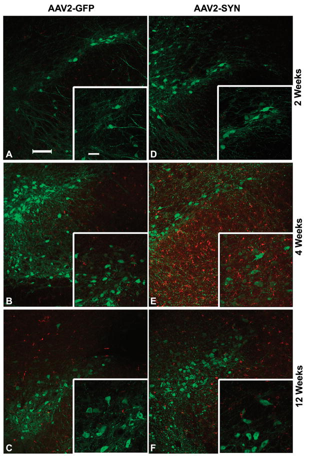
In the animals injected with AAV2-SYN, overexpression of human αSYN resulted in a marked increase in cells stained for CD68 in the SN (Fig. 1). Most of the cells stained were small in size and had the morphological features of activated microglia, with abundant cytoplasm and swelling of the processes proximally. These CD68-positive cells were found throughout the rostro-caudal extent of SN and in the adjacent pars reticulata. Almost all of the CD68-positive cells were found on the injected side; only rare positive cells were found on the contralateral side, usually near the midline. There were also CD68-positive cells extending rostrally along the labeled fibers in the medial forebrain bundle. In many cases, there was an additional cluster of CD68-positive cells dorsal to the SN in a line along the track of the injection needle. These were considered to be a result of mechanical injury related to the injection procedure. In animals injected with AAV2-GFP, CD68-positive cells were rare, except along the track of the injection.
Figure 1.
Effect of overexpression of human αSYN on CD68 immunoreactivity in the mouse SN. AAV2-SYN and AAV2-GFP (control) mice were injected into the right SN and the tissue was processed for CD68 staining using a rat monoclonal antibody against mouse CD68 at 2, 4 and 12 weeks post-injection. AAV2-SYN-injected SN displayed increased staining for CD68 (red) in close proximity to αSYN expressing neurons (green) at 4 weeks compared to controls; at 2 and 12 weeks there was less CD68 staining. Panels A–C are from the AAV2-GFP group at 2, 4 and 12 weeks respectively; panels D–F are from the AAV2-SYN group at 2, 4 and 12 weeks respectively. Insets in all sections show high magnification images of the same sections. Scale bar, 100 μm for the low-magnification images and 40 μm for the inset.
To compare the intensity of the CD68-positive staining in the α-SYN and GFP injected animals, the slides were coded and examined by an observer unaware of which viral vector was injected. This analysis showed that there was a marked difference in the intensity of staining 4 weeks after injection, with much stronger CD68 staining in the group injected with AAV2-SYN (Fig. 1; Table 2). There was also a trend towards an increase at both the 2 week and 12 weeks time points but this did not reach statistical significance, most likely because of the small group sizes and the imprecision of the subjective rating scale.
Table 2.
Semiquantitative Analysis of Effects of AAV2-SYN on IgG and CD68 Immunoreactivity in the Mouse SN*
| Group | Mouse IgG | CD68 | ||||
|---|---|---|---|---|---|---|
| 2 Wk | 4 Wk | 12 Wk | 2 Wk | 4 Wk | 12 Wk | |
| AAV2-GFP | 0.66 ± 0.54 | 0.5 ± 0.24 | 0.66 ± 0.36 | 0.66 ± 0.37 | 1.33 ± 0.23 | 1.17 ± 0.52 |
| AAV2-SYN | 2.5 ± 0.73* | 3.2 ± 0.18** | 2.33 ± 0.23** | 1.5 ± 0.38 | 2.5 ± 0.37* | 1.33 ± 0.37 |
Scoring of immunostaining was performed as described in Materials and Methods on a scale of 0 to 4. Compared to AAV2-GFP injected tissues, AAV2-SYN treatment caused a significant increase in IgG immunoreactivity at all the time points and a significant increase in CD68 only at 4 weeks. Values in the table represent mean ± SE.
p < 0.05 and
p < 0.01, AAV2-SYN versus AAV2-GFP using the Mann-Whitney U test (n = 6/group).
Human αSYN Overexpression in the Mouse SN Leads to IgG Deposition in Brain
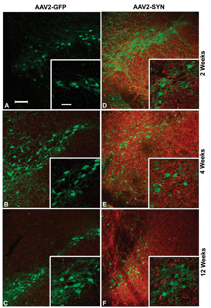
We studied the effect of overexpression of human αSYN in SN neurons on humoral components of the immune response by immunostaining for mouse IgG with a Cy3-conjugated goat anti-mouse IgG. In animals injected with the control AAV2-GFP vector, this antibody produced little or no staining of the brain. In animals injected with AAV2-SYN, there was strong immunostaining for IgG in all of the animals (Fig. 2). This typically was diffuse and included labeling of neurons, glial elements and neuropil. It was largely confined to the injected side of the brain but was not restricted to the SN and often spread to involve adjacent structures including the thalamus and adjacent cortex in some animals. Evaluation of the sections by an observer unaware of the treatments confirmed the striking difference in the intensity of staining, which was statistically significant at 2, 4, and 12 weeks (Fig. 2; Table 2).
Figure 2.
Effect of overexpression of human αSYN on IgG immunostaining in the mouse SN. AAV2-SYN or AAV2-GFP (control) mice were injected into the right SN and at 2, 4, and 12 weeks post-injection the tissue was processed for IgG staining using a goat antibody against mouse IgG. Compared to AAV2-GFP-injected tissues (A–C), AAV2-SYN-injected SN (D–F) display intense staining for IgG (red) close to the αSYN-expressing neurons (green) at each time point. IgG immunostaining is particularly intense on cells near the SN neurons. Panels A–C are from the AAV2-GFP group at 2, 4 and 12 weeks, respectively; Panels D–F are from the AAV2-SYN group at 2, 4 and 12 weeks, respectively. Insets in all sections show high magnification images of the same sections. Scale bar, 100 μm for the low-magnification images and 40 μm for the inset.
Overexpression of Human αSYN Stimulates Expression of Markers of Neuroinflammation in the Mouse SN
The expression of mRNA encoding a panel of markers of neuroinflammation (Table 1) was examined in animals injected with AAV2-SYN or AAV2-GFP. Because of the differing requirements for tissue preparation, this analysis was performed on a different set of animals from those studied with immunohistochemistry. Two or four weeks after injection of the vectors, tissue from the SN was harvested and used to prepare cDNA. Gene expression was analyzed by quantitative PCR using the SYBR Green method, as previously described (22, 26). Data were normalized to expression of the housekeeping gene GAPDH in the same samples. These experiments included tissue from naive animals as well as from animals injected with AAV2-GFP; no significant differences were observed in the expression of any of the markers studied between these two different control groups.
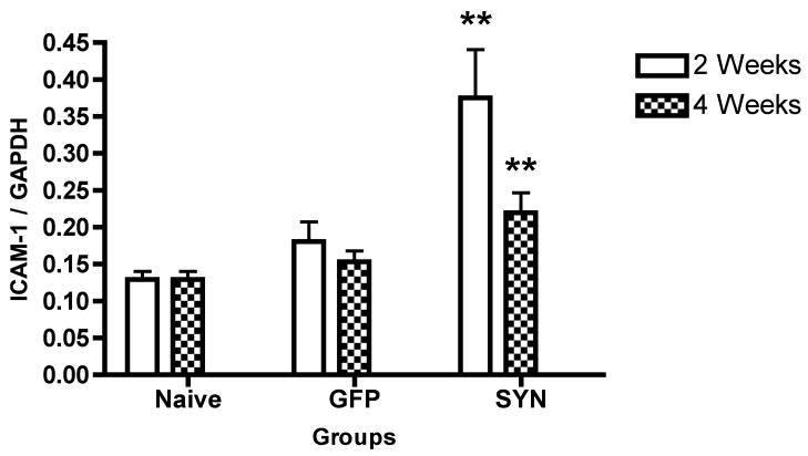
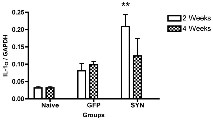
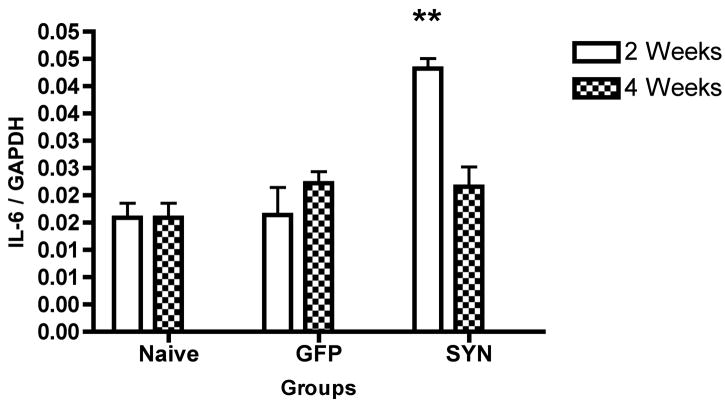
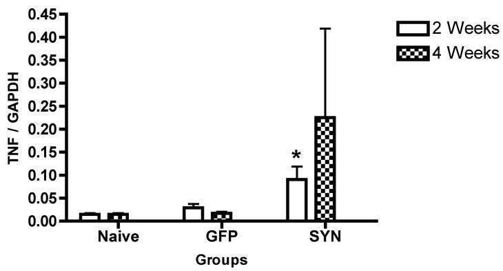
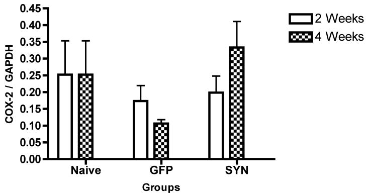
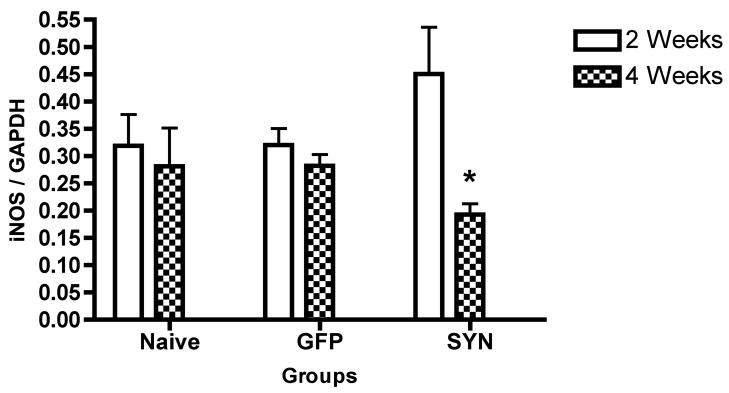
At the earliest time point examined (i.e. 2 weeks after injection of AAV2-SYN), we observed significant increases in the expression of ICAM-1, interleukin (IL)-1α, IL-6 and TNF (Fig. 3A–D), whereas no difference was found in the expression of cyclo- oxygenase-2 (COX-2) or inducible nitric oxide synthase (iNOS) (Fig. 3E, F). At 4 weeks, ICAM-1 levels remained significantly elevated (Fig. 3A), whereas IL-1α, IL-6 and TNF were no longer significantly elevated (Fig. 3B–D) and COX-2 remained unchanged (Fig. 3E). Interestingly, iNOS expression was significantly reduced compared to the AAV2-SYN group at this later time point (Fig. 3F).
Figure 3.
Effects of AAV2-SYN on the expression levels of neuroinflammatory markers in the SN. Compared to the naïve control group, the AAV2-GFP group did not have significant change in any marker expression. Compared to the AAV2-GFP group, there were significant increases in the levels of ICAM-1 (A), IL-1α (B), IL-6 (C) and TNF (D) at 2 weeks post-treatment in the AAV2-SYN group. A difference was also present for ICAM-1 at 4 weeks, whereas no significant differences for the other markers between the groups were observed at this time point. COX-2 levels did not differ between the two groups at either time point (E). iNOS expression was decreased significantly in the AAV2-SYN group compared to the naïve control group at 4 weeks (F). *, p < 0.05 and **, p < 0.01, AAV2-SYN versus AAV2-GFP for all markers except iNOS; for iNOS, *, p < 0.05, naïve control versus AAV2-SYN. One-Way ANOVA with Fisher PLSD post-hoc test, n = 6/group.
We also examined the possibility of so-called “alternative activation” of microglia in this model, since this process has been shown to result in tissue repair and healing (25) and examined the expression of 3 markers linked to alternative activation, Arginase I, IL-4, and IL-13 using the same procedure. We did not observe any significant change in the expression of any of the markers for alternative activation of microglia at either time point tested (Table 3).
Table 3.
Effect of Human α-SYN Expression on Markers of Alternative Activation in the Mouse SN*
| Markers | Naive | AAV2-GFP | AAV2-SYN | ||
|---|---|---|---|---|---|
| 2 wks | 4 wks | 2 wks | 4 wks | ||
| Arginase-1 | 1.13 ± 0.27 | 1.04 ± 0.35 | 1.18 ± 0.16 | 1.99± 0.29 | 0.83 ± 0.06 |
| IL-4 | 1.55 ± 0.46 | 0.5 ± 0.22 | 1.07 ± 0.18 | 1.01± 0.15 | 1.31 ± 0.18 |
| IL-13 | 2.26 ± 0.30 | 0.96 ± 0.51 | 1.16 ± 0.18 | 1.33 ± 0.30 | 1.82 ± 0.57 |
Real time-PCR was used to analyze expression levels of arginase 1, IL-4 and IL-13 in the SN of mice (n = 6/group) treated with AAV2-GFP or AAV2-SYN and killed at 2 weeks or 4 weeks. Data are analyzed by one-way ANOVA. Compared to the naive control group, there was no significant change in the expression of these markers was noticed in either the AAV2-GFP or AAV2-SYN groups and there were no significant differences in expression between AAV2-SYN and AAV2-GFP groups at either time point. The values are expressed as mean ± SE of ratios of expression of the markers to that of the housekeeping gene GAPDH.
B and T Cell Infiltration After Human αSYN Overexpression in the Mouse SN
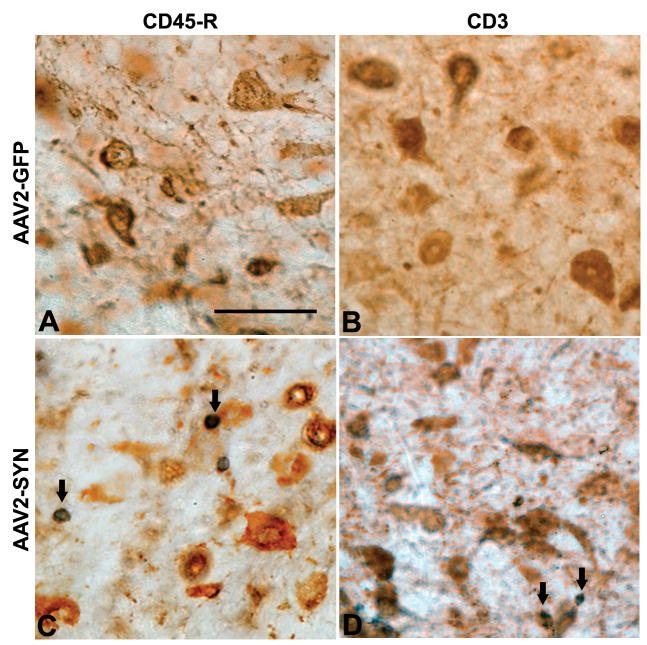
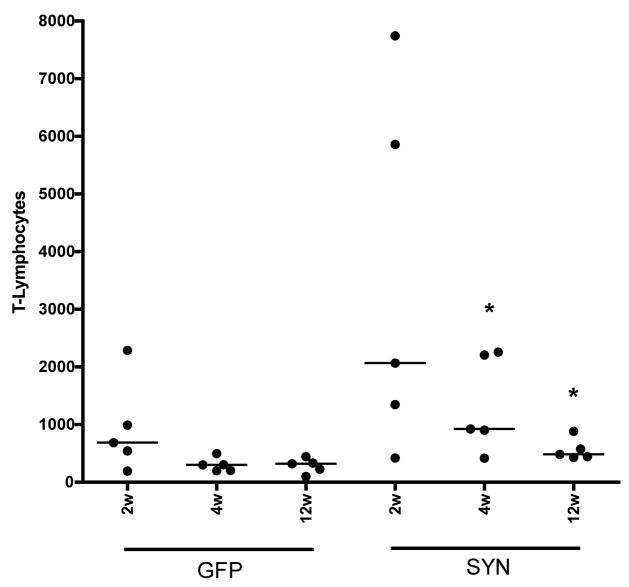
We evaluated the presence of T lymphocytes using immunostaining for CD3. Expression of human αSYN in the SN resulted in a marked increase in CD3-positive cells in the injected SN. These were present throughout the SN; they were often close to labeled neurons and extended into the adjacent pars reticulata (Fig. 4D). Animals injected with AAV2-GFP had only rare CD3-positive cells (Fig. 4B). The numbers of CD3-positive cells in the SN were determined using unbiased stereology with an optical dissector technique. This confirmed the impression of a large increase in the number of CD3-positive cells. The difference from controls was most striking at 4 weeks after injection, where the median number of CD3 positive cells was increased almost 10-fold (Fig. 5A). There were also increased numbers of CD3-positive cells in some animals at 2 weeks and 12 weeks, but there was considerable variation at these time points and only the 12 week difference was statistically significant (Fig. 5A).
Figure 4.
Infiltration of lymphocytes into the mouse SN in response to AAV2-SYN. Infiltration of B and T lymphocytes into the SN was examined using antibodies against the markers CD45R and CD3, respectively. Representative images from AAV2-GFP treated (A, B) and AAV2-SYN treated (C, D) mice at the 12 weeks time point show B and T lymphocyte infiltration in the SN. Solid arrows indicate B lymphocytes (C) and T lymphocytes (D) in the SN, close to human αSYN-expressing neurons (brown). Scale bar: 100 μm.
Figure 5.
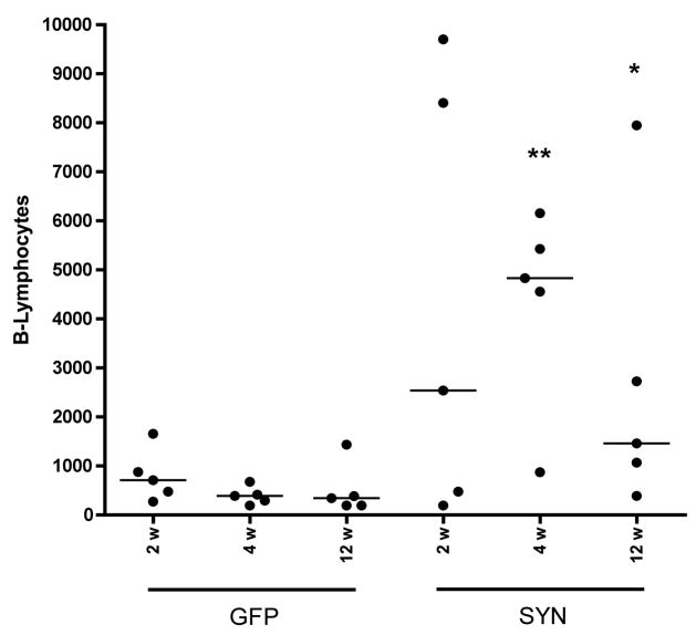
Stereological estimation of T and B lymphocyte infiltration in the SN of AAV2-SYN and AAV2-GFP injected mice. Unbiased stereological estimates of T lymphocytes (A) and B lymphocytes (B) are shown. Compared to the AAV2-GFP group, the AAV2-SYN-treated group showed a trend towards increased infiltration of both B and T cells in the SN at 2 weeks. At 4 and 12 weeks, B and T lymphocyte infiltration was significantly elevated in the AAV2-SYN group compared to the AAV2-GFP group. T lymphocytes: *, p < 0.05, AAV2-SYN, 1342.78 ± 418.59 versus AAV2-GFP, 301.35 ± 60.44 at 4 weeks and *, p < 0.05, AAV2-SYN, 564.71 ± 94.39 versus AAV2-GFP, 285.79 ± 63.85 at 12 weeks; Wilcoxon Sign-rank test, n=5/group. B lymphocytes: **, p < 0.01, AAV2-SYN, 4367.6 ± 1024.39 versus AAV2-GFP, 393.42 ± 90.57 at 4 weeks and *, p < 0.05, AAV2-SYN, 2718.98 ± 1522.31 versus AAV2-GFP, 510.56 ± 261.79 at 12 weeks; Wilcoxon Sign-rank test, n = 5/group.
We used a similar approach with immunostaining for CD45R to evaluate the number of B lymphocytes in the SN after AAV2-SYN injection. As for staining for CD3, injection with AAV2-SYN resulted in a marked increase in the number of CD45R-positive cells (Fig. 4C) confined to the injected side. The most consistent difference was observed at 4 weeks, when the median number of cells was increased nearly 5-fold (Fig. 5B). Some animals had increased numbers at 2 weeks and 12 weeks, but the effect at these time points was more variable and statistically significant only at 12 weeks (Fig. 5B).
DISCUSSION
In this study we have examined the question of whether overexpression of αSYN, in the absence of neurodegenerative cell loss or oxidative injury, is sufficient to trigger immune activation in a model of PD. We found that overexpression of human αSYN in mouse SN neurons, induced by a AAV vector, does indeed lead to activation of microglia, production of inflammatory cytokines, and stimulation of an adaptive immune response. These events occur early (i.e. 2 to 4 weeks after injection of the virus), at a time when there is little or no loss of dopaminergic neurons. No significant microglial activation or humoral response was observed after injection of control AAV2 virus expressing GFP. Both the microglial activation and the adaptive immune response are localized and concentrated in the region of human α-SYN overexpression. Together, these observations suggest that the α-SYN protein has a direct role in the activation of microglia in vivo and the stimulation of the adaptive immune system, and that cell death is not required to trigger these processes.
The most important difference between this study and earlier reports examining immune activation in PD is the nature of the model. Previous studies have employed the MPTP model, and have shown clearly that this neurotoxin leads to microglial activation and a humoral response to nitrated αSYN (4, 18, 19). In the MPTP model, however, dopamine neuronal injury is extensive and there is rapid necrotic cell death. Thus, there may be a variety of other immunologically active proteins and other molecules released in addition to nitrated αSYN. In contrast, the AAV construct used here codes for αSYN and/or GFP and does not contain any sequence coding for viral proteins. We and others have observed that expression of αSYN using AAV leads to gradual degeneration of dopamine neurons (22, 27, 28); this differs from conventional αSYN transgenic models, which for the most part show no loss of dopamine neurons and may even exhibit protection against some neurotoxic lesions (29–31). The effects we have observed are unlikely to have arisen from the AAV vector itself. A short-term humoral immune response to AAV is observed in the peripheral immune system (32), but in the brain humoral immune and inflammatory responses to a single administration of AAV are very low (33, 34). We included a control vector expressing GFP alone in all experiments, and found that the AAV2-GFP led to robust transfection of neurons and high-level expression of the GFP protein, but did not produce microglial activation, cytokine production, B or T cell infiltration, or IgG labeling. As others have observed (35–37), we found that when these genes were delivered by AAV to adult mouse brain they were expressed exclusively in neurons, implying that the microglial activation is an indirect response to the neuronal expression of αSYN. The AAV-Syn vector we used produces very long-lasting expression of αSYN in neurons; in this study, expression was observed for 12 weeks and we previously reported vigorous expression of αSYN as long as 24 weeks after administration (22). In contrast, some of the markers of immune activation that we observed were transient, while others persisted for at least 12 weeks (including IgG staining, and B and T cell infiltration). These temporal differences likely reflect the dynamic nature of the immune response rather than any change in the underlying expression of α-SYN.
Microglial activation is a common feature of PD brains (1, 6, 17, 38) as well as in animal models of PD (3, 39). HLA-DR-positive activated microglia are observed in both human PD brains and in MPTP-treated monkeys (1, 2). There is also evidence that microglial activation occurs early in the course of the disease in humans. Using positron emission tomography with the radiotracer [11C](R)-PK11195 for activated microglia, two independent groups provided evidence for increased levels of activated microglia in the SN of early stage PD patients (38, 40). Microglial activation is also observed early after treatment with MPTP in the PD rodent models (39, 41) and microglial activation precedes dopamine neuronal injury in the rodent paraquat/maneb co-administration PD model (42). Early activation of microglia is also present in a transgenic mouse model of PD in which wild-type human αSYN is expressed under a tyrosine hydroxylase promoter (31). The early microglial response we observed in the present study is consistent with these pathological findings in human PD and other animal models.
In this study, we also demonstrated that the levels of markers of neuroinflammation, TNF, ICAM-1, IL-6 and IL-1α, were elevated early in response to human αSYN overexpression in the mouse SN. There is also elevated expression of the these cytokines in the SN of PD patients as well as in the animal PD models (43–49) and an increase in circulating levels of the cytokines TNF and IL-6 have also been observed in PD patients (50). Similar to our study, an early increase in the these cytokines occurs in the MPTP model of PD in mice (4, 51, 52). In addition, in the MPTP mouse model of PD, genetic ablation of receptors for TNF confers dopaminergic neuroprotection (53).
We also explored the possibility of an alternative activation state of microglia. Alternative activation of macrophages and microglia is a response to tissue injury and is believed to play a role in tissue repair and restoration (54). Increased expression of genes associated with alternative activation has been observed in Alzheimer disease (AD) brains and in a transgenic mouse model of AD (55). To evaluate the possible involvement of alternative activation in our PD model, we measured the expression of cytokines IL-4 and IL-13 and arginase-1 in the SN of mice treated with AAV2-SYN for 2 and 4 weeks. We did not find any significant change in the levels of these markers at either time point. These data suggest that alternative activation is not a component of our model at the early time points, but it will be interesting to examine this phenomenon at later time points when neurodegeneration becomes apparent.
Human αSYN overexpression in the SN resulted in a significant increase in IgG staining at each of the early points examined. Similarly, in human PD brains there is evidence for accumulation of IgG in the SN and an increased expression of IgG binding receptors on activated microglia in the SN (6). Moreover, in rodents and in cell culture, serum from PD patients is toxic to dopaminergic neurons and this neurotoxicity is mediated through IgG binding receptors (FcγRI) on activated microglia (8, 9). There is at least one report of circulating autoantibodies to human α-SYN in patients with inherited PD (56). The source of the IgG observed in our model, as well as in human brain, is unclear; it may result either from local interruption of the blood-brain barrier and infiltration of circulating antibodies, or from local release by activated B lymphocytes.
We observed very striking B and T lymphocyte infiltrates in animals injected with AAV2-SYN. The levels of both B and T cells were highest at 4 weeks and declined at 12 weeks, but at this late time point remained significantly elevated in the AAV2-SYN group versus the control group. Both CD8-positive T lymphocytes in the SN of PD patients (1) and alterations in circulating T lymphocytes have been reported in PD patients (57). In MPTP-treated mice, marked T lymphocyte infiltration and increased MHC II immunoreactivity were found but no B lymphocyte infiltration was observed (58). In addition, neurotoxicity to MPTP is attenuated by administration of regulatory T cells (5). In contrast to the clear involvement of T lymphocytes in human PD there is little evidence pertaining to the presence of B lymphocytes in the SN in PD or in animal models of PD. It is interesting to note that, in a recent study using the MPTP mouse model of PD, animals lacking both B and T lymphocytes were resistant to MPTP toxicity (19). Because we observe increased infiltration of both B and T lymphocytes in the SN, it will be important to examine the role of these distinct populations of lymphocytes separately in our model.
An important unresolved question relates to the temporal sequence of events leading to the cellular and humoral response that we have observed. We studied early time points after AAV2-SYN injection, but even at 2 weeks we found evidence for a combined humoral and cellular response. Although some aspects of the response did not reach peak intensity until 4 weeks, the differences are not sufficient to infer a clear temporal relationship of these events. A complicating factor in interpreting the sequence is that the expression of protein by the viral vectors does not begin immediately and may require several weeks to reach peak intensity (59, 60).
Our data suggest the intriguing possibility that the initiating event in PD-associated inflammation is related to αSYN itself, rather than to neurodegeneration followed by release of modified αSYN. Several lines of evidence support the idea that αSYN may lead directly to activation of microglia. In vitro studies have shown that overexpression of human αSYN or exposure to exogenous human αSYN is sufficient to directly activate microglia and lead to proinflammatory cytokine production (32, 61–63). Exposure of microglia to nitrated αSYN, a covalently modified form of αSYN, results in enhanced expression of proinflammatory cytokines (18). The expression of αSYN induced by the AAV vector is restricted to neurons, suggesting there must be an interposed step of αSYN antigen presentation or release. The degree of αSYN overexpression induced by the AAV-SYN vector in dopaminergic cells is vigorous, and it is likely that are a variety of oligomeric, aggregated, and other modified forms of αSYN present, but which of these is directly responsible for the immune activation we observed is still uncertain. Since it is likely that neuroinflammation is important in the onset or progression of human PD, identifying the mechanisms of immune activation in this model may lead to novel therapies for the disease.
Acknowledgments
This work was supported by NIH P50 NS38372 and American Parkinson’s Disease Association.
The authors thank Dr. Selvarangan Ponnazhagan at the Department of Pathology, University of Alabama at Birmingham, for advice on AAV2 packaging and purification, Dr. Etty (Tika) Benveniste, Professor and Chairman, Department of Cell Biology at The University of Alabama at Birmingham for critical reading of the manuscript, and Dr. Buffie Clodfelder-Miller at the Alabama Neuroscience Blueprint Core Center, UAB, for help with the microbrightfield microscope for stereology.
References
- 1.McGeer PL, Itagaki S, Boyes BE, McGeer EG. Reactive microglia are positive for HLA-DR in the substantia nigra of Parkinson’s and Alzheimer’s disease brains. Neurology. 1988;38:1285–91. doi: 10.1212/wnl.38.8.1285. [DOI] [PubMed] [Google Scholar]
- 2.McGeer PL, Schwab C, Parent A, Doudet D. Presence of reactive microglia in monkey substantia nigra years after 1-methyl-4-phenyl-1,2,3,6-tetrahydropyridine administration. Ann Neurol. 2003;54:599–604. doi: 10.1002/ana.10728. [DOI] [PubMed] [Google Scholar]
- 3.Sherer TB, Betarbet R, Kim JH, Greenamyre JT. Selective microglial activation in the rat rotenone model of Parkinson’s disease. Neurosci Lett. 2003;341:87–90. doi: 10.1016/s0304-3940(03)00172-1. [DOI] [PubMed] [Google Scholar]
- 4.Kurkowska-Jastrzebska I, Wronska A, Kohutnicka M, Czlonkowski A, Czlonkowska A. The inflammatory reaction following 1-methyl-4-phenyl-1,2,3, 6-tetrahydropyridine intoxication in mouse. Exp Neurol. 1999;156:50–61. doi: 10.1006/exnr.1998.6993. [DOI] [PubMed] [Google Scholar]
- 5.Reynolds AD, Banerjee R, Liu J, Gendelman HE, Mosley RL. Neuroprotective activities of CD4+CD25+ regulatory T cells in an animal model of Parkinson’s disease. J Leukoc Biol. 2007;82:1083–94. doi: 10.1189/jlb.0507296. [DOI] [PubMed] [Google Scholar]
- 6.Orr CF, Rowe DB, Mizuno Y, Mori H, Halliday GM. A possible role for humoral immunity in the pathogenesis of Parkinson’s disease. Brain. 2005;128:2665–74. doi: 10.1093/brain/awh625. [DOI] [PubMed] [Google Scholar]
- 7.Hisanaga K, Asagi M, Itoyama Y, Iwasaki Y. Increase in peripheral CD4 bright+ CD8 dull+ T cells in Parkinson disease. Arch Neurol. 2001;58:1580–83. doi: 10.1001/archneur.58.10.1580. [DOI] [PubMed] [Google Scholar]
- 8.He Y, Le WD, Appel SH. Role of Fcgamma receptors in nigral cell injury induced by Parkinson disease immunoglobulin injection into mouse substantia nigra. Exp Neurol. 2002;176:322–27. doi: 10.1006/exnr.2002.7946. [DOI] [PubMed] [Google Scholar]
- 9.Le W, Rowe D, Xie W, Ortiz I, He Y, Appel SH. Microglial activation and dopaminergic cell injury: An in vitro model relevant to Parkinson’s disease. J Neurosci. 2001;21:8447–55. doi: 10.1523/JNEUROSCI.21-21-08447.2001. [DOI] [PMC free article] [PubMed] [Google Scholar]
- 10.Chen S, Le WD, Xie WJ, et al. Experimental destruction of substantia nigra initiated by Parkinson disease immunoglobulins. Arch Neurol. 1998;55:1075–80. doi: 10.1001/archneur.55.8.1075. [DOI] [PubMed] [Google Scholar]
- 11.Le WD, Rowe DB, Jankovic J, Xie W, Appel SH. Effects of cerebrospinal fluid from patients with Parkinson disease on dopaminergic cells. Arch Neurol. 1999;56:194–200. doi: 10.1001/archneur.56.2.194. [DOI] [PubMed] [Google Scholar]
- 12.Spillantini MG, Schmidt ML, Lee VM, Trojanowski JQ, Jakes R, Goedert M. Alpha-synuclein in Lewy bodies. Nature. 1997;388:839–40. doi: 10.1038/42166. [DOI] [PubMed] [Google Scholar]
- 13.Hardy J, Cai H, Cookson MR, Gwinn-Hardy K, Singleton A. Genetics of Parkinson’s disease and parkinsonism. Ann Neurol. 2006;60:389–98. doi: 10.1002/ana.21022. [DOI] [PubMed] [Google Scholar]
- 14.Kruger R, Vieira-Saecker AM, Kuhn W, et al. Increased susceptibility to sporadic Parkinson’s disease by a certain combined alpha-synuclein/apolipoprotein E genotype. Ann Neurol. 1999;45:611–17. doi: 10.1002/1531-8249(199905)45:5<611::aid-ana9>3.0.co;2-x. [DOI] [PubMed] [Google Scholar]
- 15.Polymeropoulos MH, Lavedan C, Leroy E, et al. Mutation in the alpha-synuclein gene identified in families with Parkinson’s disease. Science. 1997;276:2045–47. doi: 10.1126/science.276.5321.2045. [DOI] [PubMed] [Google Scholar]
- 16.Cantuti-Castelvetri I, Klucken J, Ingelsson M, et al. Alpha-synuclein and chaperones in dementia with Lewy bodies. J Neuropathol Exp Neurol. 2005;64:1058–66. doi: 10.1097/01.jnen.0000190063.90440.69. [DOI] [PubMed] [Google Scholar]
- 17.Croisier E, Moran LB, Dexter DT, Pearce RK, Graeber MB. Microglial inflammation in the parkinsonian substantia nigra: Relationship to alpha-synuclein deposition. J Neuroinflammation. 2005;2:14. doi: 10.1186/1742-2094-2-14. [DOI] [PMC free article] [PubMed] [Google Scholar]
- 18.Reynolds AD, Glanzer JG, Kadiu I, et al. Nitrated alpha-synuclein-activated microglial profiling for Parkinson’s disease. J Neurochem. 2008;104:1504–25. doi: 10.1111/j.1471-4159.2007.05087.x. [DOI] [PubMed] [Google Scholar]
- 19.Benner EJ, Banerjee R, Reynolds AD, et al. Nitrated alpha-synuclein immunity accelerates degeneration of nigral dopaminergic neurons. PLoS ONE. 2008;3:e1376. doi: 10.1371/journal.pone.0001376. [DOI] [PMC free article] [PubMed] [Google Scholar]
- 20.Mandir AS, Przedborski S, Jackson-Lewis V, et al. Poly(ADP-ribose) polymerase activation mediates 1-methyl-4-phenyl-1, 2,3,6-tetrahydropyridine (MPTP)-induced parkinsonism. Proc Natl Acad Sci U S A. 1999;96:5774–79. doi: 10.1073/pnas.96.10.5774. [DOI] [PMC free article] [PubMed] [Google Scholar]
- 21.Przedborski S, Ischiropoulos H. Reactive oxygen and nitrogen species: Weapons of neuronal destruction in models of Parkinson’s disease. Antioxid Redox Signal. 2005;7:685–93. doi: 10.1089/ars.2005.7.685. [DOI] [PubMed] [Google Scholar]
- 22.St Martin JL, Klucken J, Outeiro TF, et al. Dopaminergic neuron loss and up-regulation of chaperone protein mRNA induced by targeted over-expression of alpha-synuclein in mouse substantia nigra. J Neurochem. 2007;100:1449–57. doi: 10.1111/j.1471-4159.2006.04310.x. [DOI] [PubMed] [Google Scholar]
- 23.Zolotukhin S, Byrne BJ, Mason E, et al. Recombinant adeno-associated virus purification using novel methods improves infectious titer and yield. Gene Ther. 1999;6:973–85. doi: 10.1038/sj.gt.3300938. [DOI] [PubMed] [Google Scholar]
- 24.Auricchio A, Hildinger M, O’Connor E, Gao GP, Wilson JM. Isolation of highly infectious and pure adeno-associated virus type 2 vectors with a single-step gravity-flow column. Hum Gene Ther. 2001;12:71–76. doi: 10.1089/104303401450988. [DOI] [PubMed] [Google Scholar]
- 25.McGeer PL, McGeer EG. Glial reactions in Parkinson’s disease. Mov Disord. 2008;15:474–83. doi: 10.1002/mds.21751. [DOI] [PubMed] [Google Scholar]
- 26.Cantuti-Castelvetri I, Keller-McGandy C, Bouzou B, et al. Effects of gender on nigral gene expression and parkinson disease. Neurobiol Dis. 2007;26:606–14. doi: 10.1016/j.nbd.2007.02.009. [DOI] [PMC free article] [PubMed] [Google Scholar]
- 27.Kirik D, Rosenblad C, Burger C, et al. Parkinson-like neurodegeneration induced by targeted overexpression of alpha-synuclein in the nigrostriatal system. J Neurosci. 2002;22:2780–91. doi: 10.1523/JNEUROSCI.22-07-02780.2002. [DOI] [PMC free article] [PubMed] [Google Scholar]
- 28.Yamada M, Iwatsubo T, Mizuno Y, Mochizuki H. Overexpression of alpha-synuclein in rat substantia nigra results in loss of dopaminergic neurons, phosphorylation of alpha-synuclein and activation of caspase-9: resemblance to pathogenetic changes in Parkinson’s disease. J Neurochem. 2004;91:451–61. doi: 10.1111/j.1471-4159.2004.02728.x. [DOI] [PubMed] [Google Scholar]
- 29.Manning-Bog AB, McCormack AL, Purisai MG, Bolin LM, Di Monte DA. Alpha-synuclein overexpression protects against paraquat-induced neurodegeneration. J Neurosci. 2003;23:3095–99. doi: 10.1523/JNEUROSCI.23-08-03095.2003. [DOI] [PMC free article] [PubMed] [Google Scholar]
- 30.Masliah E, Rockenstein E, Veinbergs I, et al. Dopaminergic loss and inclusion body formation in alpha-synuclein mice: Implications for neurodegenerative disorders. Science. 2000;287:1265–69. doi: 10.1126/science.287.5456.1265. [DOI] [PubMed] [Google Scholar]
- 31.Su X, Maguire-Zeiss KA, Giuliano R, Prifti L, Venkatesh K, Federoff HJ. Synuclein activates microglia in a model of Parkinson’s disease. Neurobiol Aging. 2007 May 28; doi: 10.1016/j.neurobiolaging.2007.04.006. [Epub ahead of print] [DOI] [PMC free article] [PubMed] [Google Scholar]
- 32.Mingozzi F, Hasbrouck NC, Basner-Tschakarjan E, et al. Modulation of tolerance to the transgene product in a nonhuman primate model of AAV-mediated gene transfer to liver. Blood. 2007;110:2334–41. doi: 10.1182/blood-2007-03-080093. [DOI] [PMC free article] [PubMed] [Google Scholar]
- 33.Mandel RJ, Rendahl KG, Spratt SK, Snyder RO, Cohen LK, Leff SE. Characterization of intrastriatal recombinant adeno-associated virus-mediated gene transfer of human tyrosine hydroxylase and human GTP-cyclohydrolase I in a rat model of Parkinson’s disease. J Neurosci. 1998;18:4271–84. doi: 10.1523/JNEUROSCI.18-11-04271.1998. [DOI] [PMC free article] [PubMed] [Google Scholar]
- 34.Mastakov MY, Baer K, Symes CW, Leichtlein CB, Kotin RM, During MJ. Immunological aspects of recombinant adeno-associated virus delivery to the mammalian brain. J Virol. 2002;76:8446–54. doi: 10.1128/JVI.76.16.8446-8454.2002. [DOI] [PMC free article] [PubMed] [Google Scholar]
- 35.Bartlett JS, Samulski RJ, McCown TJ. Selective and rapid uptake of adeno-associated virus type 2 in brain. Hum Gene Ther. 1998;9:1181–86. doi: 10.1089/hum.1998.9.8-1181. [DOI] [PubMed] [Google Scholar]
- 36.Burger C, Gorbatyuk OS, Velardo MJ, et al. Recombinant AAV viral vectors pseudotyped with viral capsids from serotypes 1, 2, and 5 display differential efficiency and cell tropism after delivery to different regions of the central nervous system. Mol Ther. 2004;10:302–17. doi: 10.1016/j.ymthe.2004.05.024. [DOI] [PubMed] [Google Scholar]
- 37.Tenenbaum L, Chtarto A, Lehtonen E, Velu T, Brotchi J, Levivier M. Recombinant AAV-mediated gene delivery to the central nervous system. J Gene Med. 2004;6 (Suppl 1):S212–22. doi: 10.1002/jgm.506. [DOI] [PubMed] [Google Scholar]
- 38.Ouchi Y, Yoshikawa E, Sekine Y, et al. Microglial activation and dopamine terminal loss in early Parkinson’s disease. Ann Neurol. 2005;57:168–75. doi: 10.1002/ana.20338. [DOI] [PubMed] [Google Scholar]
- 39.Wu DC, Jackson-Lewis V, Vila M, et al. Blockade of microglial activation is neuroprotective in the 1-methyl-4-phenyl-1,2,3,6-tetrahydropyridine mouse model of Parkinson disease. J Neurosci. 2002;22:1763–71. doi: 10.1523/JNEUROSCI.22-05-01763.2002. [DOI] [PMC free article] [PubMed] [Google Scholar]
- 40.Gerhard A, Pavese N, Hotton G, et al. In vivo imaging of microglial activation with [11C](R)-PK11195 PET in idiopathic Parkinson’s disease. Neurobiol Dis. 2006;21:404–12. doi: 10.1016/j.nbd.2005.08.002. [DOI] [PubMed] [Google Scholar]
- 41.Mount MP, Lira A, Grimes D, et al. Involvement of interferon-gamma in microglial-mediated loss of dopaminergic neurons. J Neurosci. 2007;27:3328–37. doi: 10.1523/JNEUROSCI.5321-06.2007. [DOI] [PMC free article] [PubMed] [Google Scholar]
- 42.Saint-Pierre M, Tremblay ME, Sik A, Gross RE, Cicchetti F. Temporal effects of paraquat/maneb on microglial activation and dopamine neuronal loss in older rats. J Neurochem. 2006;98:760–72. doi: 10.1111/j.1471-4159.2006.03923.x. [DOI] [PubMed] [Google Scholar]
- 43.Lee DY, Oh YJ, Jin BK. Thrombin-activated microglia contribute to death of dopaminergic neurons in rat mesencephalic cultures: Dual roles of mitogen-activated protein kinase signaling pathways. Glia. 2005;51:98–110. doi: 10.1002/glia.20190. [DOI] [PubMed] [Google Scholar]
- 44.Klegeris A, McGeer PL. Rat brain microglia and peritoneal macrophages show similar responses to respiratory burst stimulants. J Neuroimmunol. 1994;53:83–90. doi: 10.1016/0165-5728(94)90067-1. [DOI] [PubMed] [Google Scholar]
- 45.Sawada M, Imamura K, Nagatsu T. Role of cytokines in inflammatory process in Parkinson’s disease. J Neural Transm Suppl. 2006;70:373–81. doi: 10.1007/978-3-211-45295-0_57. [DOI] [PubMed] [Google Scholar]
- 46.Imamura K, Hishikawa N, Sawada M, Nagatsu T, Yoshida M, Hashizume Y. Distribution of major histocompatibility complex class II-positive microglia and cytokine profile of Parkinson’s disease brains. Acta Neuropathol. 2003;106:518–26. doi: 10.1007/s00401-003-0766-2. [DOI] [PubMed] [Google Scholar]
- 47.Wilms H, Rosenstiel P, Sievers J, Deuschl G, Zecca L, Lucius R. Activation of microglia by human neuromelanin is NF-kappaB dependent and involves p38 mitogen-activated protein kinase: implications for Parkinson’s disease. Faseb J. 2003;17:500–2. doi: 10.1096/fj.02-0314fje. [DOI] [PubMed] [Google Scholar]
- 48.Depino AM, Earl C, Kaczmarczyk E, et al. Microglial activation with atypical proinflammatory cytokine expression in a rat model of Parkinson’s disease. Eur J Neurosci. 2003;18:2731–42. doi: 10.1111/j.1460-9568.2003.03014.x. [DOI] [PubMed] [Google Scholar]
- 49.Teismann P, Tieu K, Choi DK, et al. Cyclooxygenase-2 is instrumental in Parkinson’s disease neurodegeneration. Proc Natl Acad Sci U S A. 2003;100:5473–78. doi: 10.1073/pnas.0837397100. [DOI] [PMC free article] [PubMed] [Google Scholar]
- 50.Dobbs RJ, Charlett A, Purkiss AG, Dobbs SM, Weller C, Peterson DW. Association of circulating TNF-alpha and IL-6 with ageing and parkinsonism. Acta Neurol Scand. 1999;100:34–41. doi: 10.1111/j.1600-0404.1999.tb00721.x. [DOI] [PubMed] [Google Scholar]
- 51.Pattarini R, Smeyne RJ, Morgan JI. Temporal mRNA profiles of inflammatory mediators in the murine 1-methyl-4-phenyl-1,2,3,6-tetrahydropyrimidine model of Parkinson’s disease. Neuroscience. 2007;145:654–68. doi: 10.1016/j.neuroscience.2006.12.030. [DOI] [PMC free article] [PubMed] [Google Scholar]
- 52.Hebert G, Arsaut J, Dantzer R, Demotes-Mainard J. Time-course of the expression of inflammatory cytokines and matrix metalloproteinases in the striatum and mesencephalon of mice injected with 1-methyl-4-phenyl-1,2,3,6-tetrahydropyridine, a dopaminergic neurotoxin. Neurosci Lett. 2003;349:191–95. doi: 10.1016/s0304-3940(03)00832-2. [DOI] [PubMed] [Google Scholar]
- 53.Sriram K, Matheson JM, Benkovic SA, Miller DB, Luster MI, O’Callaghan JP. Mice deficient in TNF receptors are protected against dopaminergic neurotoxicity: Implications for Parkinson’s disease. Faseb J. 2002;16:1474–76. doi: 10.1096/fj.02-0216fje. [DOI] [PubMed] [Google Scholar]
- 54.Ponomarev ED, Maresz K, Tan Y, Dittel BN. CNS-derived interleukin-4 is essential for the regulation of autoimmune inflammation and induces a state of alternative activation in microglial cells. J Neurosci. 2007;27:10714–21. doi: 10.1523/JNEUROSCI.1922-07.2007. [DOI] [PMC free article] [PubMed] [Google Scholar]
- 55.Colton CA, Mott RT, Sharpe H, Xu Q, Van Nostrand WE, Vitek MP. Expression profiles for macrophage alternative activation genes in AD and in mouse models of AD. J Neuroinflammation. 2006;3:27. doi: 10.1186/1742-2094-3-27. [DOI] [PMC free article] [PubMed] [Google Scholar]
- 56.Papachroni KK, Ninkina N, Papapanagiotou A, et al. Autoantibodies to alpha-synuclein in inherited Parkinson’s disease. J Neurochem. 2007;101:749–56. doi: 10.1111/j.1471-4159.2006.04365.x. [DOI] [PMC free article] [PubMed] [Google Scholar]
- 57.Baba Y, Kuroiwa A, Uitti RJ, Wszolek ZK, Yamada T. Alterations of T-lymphocyte populations in Parkinson disease. Parkinsonism Relat Disord. 2005;11:493–98. doi: 10.1016/j.parkreldis.2005.07.005. [DOI] [PubMed] [Google Scholar]
- 58.Kurkowska-Jastrzebska I, Wronska A, Kohutnicka M, Czlonkowski A, Czlonkowska A. MHC class II positive microglia and lymphocytic infiltration are present in the substantia nigra and striatum in mouse model of Parkinson’s disease. Acta Neurobiol Exp (Wars) 1999;59:1–8. doi: 10.55782/ane-1999-1289. [DOI] [PubMed] [Google Scholar]
- 59.Afione SA, Wang J, Walsh S, Guggino WB, Flotte TR. Delayed expression of adeno-associated virus vector DNA. Intervirology. 1999;42:213–20. doi: 10.1159/000024980. [DOI] [PubMed] [Google Scholar]
- 60.Ferrari FK, Samulski T, Shenk T, Samulski RJ. Second-strand synthesis is a rate-limiting step for efficient transduction by recombinant adeno-associated virus vectors. J Virol. 1996;70:3227–34. doi: 10.1128/jvi.70.5.3227-3234.1996. [DOI] [PMC free article] [PubMed] [Google Scholar]
- 61.Jin J, Shie FS, Liu J, Wang Y, Davis J, Schantz AM, Montine KS, Montine TJ, Zhang J. Prostaglandin E2 receptor subtype 2 (EP2) regulates microglial activation and associated neurotoxicity induced by aggregated alpha-synuclein. J Neuroinflammation. 2007;4:2. doi: 10.1186/1742-2094-4-2. [DOI] [PMC free article] [PubMed] [Google Scholar]
- 62.Klegeris A, Pelech S, Giasson BI, et al. alpha-Synuclein activates stress signaling protein kinases in THP-1 cells and microglia. Neurobiol Aging. 2008;29:739–52. doi: 10.1016/j.neurobiolaging.2006.11.013. [DOI] [PubMed] [Google Scholar]
- 63.Zhang W, Wang T, Pei Z, et al. Aggregated alpha-synuclein activates microglia: A process leading to disease progression in Parkinson’s disease. Faseb J. 2005;19:533–42. doi: 10.1096/fj.04-2751com. [DOI] [PubMed] [Google Scholar]


