
Figure 5.
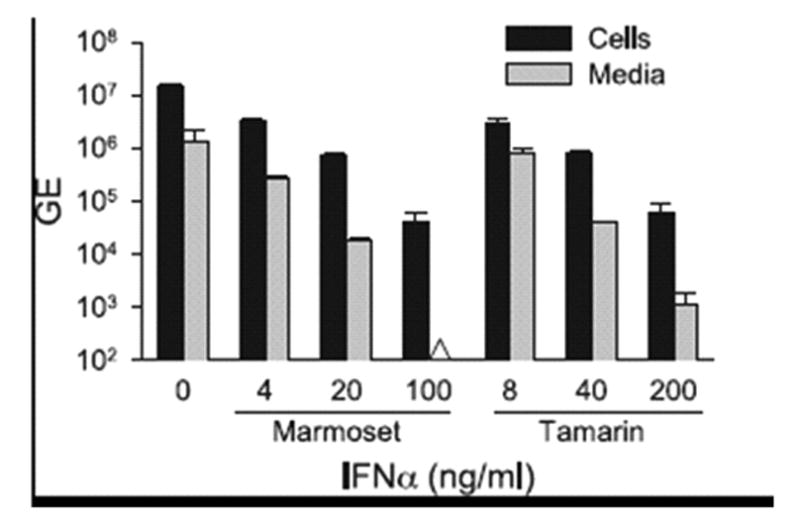
Comparison of antiviral activity of marmoset and tamarin IFNα2 on marmoset hepatocytes infected with GBV-B. Primary marmoset hepatocytes were treated with either marmoset or tamarin IFNα2 at concentrations of 0 to 200 ng/ml for 24 hr prior to infection with GBV-B and for another 72 hr post-infection. Cell and media were analyzed for GBV-B RNA as described for Figure 4. Black bars = GBV-B RNA GE in cell-associated viral RNA, grey bars = GE of GBV-B secreted into media. The Δ symbol denotes that the sample was below the level of detection. Error bars indicate the variation in values from duplicate cultures. The level of variation was not sufficient to yield visible error bars on those were none appear.