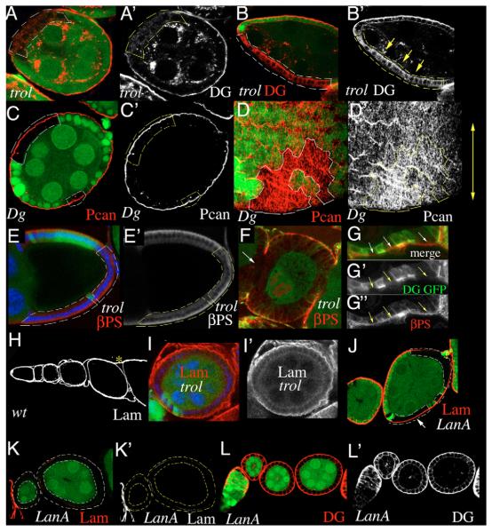
Fig. 4. Perlecan is required for basal localization of Dystroglycan.
(A,B) Dg (red) is no longer present in the basal cell membrane of trol clones. Occasionally, Dg appears to be redistributed to the apical cell membrane (yellow arrows in B’). (C,D) Pcan localization in the ECM is unchanged in a Dg323 clone (C) and orientation of Pcan-stripes appears normal (D). (E) βPS Int (red) is normally expressed in trol clones. Blue indicates DNA. (F) In a FCE entirely composed of mutant cells, gaps in βPS staining can be seen. Green indicates GFP and Dg; arrow indicates gap. (G) βPS Int (red) is still present (arrows) in the basal membrane of a trol clone that has lost Dg (green). (G’) Green channel only, showing both GFP and anti-Dgcyto staining. (G”) Red channel only. (H) Expression of Lam in a wild-type ovariole. Asterisk indicates muscular sheath. (I) A very large trol clone showing fuzzy Lam (red) distribution. Blue indicates DNA. (J) Lam (red) is still present at the basal side of a medium sized lanA clone (arrow). (K) Two egg chambers whose FCE are entirely composed of lanA mutant cells. Lam (red) is almost completely absent. (L) Three egg chambers whose FCE are entirely composed of lanA mutant cells. Dg (red) is still localized at the basal membrane. Clones are marked by the loss of GFP (green) and indicated by broken lines. (A’- L’) Red channels only.

