Abstract
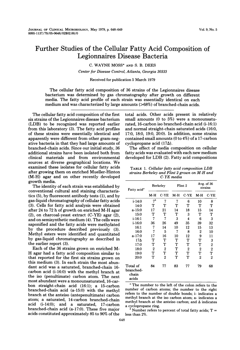
The cellular fatty acid composition of 36 strains of the Legionnaires disease bacterium was determined by gas chromatography after growth on different media. The fatty acid profile of each strain was essentially identical on each medium and was characterized by large amounts (greater than 68%) of branched-chain acids.
Full text
PDF

Selected References
These references are in PubMed. This may not be the complete list of references from this article.
- Cherry W. B., Pittman B., Harris P. P., Hebert G. A., Thomason B. M., Thacker L., Weaver R. E. Detection of Legionnaires disease bacteria by direct immunofluorescent staining. J Clin Microbiol. 1978 Sep;8(3):329–338. doi: 10.1128/jcm.8.3.329-338.1978. [DOI] [PMC free article] [PubMed] [Google Scholar]
- Moss C. W., Weaver R. E., Dees S. B., Cherry W. B. Cellular fatty acid composition of isolates from Legionnaires disease. J Clin Microbiol. 1977 Aug;6(2):140–143. doi: 10.1128/jcm.6.2.140-143.1977. [DOI] [PMC free article] [PubMed] [Google Scholar]
- Pine L., George J. R., Reeves M. W., Harrell W. K. Development of a chemically defined liquid medium for growth of Legionella pneumophila. J Clin Microbiol. 1979 May;9(5):615–626. doi: 10.1128/jcm.9.5.615-626.1979. [DOI] [PMC free article] [PubMed] [Google Scholar]


