Abstract
Background
Because of the changing dietary habits of an aging population, hyperuricemia is frequently found in combination with other metabolic disorders. Longstanding elevation of the serum uric acid level can lead to the deposition of monosodium urate crystals, causing gout (arthritis, urate nephropathy, tophi). In Germany, the prevalence of gouty arthritis is estimated at 1.4%, higher than that of rheumatoid arthritis. There are no German guidelines to date for the treatment of gout. Its current treatment is based largely on expert opinion.
Methods
Selective literature review on the diagnosis and treatment of gout.
Results and conclusions
Asymptomatic hyperuricemia is generally not an indication for pharmacological intervention to lower the uric acid level. When gout is clinically manifest, however, acute treatment of gouty arthritis should be followed by determination of the cause of hyperuricemia, and long-term treatment to lower the uric acid level is usually necessary. The goal of treatment is to diminish the body‘s stores of uric acid crystal deposits (the intrinsic uric acid pool) and thereby to prevent the inflammatory processes that they cause, which lead to structural alterations. In the long term, serum uric acid levels should be kept below 360 µmol/L (6 mg/dL). The available medications for this purpose are allopurinol and various uricosuric agents, e.g., benzbromarone. There is good evidence to support the treatment of gouty attacks by the timely, short-term use of non-steroidal anti-inflammatory drugs (NSAID), colchicine, and glucocorticosteroids.
Keywords: allopurinol; gout; hyperuricemia,deposits of uric acid; treatment
Gout, a result of hyperuricemia above 390 µmol/L (6.5 mg/dL), is often associated with other metabolic disorders such as obesity, diabetes mellitus, and hypertonia, and carries an increased risk of cardiovascular problems (1– 3, e1). Because of changing dietary and other lifestyle habits, at least 1% to 2% of all adults in the industrialized nations are now affected by gout. In the Framingham Study, 9.2% of men and 0.4% of women had hyperuricemia, and 19% of these suffered from gout (e2).
Gout is the name given to the condition when an excess of uric acid (urate) in the body (hyperuricemia) leads to the formation in various tissues of crystals of monosodium urate. The result is attacks of gout, urate nephropathy, and/or tophi. Apart from hereditary disorders of uric acid excretion and purine metabolism, the main causes of gout are purine-rich food, alcohol consumption, and overweight (4, 5, e3). The incidence of gout correlates strongly with serum urate concentrations, increasing markedly when these exceed 480 µmol/L (8.0 mg/dL) (3, e4).
According to recent studies, gouty arthritis (as an indicator of gout) is the most common form of arthritis seen in general practice in adults, with a prevalence of about 1.4%; this figure rises markedly with age. In comparison, the prevalence of rheumatoid arthritis is about 0.5% to 1% (1, 2, 6). The causes of the observed rising lifetime prevalence of gout are not only increasing life expectancy with the accompanying increase in co-morbid conditions such as kidney failure, but also the consumption of drugs that inhibit uric acid excretion, e.g. thiazide (e5– e7).
Compared with women, men have a four- to nine-fold increased risk of developing gout. Women often do not develop gout until they reach menopause, when the uricosuric action of estrogens is lost. As a rule, in Germany gout is treated primarily by primary care physicians and internists. Patients with persistent disease, those with an atypical course with polyarticular gout or joint destruction, or those whose cases are complicated by progressive kidney failure or allopurinol intolerance are treated by rheumatologists or nephrologists (2).
Unfortunately no guidelines exist in Germany for the diagnosis and treatment of gout; all that have been published are recommendations on the basis of experience and expert opinion (7). European recommendations for the management of gout were published in 2006 by the European League Against Rheumatism (EULAR), and the British Society for Rheumatology (BSR) published its guidelines in 2007 (8, 9). Each of these was produced by a professional rheumatological body and was based on a comprehensive analysis of the evidence base as represented by the current international literature. Although they may be regarded as high-quality guidelines, because other specialities involved in the treatment of gout, such as general medicine, were not involved in their production, they cannot by definition be equated with S3 guidelines.
On the basis of these publications, an additional PubMed literature search was carried out using the earch terms "gout" and "randomised trial" and covering the dates from 1980 to the first quarter of 2008. This resulted in the identification of a further 14 randomized, controlled studies of treatment and its results which were included in the present article. This review article aims to shed light on the subject of the diagnosis and treatment of gout and present guidelines for medical practice with defined levels of evidence (EL) (Tables 1, 2).
Table 1. Long-term medical treatment to reduce uric acid. Evidence levels (EL) are shown in parentheses.
| Active substance/dosages (evidence level) | Comments/concerns |
| Allopurinol | Renal function (dose reduction) |
| 100–300 mg/d | Drug interactions (metabolization of azathioprine and 6-mercaptopurine is inhibited, leading to serious neutropenia) |
| (short-term up to max. 600–800 mg/d) | Target serum uric acid levels are not always achieved |
| (EL Ib) | Hypersensitivity reactions in 1 case in 300 (very rarely fatal; often start after latent period of weeks or months) |
| Nonselective inhibition of xanthine oxidase | |
| Benzbromarone | Liver toxicity |
| 20–100 mg/d | Risk of uric acid stone formation |
| (EL Ib) | |
| Probenecid | Only when renal function is normal |
| 1–3 g/d as separate doses | Drug interactions |
| (EL Ib) | Target serum uric acid levels are not always achieved |
| Requirement: several doses must be spread throughout the day | |
| Urinary alkalizing substances | Check urinary pH every 2 hours; uric acid excretion is improved at low urinary pH. |
| (citrate) | To prevent kidney stones, take 3–4 times daily alongside treatment with uricosuric drugs. |
| Blemaren 3 × 1–2 dispersible tablets | |
| (EL III) | Costs often not reimbursed |
Table 2. Evidence level.
| Evidence level | Underlying evidence/explanation |
| Ia | Meta-analysis of randomized controlled studies |
| Ib | At least one randomized controlled study |
| IIa | At least one controlled study, without randomization |
| IIb | At least one experimental study |
| III | At least one nonexperimental, descriptive study |
| (e.g., comparative or case-control study) | |
| IV | Expert reports and opinions and/or experience of respected authorities |
Pathophysiology and clinical features of gout
Urate is the end product of purine metabolism. Important steps in this are the degradation of xanthine and hypoxanthine by the enzyme xanthine oxidase. Urate is excreted primarily via the kidneys. In recent years important urate transport proteins such as the human URAT1 transporter (hURAT1) and the fructose transporter SCL2A9 have been characterized (10, 11). Polymorphisms in the corresponding genes lead to a disturbance in the function of the transporters, with reduced renal urate excretion and consequent accumulation of urate, and are often associated with gout (12, e8, e9). The transport function is also affected by various drugs: for example, low-dose aspirin treatment and diuretics reduce urate excretion by inhibiting hURAT1 (10). In practice, these conditions in which the excretion of urate is reduced can be distinguished from other, rarer causes of hyperuricemia in which the production of urate is increased, e.g., in hematological diseases with increased cell turnover (table 3).
Table 3. Clinical causes of increased urate production and/or reduced urate excretion, modified from (14).
| Causes of increased urate production | |
| Dietary | Purine-rich and fructose-rich foods, weight loss (fasting) |
| Hematological | Myeloproliferative and lymphoproliferative diseases |
| Other | Psoriasis, tumor lysis syndrome |
| Causes of reduced renal urate excretion | |
| Drugs | Cyclosporine, thiazides, loop diuretics, aspirin (500–1000 mg/d) |
| Renal | Hypertension, polycystic kidney disease, chronic renal failure of various etiologies |
| Metabolic/endocrinological | Dehydration (often associated with surgery), lactic acidosis, ketosis, hypothyroidism |
| Other | Obesity |
| Combined mechanisms | |
| Alcohol, shock, metabolic syndrome (obesity, hypertriglyceridemia) | |
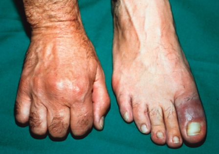
In accordance with physicochemical laws, once uric acid has passed its saturation point of 400 µmol/L (6.8 mg/dL; at 37 °C, pH 7.4), it starts to precipitate out in the form of monosodium urate crystals (13). Sites of predilection are peripheral regions of the body (e.g., the joints of the extremities) when ambient temperatures are low and inflamed joints (14) (figure 1). Urate crystals lead to activation of the NALP3 inflammasome with release of proinflammatory cytokines, among them interleukins 1, 18, and 8, and of tumor necrosis factor, attracting more polymorphonuclear neutrophilic granulocytes (e10– e12).
Figure 1.
Acute gout attack with classic podagra and synovitis in the second metacarpophalangeal joint
The usual triggers of gout attacks are a sudden rise in serum urate, e.g., after excessive eating and alcohol intake (4, 5). A rapid drop in serum urate, as for example at the start of urate-lowering therapy, can also trigger an attack of gout. In this case the release of urate from the margins of crystal deposits as a result of the concentration gradient between serum and tissue seems to stimulate an immune response (15, e13). The typical first manifestation of gout is an acute episode of monoarticular arthritis at the metatarsophalangeal joint of the large toe (podagra) that is very painful, starts at night, lasts around a week, and in many cases is self-limiting (e14) (figure 1).
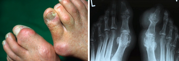
The deposition of urate crystals in various tissues such as joints, connective tissue, and kidneys explains the chronic character of the gout. Almost 90% of patients who have suffered an attack of gout experience repeat episodes during the following 5 years (16). In the course of the disease atypical manifestations may be seen: other joints (e.g., finger joints) may be affected, and oligoarticular or polyarticular arthritis may develop (figure 2). The differential diagnosis includes other crystal-induced forms of arthritis such as pseudogout/chondrocalcinosis with deposition of calcium pyrophosphate dihydrate crystals, and oxalosis arthropathies (e.g., secondary calcium oxalate deposits in patients on long-term dialysis). In addition, septic arthritis, psoriatic arthritis, and hemochromatosis should be ruled out (17).
Figure 2.
Chronic gout.
a) Tophaceous gout with destructive joint changes and subcutaneous deposits of uric acid,
b) radiological changes in tophaceous gout
Diagnosing gout
A suspected diagnosis of gout may safely be made on the basis of an episode of excessive eating and/or drinking (of alcohol) in the recent history—e.g., a barbecue—when the large toe shows the typical signs of a gout attack and the serum concentration of urate is raised. It is quite common for the serum urate level to be normal or low during an attack, so the best time to measure it is 2 to 3 weeks after an attack (evidence level [EL] IV) (15, 18, 19). If the manifestation is atypical and serum urate normal, joint puncture to demonstrate the presence of crystals is highly desirable (EL IIb); the differential diagnosis in such a case includes septic arthritis (8, 20). The important thing here is to examine the untreated crystals (urate crystals dissolve in formalin) under a polarization microscope. The crystals appear as birefringent intra- and extracellular needles 10 to 20 µm in length.
Once gout has been diagnosed, the possible causes need to be identified. Since, given the appropriate genetic predisposition, it is possible that, in addition to the increased urate (often promoted by diet), cell turnover may in rare cases be increased due to the presence of occult neoplastic disease (e.g., leukemia or plasmacytosis), cell count, differential cell count, and erythrocyte sedimentation rate should be carried out, together with determination of lactate dehydrogenase and possibly serum albumin electrophoresis (EL IV) (13, e15).
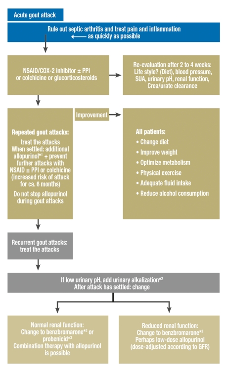
If no explanation for the gout attack is found, especially in younger patients with a family history of gout, then owing to the frequent association with impaired renal function, serum creatinine should be determined, as should 12- or 24-hour urinary clearance of creatinine and urate, and a urinary pH strip test should be performed (EL IIb) (7, 8, 11) (figure 3). Since patients with gout have an up to 2.5-times increased risk of developing urate stones, leading to urate nephropathy, the kidneys should be examined by ultrasound to rule out the presence of stones (21, 22). Because of the frequent association with other metabolic and endocrine diseases—over 50% of patients have a metabolic syndrome—the guidelines for risk stratification recommend determination of fasting blood sugar, and possibly of HbA1c, fasting blood lipids/cholesterol, and thyroid parameters (EL IIa to IIb) (1, 2, 9).
Figure 3.
Treatment algorithm for gout
*1 Dose titration of allopurinol depending on serum urate level, up to a maximum of 800 mg/d with monitoring of renal function; *2 urinary alkalization using citrate compounds to prevent urate stones (EL III); *3 not available in Austria and Switzerland; COX-2, cyclooxygenase-2; PPI, proton pump inhibitor; SUA, serum urate; Crea, creatinine; GFR, glomerular filtration rate; modified from (19)
In the early stages of gouty arthritis, erosive joint changes are only rarely seen on radiographs. Despite this, in case of doubt the affected joints should still undergo x-ray in order to rule out other causes such as osteoarthritis of the big toe MTP joint or psoriatic arthritis (EL IIb) (figure 2) (8). Joint effusions and tophi can be well demonstrated by joint ultrasonography, and this is often helpful before joint puncture or to monitor the course of the disease (EL III) (e16, e17).
Treatment of gout
The first therapeutic goal is acute treatment of the gout attack with rapid alleviation of pain and inhibition of the inflammation. A longer-term goal is to prevent further attacks, eliminate tophi, and prevent joint destruction, by consistently reducing the level of urate (9). It is postulated that gout is "curable" if existing deposits of urate crystals can be successfully removed and the formation of new precipitates prevented (16). To achieve this, according to international recommendations, serum urate levels in patients with recurring attacks of gout should if possible be kept below 360 µmol/L (6.0 mg/dL) (EL III) (8, 9, e18, e19, e20).
Acute treatment of the gout attack
In addition to nonspecific measures (EN III) such as resting and cooling the affected limb, nonsteroidal anti-inflammatory drugs (NSAIDs) are used (19). The first line of treatment is early, short-term treatment with an NSAID such as diclofenac (up to 250 mg/d) or ibuprofen (up to 2400 mg/d) or indomethacin (up to 150 mg/d), and the cyclooxygenase-2 inhibitor etoricoxib (120 mg/d) (EL Ib) (e21). If the patient has a history of gastrointestinal ulcers or bleeding, proton pump inhibitors should be given in addition (e22). Alternatively, so long as renal function is normal, colchicine may be given; at a dosage of 0.5 mg every 2 hours, this settles the gout attack within 1 day for 80% of patients (e23). However, high doses often lead to nausea and diarrhea. Given up to three times at a dose of 0.5 mg/d, it is usually well tolerated and adequately effective (EL Ib) (9, 18, e24, e25). As a further option, especially where there are contraindications, intolerance, or advanced kidney failure, glucocorticosteroids (20 to 40 mg prednisolone equivalent/d) may be given (EL Ib) (table 4) (7, 19, e26).
Table 4. Treatment for acute gout attack. Evidence levels (EL) are shown in parentheses.
| Therapeutic option (evidence level) | Comments/concerns | Complications of long-term continuation of treatment |
| Nonpharmacological | Rest, raise the limb, | |
| (EL III) | topical application of ice | |
| NSAID/COX-2 inhibitor | Not for patients with gastrointestinal ulcer or bleeding, | Gastrointestinal side effects, renal effects |
| (± PPI) | NSAID-induced asthma or impaired renal function | |
| (EL Ib) | Interaction with coumarin/warfarin | |
| Colchicine | Care is needed in patients with hepatobiliary dysfunction, acute infections, or age over 70 years | Potential for serious side effects |
| (EL Ib) | ||
| Drug interactions | ||
| Watch out for: diarrhea, gastrointestinal intolerance, and local tissue necrosis | ||
| Glucocorticosteroids | Deterioration of diabetic metabolism in patients with diabetes mellitus | High blood pressure Raised blood sugar Osteoporosis Gastrointestinal side effects |
| (EL Ib) | Possible need for additional anti-inflammatory substances/analgesia or use of moderate to high doses |
Long-term treatment to lower uric acid levels
As a general measure, the patient should be given advice on possible changes of life habits that could lead to an improvement in his or her overall metabolic profile (19). An extensive interview at the start of therapy often improves the patient‘s understanding and therefore also the compliance. Many patients fail to understand that the treatment for the acute gout attack is not treatment for the actual cause of the gout. As has been shown recently, in Germany patients often stop taking their urate-lowering medication after 3 months if they have no symptoms; at the end of a year only 30% of gout patients are still receiving allopurinol (2).
In addition to reduced consumption of purine-rich foods such as offal and seafood, patients should also limit their consumption of fructose-containing drinks (i.e., sugar-sweetened soft drinks) as these reduce the excretion of uric acid. The diet should be rich in milk and skimmed milk products and in vegetable protein (4). An important element is limited consumption of alcohol: There should be at least three alcohol-free days a week. Beer should be avoided because of its high purine content, but a glass of wine is regarded as harmless in gout (5). Careful weight loss of less than 1 kg/month with light physical exercise is desirable (e27); more rapid loss of weight could lead to ketoacidosis, provoking gout attacks. Patients with a history of kidney stones are recommended to drink more than 2 L/d. These changes in life habits (EL IIb to IV), however, usually make only a moderate contribution to urate reduction. A consistently purine-poor diet, for example, may be expected to result in a 10% to 15% reduction in serum urate concentration (4). Given that for many gout patients the serum urate concentration measured before the start of treatment is 480 µmol/L (8 mg/dL), such a reduction would result in a value of 400 µmol/L (6.7 mg/dL)—still above the required target range for gout patients (19).
Indications for and timing of additional urate-lowering drug therapy
Although it may be difficult to implement in practice, it is advisable to start patients who have suffered a second attack of gout within a year on a course of drug therapy to increase the excretion of uric acid in the urine. Other indications for urate-lowering drug therapy are destructive joint changes and/or tophi and the presence of gout-related kidney failure (urate nephropathy) with or without uric acid stones (EL IIb to IV) (19, e28, e29).
If at all possible, the treatment should not start during an acute attack, since the dissolution of crystal deposits increases the risk of gout attacks (EL IV) (e30). If possible, it is advisable to provide the patient with NSAIDs during the attack, and to determine urate levels at a follow-up visit 2 to 3 weeks later before starting treatment. The inexpensive xanthine oxidase inhibitor allopurinol has been available for urate-lowering therapy in Germany since 1964 (e31). Allopurinol should be gradually titrated up with the serum urate level being monitored. Treatment starts with 100 mg and depending on the urate value is increased by 50 to 100 mg per week until a maximum of 800 mg is reached (EL III) (19). In a patient with reduced kidney function, the dosage must be matched to the glomerular filtration rate (GFR < 30 mL/min: 100 mg allopurinol every second day) (23). This is important to prevent toxic accumulation of oxipurinol, a long-acting metabolite of allopurinol, and the risk of development of a hypersensitivity to allopurinol (23, e32, e33). Although 2% of patients given allopurinol have an uncomplicated skin reaction (rash and itching), in 1 out of 300 a hypersensitivity reaction can occur which in 20% of cases is fatal (e34). Typical features are generalized itching with eczema-type skin changes, raised liver values, and eosinophilia, which can occur after a latency period of weeks or months after the first administration of allopurinol (24, e33). Patients with allopurinol intolerance, inadequate reduction of urate, and/or primary hyperuricemia with impaired renal excretion of urate (<800 mg/24 h) but otherwise normal renal function, can be treated with the uricosuric drugs benzbromarone (20 to 100 mg/d) or probenecid (1 to 3 g/d in three separate doses); liver values must be monitored. For kidney stone prophylaxis and additional improvement of urate excretion, patients with low urinary pH may be given urine-alkalizing substances (EL Ia to III) (7, e35) (table 1).
Some interesting results have been recently published from a randomized study comparing the three available substances in relation to their urate-lowering effect, with the target value for serum urate set at below 300 µmol/L (5.0 mg/dL). Among the patients given 300 mg/d allopurinol, only 24% achieved optimal urate reduction; the comparable figures were 92% of those given 200 mg/d benzbromarone and 65% of those given 2000 mg/d probenecid (EL Ib) (e36). If more gout attacks occur during urate-lowering treatment, colchicine (0.5 mg/d) is recommended for the first 6 weeks to 6 months for prophylaxis (EL Ib). In our experience, however, this is very rarely required (7, 9, 19, e37).
As a rule, urate reduction needs to be continued for several years, often life-long; however, this is a decision that needs to be made individually in each case (e18, e38, e39). Controlled urate-lowering therapy carried out over a long period results in patients remaining free of attacks for a long time after treatment has ceased (EL III) (25, e40). It is important to monitor serum urate concentrations regularly during the treatment (EL IV) (7, 19).
Future prospects
The recently licensed xanthine oxidase inhibitor febuxostat appears to be an alternative treatment for example in patients with allopurinol intolerance, contraindications for uricosurics, or when uricosurics are unavailable (benzbromarone and probenecid are not available in Austria and Switzerland). An advantage is that febuxostat can be used in patients with renal failure as it is metabolized in the liver. Current studies have shown that with a comparable side-effects profile, more effective urate reduction is achieved: 53% of patients given febuxostat 80 mg/d and 62% of those given 120 mg/d had serum urate concentrations below 360 µmol/L (6.0 mg/dL), compared to 21% of those treated with 300 mg/d allopurinol. However, it could not be shown that febuxostat was superior to allopurinol in reducing gout attacks as a clinical parameter (e41, e42). Febuxostat would therefore be a candidate alternative drug for use when allopurinol cannot be used because of intolerance or other contraindications, or when uricosuric treatment has reached its limits or is not possible. As a novel drug, the daily treatment costs will no doubt be much higher than those of allopurinol (which are a matter of cents).
Therapy using recombinant uratoxidase or its longer-acting pegylated form to degrade urate into the easily water-soluble allantoin is at present licensed only for use in tumor lysis syndrome. Partly because of large numbers of severe anaphylactic reactions due to possible antigenicity caused by an animal element, and because of high costs amounting to around 10 000 euros/year, this treatment can only be considered off-label in rare cases of severe tophaceous gout. First results of phase 2 studies have been published (e43, e44).
Acknowledgments
Translated from the original German by Kersti Wagstaff, MA.
Footnotes
Conflict of interest statement
Dr. Tausche and Prof. Dr. Müller-Ladner have received lecture and consultancy fees from Ipsen Pharma S.A., France.
Dr. Jansen, Prof. Schröder, Prof. Bornstein, and Prof. Aringer declare that they have no conflict of interest as defined by the guidelines of the International Committee of Medical Journal Editors.
References
- 1.Mikuls TR, Farrar JT, Bilker WB, Fernandes S, Schumacher HR, Jr, Saag KG. Gout epidemiology: results from the UK General Practice Research Database, 1990-1999. Ann Rheum Dis. 2005;64:267–272. doi: 10.1136/ard.2004.024091. [DOI] [PMC free article] [PubMed] [Google Scholar]
- 2.Annemans L, Spaepen E, Gaskin M, et al. Gout in the UK and Germany: prevalence, comorbidities and management in general practice 2000-2005. Ann Rheum Dis. 2008;67:960–966. doi: 10.1136/ard.2007.076232. [DOI] [PMC free article] [PubMed] [Google Scholar]
- 3.Campion EW, Glynn RJ, DeLabry LO. Asymptomatic hyperuricemia. Risks and consequences in the normative aging study. Am J Med. 1987;82:421–426. doi: 10.1016/0002-9343(87)90441-4. [DOI] [PubMed] [Google Scholar]
- 4.Choi HK, Atkinson K, Karlson EW, Willett W, Curhan G. Purine-rich foods, dairy and protein intake, and the risk of gout in men. N Engl J Med. 2004;350:1093–1103. doi: 10.1056/NEJMoa035700. [DOI] [PubMed] [Google Scholar]
- 5.Choi HK, Atkinson K, Karlson EW, Willett W, Curhan G. Alcohol intake and risk of incident gout in men: a prospective study. Lancet. 2004;363:1277–1281. doi: 10.1016/S0140-6736(04)16000-5. [DOI] [PubMed] [Google Scholar]
- 6.Alamanos Y, Voulgari PV, Drosos AA. Incidence and prevalence of rheumatoid arthritis, based on the 1987 American College of Rheumatology criteria: a systematic review. Semin Arthritis Rheum. 2006;36:182–188. doi: 10.1016/j.semarthrit.2006.08.006. [DOI] [PubMed] [Google Scholar]
- 7.Tausche AK, Unger S, Richter K, et al. Hyperuricemia and gout: diagnosis and therapy. Internist (Berl) 2006;47:509–522. doi: 10.1007/s00108-006-1578-y. [DOI] [PubMed] [Google Scholar]
- 8.Zhang W, Doherty M, Pascual E, et al. EULAR evidence based recommendations for gout. Part I: Diagnosis. Report of a task force of the Standing Committee for International Clinical Studies Including Therapeutics (ESCISIT) Ann Rheum Dis. 2006;65:1301–1311. doi: 10.1136/ard.2006.055251. [DOI] [PMC free article] [PubMed] [Google Scholar]
- 9.Zhang W, Doherty M, Bardin T, et al. EULAR evidence based recommendations for gout. Part II: Management. Report of a task force of the EULAR Standing Committee for International Clinical Studies Including Therapeutics (ESCISIT) Ann Rheum Dis. 2006;65:1312–1324. doi: 10.1136/ard.2006.055269. [DOI] [PMC free article] [PubMed] [Google Scholar]
- 10.Unger S, Tausche AK, Kopprasch S, Bornstein SR, Aringer M, Grässler J. Molecular basis of primary renal hyperuricemia: role of the human urate transporter hURAT1. Z Rheumatol. 2007;66:58–61. doi: 10.1007/s00393-007-0208-y. [DOI] [PubMed] [Google Scholar]
- 11.Graessler J, Graessler A, Unger S, et al. Association of the human urate transporter 1 with reduced renal uric acid excretion and hyperuricemia in a German Caucasian population. Arthritis Rheum. 2006;54:292–300. doi: 10.1002/art.21499. [DOI] [PubMed] [Google Scholar]
- 12.Graessler J, Unger S, Tausche AK, Kopprasch S, Bornstein SR. Gout - new insights into a forgotten disease. Horm Metab Res. 2006;38:203–204. doi: 10.1055/s-2006-925203. [DOI] [PubMed] [Google Scholar]
- 13.Choi HK, Mount DB, Reginato AM. American College of Physicians; American Physiological Society: Pathogenesis of gout. Ann Intern Med. 2005;143:499–516. doi: 10.7326/0003-4819-143-7-200510040-00009. [DOI] [PubMed] [Google Scholar]
- 14.Harris MD, Siegel LB, Alloway JA. Gout and hyperuricemia. Am Fam Physician. 1999;59:925–934. [PubMed] [Google Scholar]
- 15.Urano W, Yamanaka H, Tsutani H, et al. The inflammatory process in the mechanism of decreased serum uric acid concentrations during acute gouty arthritis. J Rheumatol. 2002;29:1950–1953. [PubMed] [Google Scholar]
- 16.Wortmann RL. The management of gout: it should be crystal clear. J Rheumatol. 2006;33 [PubMed] [Google Scholar]
- 17.McGill NW. Gout and other crystal-associated arthropathies. Baillieres Best Pract Res Clin Rheumatol. 2000;14:445–460. doi: 10.1053/berh.2000.0087. [DOI] [PubMed] [Google Scholar]
- 18.Borstad GC, Bryant LR, Abel MP, Scroggie DA, Harris MD, Alloway JA. Colchicine for prophylaxis of acute flares when initiating allopurinol for chronic gouty arthritis. J Rheumatol. 2004;31:2429–2432. [PubMed] [Google Scholar]
- 19.Jordan KM, Cameron JS, Snaith M, et al. British Society for Rheumatology and British Health Professionals in Rheumatology Standards, Guidelines and Audit Working Group (SGAWG): British Society for Rheumatology and British Health Professionals in Rheumatology guideline for the management of gout. Rheumatology (Oxford) 2007;46:1372–1374. doi: 10.1093/rheumatology/kem056a. [DOI] [PubMed] [Google Scholar]
- 20.Lumbreras B, Pascual E, Frasquet J, González-Salinas J, Rodríguez E, Hernández-Aguado I. Analysis for crystals in synovial fluid: training of the analysts results in high consistency. Ann Rheum Dis. 2005;64:612–615. doi: 10.1136/ard.2004.027268. [DOI] [PMC free article] [PubMed] [Google Scholar]
- 21.Shekarriz B, Stoller ML. Uric acid nephrolithiasis: current concepts and controversies. J Urol. 2002;168:1307–1314. doi: 10.1016/S0022-5347(05)64439-4. [DOI] [PubMed] [Google Scholar]
- 22.Kramer HJ, Choi HK, Atkinson K, Stampfer M, Curhan GC. The association between gout and nephrolithiasis in men: The Health Professionals’ Follow-Up Study. Kidney Int. 2003;64:1022–1026. doi: 10.1046/j.1523-1755.2003.t01-2-00171.x. [DOI] [PubMed] [Google Scholar]
- 23.Hande KR, Noone RM, Stone WJ. Severe allopurinol toxicity: description and guidelines for prevention in patients with renal insufficiency. Am J Med. 1984;76:47–56. doi: 10.1016/0002-9343(84)90743-5. [DOI] [PubMed] [Google Scholar]
- 24.Tausche AK, Aringer M, Schroeder HE, Bornstein SR, Wunderlich C, Wozel G. The janus faces of allopurinol - allopurinol hypersensitivity syndrome. Am J Med. 2008;121:e3–e4. doi: 10.1016/j.amjmed.2007.10.028. [DOI] [PubMed] [Google Scholar]
- 25.Perez-Ruiz F, Atxotegi J, Hernando I, Calabozo M, Nolla JM. Using serum urate levels to determine the period free of gouty symptoms after withdrawal of long-term urate-lowering therapy: a prospective study. Arthritis Rheum. 2006;55:786–790. doi: 10.1002/art.22232. [DOI] [PubMed] [Google Scholar]
- e1.Feig DI, Kang DH, Johnson RJ. Uric acid and cardiovascular risk. N Engl J Med. 2008;359:1811–1821. doi: 10.1056/NEJMra0800885. [DOI] [PMC free article] [PubMed] [Google Scholar]
- e2.Brand FN, McGee DL, Kannel WB, Stokes J, 3rd, Castelli WP. Hyperuricemia as a risk factor of coronary heart disease: The Framingham Study. Am J Epidemiol. 1985;121:11–18. doi: 10.1093/oxfordjournals.aje.a113972. [DOI] [PubMed] [Google Scholar]
- e3.Julius U, Schulze J, Hanefeld M. Ätiologie und Pathogenese des Metabolischen Syndroms. Diabetes und Stoffwechsel. 2005;14:333–349. [Google Scholar]
- e4.Schröder HE. Gicht. In: Hartmann F, Philipp T, editors. Klinik der Gegenwart. München, Wien, Baltimore: Urban und Schwarzenberg; 1993. pp. 1–48. [Google Scholar]
- e5.Wallace KL, Riedel AA, Joseph-Ridge N, Wortmann R. Increasing prevalence of gout and hyperuricemia over 10 years among older adults in a managed care population. J Rheumatol. 2004;31:1582–1587. [PubMed] [Google Scholar]
- e6.Weaver AL. Epidemiology of gout. Cleve Clin J Med. 2008;75:9–12. doi: 10.3949/ccjm.75.suppl_5.s9. [DOI] [PubMed] [Google Scholar]
- e7.Saag KG, Choi H. Epidemiology, risk factors, and lifestyle modifications for gout. Arthritis Res Ther. 2006;8 doi: 10.1186/ar1907. [DOI] [PMC free article] [PubMed] [Google Scholar]
- e8.Vitart V, Rudan I, Hayward C, et al. SLC2A9 is a newly identified urate transporter influencing serum urate concentration, urate excretion and gout. Nat Genet. 2008;40:437–442. doi: 10.1038/ng.106. [DOI] [PubMed] [Google Scholar]
- e9.Aringer M, Graessler J. Understanding deficient elimination of uric acid. Lancet. 2008;372:1929–1930. doi: 10.1016/S0140-6736(08)61344-6. [DOI] [PubMed] [Google Scholar]
- e10.Martinon F, Pétrilli V, Mayor A, Tardivel A, Tschopp J. Gout-associated uric acid crystals activate the NALP3 inflammasome. Nature. 2006;440:237–241. doi: 10.1038/nature04516. [DOI] [PubMed] [Google Scholar]
- e11.Pétrilli V, Dostert C, Muruve DA, Tschopp J. The inflammasome: a danger sensing complex triggering innate immunity. Curr Opin Immunol. 2007;19:615–622. doi: 10.1016/j.coi.2007.09.002. [DOI] [PubMed] [Google Scholar]
- e12.Dalbeth N, Haskard DO. Inflammation and tissue damage in crystal deposition diseases. Curr Opin Rheumatol. 2005;17:314–318. doi: 10.1097/01.bor.0000157041.12116.69. [DOI] [PubMed] [Google Scholar]
- e13.Rose DM, Liu-Bryan R. Innate immunity in triggering and resolution of acute gouty inflammation. Curr Rheumatol Rep. 2006;8:209–214. doi: 10.1007/s11926-996-0027-1. [DOI] [PubMed] [Google Scholar]
- e14.Bellamy N, Downie WW, Buchanan WW. Observations on spontaneous improvement in patients with podagra: implications for therapeutic trials of non-steroidal anti-inflammatory drugs. Br J Clin Pharmacol. 1987;24:33–36. doi: 10.1111/j.1365-2125.1987.tb03132.x. [DOI] [PMC free article] [PubMed] [Google Scholar]
- e15.Smyth CJ. Disorders associated with hyperuricemia. Arthritis Rheum. 1975;18:713–719. doi: 10.1002/art.1780180712. [DOI] [PubMed] [Google Scholar]
- e16.Schueller-Weidekamm C, Schueller G, Aringer M, Weber M, Kainberger F. Impact of sonography in gouty arthritis: Comparison with conventional radiography, clinical examination, and laboratory findings. Eur J Radiol. 2007;62:437–442. doi: 10.1016/j.ejrad.2006.12.005. [DOI] [PubMed] [Google Scholar]
- e17.Puig JG, de Miguel E, Castillo MC, Rocha AL, Martínez MA, Torres RJ. Asymptomatic hyperuricemia: impact of ultrasonography. Nucleosides Nucleotides Nucleic Acids. 2008;27:592–595. doi: 10.1080/15257770802136040. [DOI] [PubMed] [Google Scholar]
- e18.Loebl WY, Scott JT. Withdrawal of allopurinol in patients with gout. Ann Rheum Dis. 1974;33:304–307. doi: 10.1136/ard.33.4.304. [DOI] [PMC free article] [PubMed] [Google Scholar]
- e19.Li-Yu J, Clayburne G, Sieck M, et al. Treatment of chronic gout. Can we determine when urate stores are depleted enough to prevent attacks of gout? J Rheumatol. 2001;28:577–580. [PubMed] [Google Scholar]
- e20.Shoji A, Yamanaka H, Kamatani N. A retrospective study of the relationship between serum urate level and recurrent attacks of gouty arthritis: evidence for reduction of recurrent gouty arthritis with antihyperuricemic therapy. Arthritis Rheum. 2004;51:321–325. doi: 10.1002/art.20405. [DOI] [PubMed] [Google Scholar]
- e21.Rubin BR, Burton R, Navarra S, et al. Efficacy and safety profile of treatment with etoricoxib 120 mg once daily compared with indomethacin 50 mg three times daily in acute gout: a randomized controlled trial. Arthritis Rheum. 2004;50:598–606. doi: 10.1002/art.20007. [DOI] [PubMed] [Google Scholar]
- e22.Scheiman JM. Prevention of NSAID-induced ulcers. Curr Treat Options Gastroenterol. 2008;11:125–134. doi: 10.1007/s11938-008-0025-7. [DOI] [PubMed] [Google Scholar]
- e23.Ahern MJ, Reid C, Gordon TP, McCredie M, Brooks PM, Jones M. Does colchicine work? The results of the first controlled study in acute gout. Aust N Z J Med. 1987;17:301–304. doi: 10.1111/j.1445-5994.1987.tb01232.x. [DOI] [PubMed] [Google Scholar]
- e24.Nuki G. Colchicine: its mechanism of action and efficacy in crystal-induced inflammation. Curr Rheumatol Rep. 2008;10:218–227. doi: 10.1007/s11926-008-0036-3. [DOI] [PubMed] [Google Scholar]
- e25.Sutaria S, Katbamna R, Underwood M. Effectiveness of interventions for the treatment of acute and prevention of recurrent gout—a systematic review. Rheumatology (Oxford) 2006;45:1422–1431. doi: 10.1093/rheumatology/kel071. [DOI] [PubMed] [Google Scholar]
- e26.Janssens HJ, Janssen M, van de Lisdonk EH, van Riel PL, van Weel C. Use of oral prednisolone or naproxen for the treatment of gout arthritis: a double-blind, randomised equivalence trial. Lancet. 2008;371:1854–1860. doi: 10.1016/S0140-6736(08)60799-0. [DOI] [PubMed] [Google Scholar]
- e27.Williams PT. Effects of diet, physical activity and performance, and body weight on incident gout in ostensibly healthy, vigorously active men. Am J Clin Nutr. 2008;87:1480–1487. doi: 10.1093/ajcn/87.5.1480. [DOI] [PMC free article] [PubMed] [Google Scholar]
- e28.Gibson T, Rodgers V, Potter C, Simmonds HA. Allopurinol treatment and its effect on renal function in gout: a controlled study. Ann Rheum Dis. 1982;41:59–65. doi: 10.1136/ard.41.1.59. [DOI] [PMC free article] [PubMed] [Google Scholar]
- e29.Ferraz MB, O‘Brien B. A cost effectiveness analysis of urate lowering drugs in nontophaceous recurrent gouty arthritis. J Rheumatol. 1995;22:908–914. [PubMed] [Google Scholar]
- e30.Delbarre F, Amor B, Auscher C, de Gery A. Treatment of gout with allopurinol. A study of 106 cases. Ann Rheum Dis. 1966;25:627–633. doi: 10.1136/ard.25.suppl_6.627. [DOI] [PMC free article] [PubMed] [Google Scholar]
- e31.Yu TF. The effect of allopurinol in primary and secondary gout. Arthritis Rheum. 1965;8:905–906. doi: 10.1002/art.1780080456. [DOI] [PubMed] [Google Scholar]
- e32.Lupton GP, Odom RB. The allopurinol hypersensitivity syndrome. Am Acad Dermatol. 1979;1:365–374. doi: 10.1016/s0190-9622(79)70031-4. [DOI] [PubMed] [Google Scholar]
- e33.Singer JZ, Wallace SL. The allopurinol hypersensitivity syndrome. Unnecessary morbidity and mortality. Arthritis Rheum. 1986;29:82–87. doi: 10.1002/art.1780290111. [DOI] [PubMed] [Google Scholar]
- e34.Lee HY, Ariyasinghe JT, Thirumoorthy T. Allopurinol hypersensitivity syndrome: a preventable severe cutaneous adverse reaction? Singapore Med J. 2008;49:384–387. [PubMed] [Google Scholar]
- e35.Pak CY, Sakhaee K, Fuller C. Successful management of uric acid nephrolithiasis with potassium citrate. Kidney Int. 1986;30:422–428. doi: 10.1038/ki.1986.201. [DOI] [PubMed] [Google Scholar]
- e36.Reinders MK, van Roon EN, Jansen TL, et al. Efficacy and tolerability of urate lowering drugs in gout: a randomised controlled trial of benzbromarone versus probenecid after failure of allopurinol. Ann Rheum Dis. 2009;68:51–56. doi: 10.1136/ard.2007.083071. [DOI] [PubMed] [Google Scholar]
- e37.Paulus HE, Schlosstein LH, Godfrey RG, Klinenberg JR, Bluestone R. Prophylactic colchicine therapy of intercritical gout. A placebo-controlled study of probenecid-treated patients. Arthritis Rheum. 1974;17:609–614. doi: 10.1002/art.1780170517. [DOI] [PubMed] [Google Scholar]
- e38.Pascual E, Sivera F. Time required for disappearance of urate crystals from synovial fluid after successful hypouricaemic treatment relates to the duration of gout. Ann Rheum Dis. 2007;66:1056–1058. doi: 10.1136/ard.2006.060368. [DOI] [PMC free article] [PubMed] [Google Scholar]
- e39.Gast LF. Withdrawal of longterm antihyperuricemic therapy in tophaceous gout. Clin Rheumatol. 1987;6:70–73. doi: 10.1007/BF02201004. [DOI] [PubMed] [Google Scholar]
- e40.Lieshout-Zuidema MF, Breedveld FC. Withdrawal of longterm antihyperuricemic therapy in tophaceous gout. J Rheumatol. 1993;20:1383–1385. [PubMed] [Google Scholar]
- e41.Becker MA, Schumacher HR, Wortmann RL, et al. Febuxostat compared with allopurinol in patients with hyperuricemia and gout. N Engl J Med. 2005;353:2450–2461. doi: 10.1056/NEJMoa050373. [DOI] [PubMed] [Google Scholar]
- e42.Hair PI, McCormack PL, Keating GM. Febuxostat. Drugs. 2008;68:1865–1874. doi: 10.2165/00003495-200868130-00006. [DOI] [PubMed] [Google Scholar]
- e43.Fels E, Sundy JS. Refractory gout: what is it and what to do about it? Curr Opin Rheumatol. 2008;20:198–202. doi: 10.1097/BOR.0b013e3282f4eff5. [DOI] [PubMed] [Google Scholar]
- e44.Sundy JS, Becker MA, Baraf HS, et al. Reduction of plasma urate levels following treatment with multiple doses of pegloticase (polyethylene glycol-conjugated uricase) in patients with treatment-failure gout: Results of a phase II randomized study. Arthritis Rheum. 2008;58:2882–2891. doi: 10.1002/art.23810. [DOI] [PubMed] [Google Scholar]


