Abstract
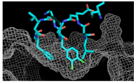
There is great interest in molecules capable of inhibiting the interactions between p53 and its negative regulators hDM2 and hDMX, as these molecules have validated potential against cancers in which one or both oncoproteins are overexpressed. We reported previously that appropriately substituted β3-peptides inhibit these interactions and, more recently, that minimally cationic β3-peptides are sufficiently cell permeable to upregulate p53-dependent genes in live cells. These observations, coupled with the known stability of β-peptides in a cellular environment, and the recently reported structures of hDM2 and hDMX, motivated us to exploit computational modeling to identify β-peptides with improved potency and/or selectivity. This exercise successfully identified a new β3-peptide, β53-16, that possesses the highly desirable attribute of high affinity for both hDM2 as well as hDMX and identifies the 3,4-dichlorophenyl moiety as a novel determinant of hDMX affinity.
There is great interest in molecules that inhibit interactions between p53 and its negative regulators hDM2 and hDMX, as these molecules have validated potential against cancers that overexpress one or both of these oncoproteins.1,2 We reported that substituted β3-peptides can inhibit these interactions3,4 and, more recently, that minimally cationic β3-peptides are sufficiently cell permeable to upregulate p53-dependent genes in live cells.5,6 These observations, coupled with the established intracellular stability of β-peptides7–9 and the recently reported structures of hDM210 and hDMX,11 motivated us to exploit computational methods to identify β-peptides with improved potency and/or selectivity. This exercise successfully identified a new β3-peptide, β53-16, that possesses the desirable attribute of high affinity for hDM2 and hDMX and identifies the 3,4-dichlorophenyl moiety as a novel determinant of hDMX affinity.
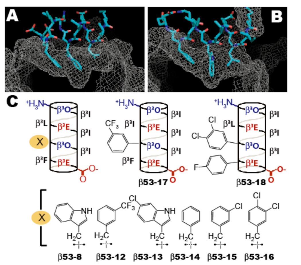
Our computational modeling began with the application of Visual Molecular Dynamics (VMD)12 to generate a model of previously reported β53-8 bound to the p53 binding site on hDM2 (Figure 1A). In this model, β53-8 is bound as a 14-helix that is slightly unwound at the C-terminus, mimicking its conformation in solution.13 The three hDM2 hydrophobic pockets occupied in the native structure by the p53 side chains of Leu26, Trp23 and Phe19 10 are occupied in the modeled complex by the corresponding β3-amino acid side chains at positions 3, 6, and 9. An analogous model of β53-8 bound to hDMX was also prepared (Figure 1B).11
Figure 1.
Computationally generated models of β53-8 (blue) in complex with (a) hDM2 and (b) hDMX illustrating differences in binding site topologies. (c) Helical net representations of β3-peptides studied herein.
We then applied a hierarchical computational strategy to search for alternative side chains that would improve packing at one or both interfaces. With the de novo design program BOMB14 we screened over ten thousand β53-8 analogs containing substituted aromatic and non-aromatic heterocycles and short hydrocarbon side chains in place of Leu26, Trp23 and Phe19.10 About 50 candidates were identified by scoring and visualization for evaluation with MCPRO.15 Binding free energies were predicted via Monte Carlo Free Energy Perturbation (MC/FEP) calculations using the OPLS-AA force field16 for the protein-ligand complex and the TIP4P model for water.17 In these simulations, the protein backbones remained fixed; the affinities of the eight most interesting and synthetically accessible compounds (Figure 1C) were subsequently reevaluated in a second round of MC/FEP calculations that permitted backbone motions.18
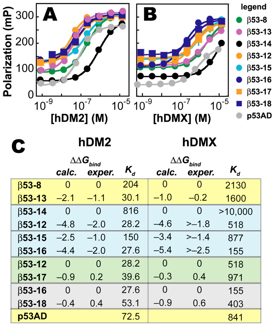
The models were first validated by evaluating whether they would predict the large increase in hDM2 affinity realized when the tryptophan side chain at position 6 is replaced by 6-chlorotryptophan (6-ClW) (compare β53-8 and β53-13,Figure 1C).19 The calculations predict that substitution of 6-ClW at position 6 should significantly improve binding to hDM2 (ΔΔG = –2.1 kcal•mol−1) but not hDMX (ΔΔG = +1.0 kcal•mol−1,Figure 2C). These predictions are fully aligned with the experimental results: the stability of the hDM2•β53-13 complex is significantly higher (Kd = 30.1 nM, ΔG = –10.25 kcal•mol−1) than that of the hDM2•β53-8 complex (Kd = 204 nM, ΔG = –9.12 kcal•mol−1), whereas the stabilities of the analogous hDMX complexes are comparable (Kd = 1.6 and 2.1 µM for β53-13 and β53-8, respectively). The improvement in hDM2 but not hDMX affinity upon substitution of 6-ClW is consistent with results observed in the context of previously reported ligands.20–23
Figure 2.
Direct fluorescence polarization analysis of the affinity of each β-peptide shown for (A) hDM2 and (B) hDMX. (C) Comparison of calculated and experimental binding free energies expressed in terms of ΔΔGbind relative to the standard shown (kcal•mol−1); Kd values in nM units.
The models were further validated by their ability to predict the large increase in hDM2 and hDMX affinity observed for β-peptides containing a central meta-trifluoromethyl phenyl substituent (CF3F) when compared with an unsubstituted phenyl ring (compare β53-12 with β53-14,Figure 1C). The calculations predict that the CF3F side chain should favor binding to both hDM2 and hDMX (ΔΔG = –4.8 and –4.6 kcal•mol−1, respectively). This increase was also realized experimentally, albeit in an attenuated way: the stability of the hDM2•β53-12 complex is significantly higher (Kd = 28.2 nM, ΔG = –10.29 kcal•mol−1) than that of the hDM2•β53-14 complex (Kd = 816 nM, ΔG = –8.3 kcal•mol−1); analogous differences are seen for the hDMX complexes (Figure 2).24
Next we examined whether the affinity of β53-12 could be increased further by substituting the leucine side chain at position 6 with one of eight cyclic and acyclic hydrocarbon alternatives. Although few promising candidates emerged from the BOMB and MC/FEP analyses, we did investigate β53-17, in which the Leu side chain is replaced by Ile. This substitution was predicted to slightly favor the binding of both hDM2 and hMDX (ΔΔGbind = –0.9 and –0.3 kcal•mol−1, respectively). However, no increase in affinity was observed, and these molecules were not studied further. We note that 53-17 is significantly less 14-helical than 53-12 as judged by circular dichroism analysis (Figure SI-1). As the computational model does not account for changes in β-peptide secondary structure, it is possible that the observed change in secondary structure accounts for the poor agreement between prediction and experiment in this case. The predictions may be also be affected by uncertainty in the structures of unliganded hDM2 and hDMX as the 23 N-terminal residues of both proteins are only partially resolved due to their flexibility.25,26
Based on these observations, we returned attention to the central side chain of the hDM2/hDMX epitope and evaluated the relative hDM2 and hDMX affinities of hundreds of β53-12 analogs containing substituted phenylalanine analogs at position 6. This analysis suggested that β-peptides containing either meta-chlorophenylalanine or para-chlorophenylalanine at this position would show improved affinity for both hDM2 and hDMX when compared with β53-14 (−3.5 kcal•mol−1 > ΔΔGbind > −2.5 kcal•mol−1). Indeed, the stability of the hDM2•β53-15 complex (meta-chloro substituent,Figure 1C) is significantly higher (Kd = 150 nM, ΔG = −9.3 kcal•mol−1) than that of hDM2·β53-14; analogous differences are observed for the hDMX complexes (Figure 2). However, as predicted, the stabilities of the β53-15 complexes were not greater than those of the β53-12 complexes. Therefore, since the gains in affinity for the para-chlorophenylalanine were predicted to be similar to those of 53-15, this additional analog was not tested experimentally.
Finally we examined the effect of a meta,para-dichlorophenylalanine side chain at the central position of the recognition epitope (β53-16,Figure 1), whose inclusion was predicted to significantly improve affinity for both hDM2 and hDMX compared to β53-14 (ΔΔGbind = −4.4 and −5.4 kcal•mol−1, respectively. Indeed, the stabilities of both hDM2•β53-16 and hDMX•β53-16 are significantly higher than the corresponding β53-14 complexes (Kd = 27.6 nM and 155 nM, respectively for β53-16,Figure 2). They also equal or exceed the stabilities of the corresponding complexes with 53-12. Competition fluorescence polarization experiments confirm that β53-16 competes with p53AD for binding to hDM2 and hDMX and shows improved inhibitory potency towards hDMX (Figure SI-2).
Thus, β53-16 offers significantly improved affinity for hDMX without loss of affinity for hDM2. Analysis of the MC/FEP simulations suggests more favorable interaction of the dichlorophenyl group with residues 50–54 in hDMX than with equivalent residues 54–58 in hDM2. We subsequently examined whether the affinity of β53-16 could be improved further upon replacement of the adjacent phenylalanine side chain with one of twelve substituted analogs. This scan failed to identify promising substitutions as the phenylalanine side chain appears to bind tightly to the hydrophobic pocket of both proteins; however minor gains in affinity for both hDM2 and hDMX were predicted for a para-fluorophenylalanine substitution (ΔΔGbind = −0.4 and −0.9 kcal•mol−1, respectively). No increase in affinity was observed experimentally with this peptide, β53-18, so further modification at this position was not pursued (Figure 1, Table 1). β53-16 represents the highest affinity β3-peptide for hDMX reported to date, with significantly higher affinity than the prototypic hDM2 ligand, Nutlin-3. Thus, β53-16 embodies the pan-specificity of well known peptidic hDM2/hDMX inhibitors27,28 without the limitations of protease sensitivity or poor uptake.
Supplementary Material
Acknowledgment
This work was supported by the NIH (GM 74756 and GM 32136) and the National Foundation for Cancer Research.
Footnotes
Supporting Information Available: Details of computational protocols, CD and binding assays and the complete citation for reference 20. This material is available free of charge via the Internet at http://pubs.acs.org
References
- 1.Chene P. Nat. Rev. Cancer. 2003;3:102–109. doi: 10.1038/nrc991. [DOI] [PubMed] [Google Scholar]
- 2.Shangary S, Wang S. Clin. Cancer Res. 2008;14:5318–5324. doi: 10.1158/1078-0432.CCR-07-5136. [DOI] [PMC free article] [PubMed] [Google Scholar]
- 3.Kritzer JA, Luedtke NW, Harker EA, Schepartz A. J. Am. Chem. Soc. 2005;127:14584–14585. doi: 10.1021/ja055050o. [DOI] [PMC free article] [PubMed] [Google Scholar]
- 4.Kritzer JA, Lear JD, Hodsdon ME, Schepartz A. J. Am. Chem. Soc. 2004;126:9468–9469. doi: 10.1021/ja031625a. [DOI] [PubMed] [Google Scholar]
- 5.Harker EA, Daniels DS, Guarracino DA, Schepartz A. Bioorg. Med. Chem. 2009;17:2038–2046. doi: 10.1016/j.bmc.2009.01.039. [DOI] [PMC free article] [PubMed] [Google Scholar]
- 6.Harker EA, Schepartz A. ChemBioChem. 2009;10:990–993. doi: 10.1002/cbic.200900049. [DOI] [PMC free article] [PubMed] [Google Scholar]
- 7.Frackenpohl J, Arvidsson PI, Schreiber JV, Seebach D. ChemBioChem. 2001;2:445–455. doi: 10.1002/1439-7633(20010601)2:6<445::aid-cbic445>3.3.co;2-i. [DOI] [PubMed] [Google Scholar]
- 8.Seebach D, Abele S, Schreiber JV, Martinoni B, Nussbaum AK, Schild H, Schulz H, Hennecke H, Woessner R, Bitsch F. Chimia. 1998;52:734–739. [Google Scholar]
- 9.Weiss HM, Wirz B, Schweitzer A, Amstutz R, Perez MIR, Andres H, Metz Y, Gardiner J, Seebach D. Chem. Biodiversity. 2007;4:1413–1437. doi: 10.1002/cbdv.200790121. [DOI] [PubMed] [Google Scholar]
- 10.Kussie PH, Gorina S, Marechal V, Elenbaas B, Moreau J, Levine AJ, Pavletich NP. Science. 1996;274:948–953. doi: 10.1126/science.274.5289.948. [DOI] [PubMed] [Google Scholar]
- 11.Popowicz GM, Czarna A, Rothweiler U, Szwagierczak A, Krajewski M, Weber L, Holak TA. Cell Cycle. 2007;6:2386–2392. doi: 10.4161/cc.6.19.4740. [DOI] [PubMed] [Google Scholar]
- 12.Humphrey W, Dalke A, Schulten K. J. Mol. Graphics. 1996;14:33–38. doi: 10.1016/0263-7855(96)00018-5. [DOI] [PubMed] [Google Scholar]
- 13.Kritzer JA, Hodsdon ME, Schepartz A. J. Am. Chem. Soc. 2005;127:4118–4119. doi: 10.1021/ja042933r. [DOI] [PMC free article] [PubMed] [Google Scholar]
- 14.Jorgensen WL. Science. 2004;303:1813–1818. doi: 10.1126/science.1096361. [DOI] [PubMed] [Google Scholar]
- 15.Jorgensen WL, Tirado-Rives J. J. Comput. Chem. 2005;26:1689–1700. doi: 10.1002/jcc.20297. [DOI] [PubMed] [Google Scholar]
- 16.Jorgensen WL, Maxwell DS, Tirado-Rives J. J. Am. Chem. Soc. 1996;118:11225–11236. [Google Scholar]
- 17.Jorgensen WL, Chandrasekhar J, Madura JD, Impey RW, Klein ML. J. Chem. Phys. 1983;79:926–935. [Google Scholar]
- 18.Ulmschneider JP, Jorgensen WL. J. Chem. Phys. 2003;118:4261–4271. [Google Scholar]
- 19.Harker EA, Daniels DS, Guarracino DA, Schepartz A. Bioorg. Med. Chem. doi: 10.1016/j.bmc.2009.01.039. In press. [DOI] [PMC free article] [PubMed] [Google Scholar]
- 20.Shangary, et al. Proc. Natl. Acad. Sci. U.S.A. 2008;105:3933–3938. doi: 10.1073/pnas.0708917105. [DOI] [PMC free article] [PubMed] [Google Scholar]
- 21.Hu B, Gilkes DM, Farooqi B, Sebti SM, Chen J. J. Biol. Chem. 2006;281:33030–33035. doi: 10.1074/jbc.C600147200. [DOI] [PubMed] [Google Scholar]
- 22.Patton JT, Mayo LD, Singhi AD, Gudkov AV, Stark GR, Jackson MW. Cancer Res. 2006;66:3169. doi: 10.1158/0008-5472.CAN-05-3832. [DOI] [PubMed] [Google Scholar]
- 23.Wade M, Wong ET, Tang M, Stommel JM, Wahl GM. J. Biol. Chem. 2006;281:33036–33044. doi: 10.1074/jbc.M605405200. [DOI] [PubMed] [Google Scholar]
- 24.The slightly larger spread of computed binding affinities has been observed previouslyJorgensen WL. Acc. Chem. Res. 2009 doi: 10.1021/ar800236t. in press.Deng Y, Roux B. J. Phys. Chem. B. 2009;113:2234–2246. doi: 10.1021/jp807701h.
- 25.Showalter SA, Bruschweiler-Li L, Johnson E, Zhang F, Bruschweiler R. J. Am. Chem. Soc. 2008;130:6472–6478. doi: 10.1021/ja800201j. [DOI] [PubMed] [Google Scholar]
- 26.Uhrinova S, Uhrin D, Powers H, Watt K, Zheleva D, Fischer P, McInnes C, Barlow PN. J. Mol. Biol. 2005;350:587–598. doi: 10.1016/j.jmb.2005.05.010. [DOI] [PubMed] [Google Scholar]
- 27.Bottger V, Bottger A, Garcia-Echeverria C, Ramos YFM, van der Eb AJ, Jochemsen AG, Lane DP. Oncogene. 1999;18:189–199. doi: 10.1038/sj.onc.1202281. [DOI] [PubMed] [Google Scholar]
- 28.Hu B, Gilkes DM, Chen J. Cancer Res. 2007;67:8810–8817. doi: 10.1158/0008-5472.CAN-07-1140. [DOI] [PubMed] [Google Scholar]
Associated Data
This section collects any data citations, data availability statements, or supplementary materials included in this article.


