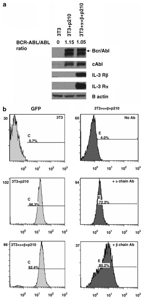
Figure 1.
Expression of human interleukin (IL)-3 Rα and IL-3 Rβ in 3T3+ α + β + P210 cells. (a) Western blotting results show the expression of Bcr-Abl, IL-3 Rα and IL-3 Rβ chains in NIH 3T3 cell transfectants. Whole cell lysates were blotted with anti-Abl antibody 8E9, or human anti-IL-3 receptors antibodies. (b) Flow cytometry assay of NIH 3T3, 3T3 +P210 and 3T3+ α + β + P210 cells after staining with conjugated mouse anti-human IL-3 Rα or IL-3 Rβ antibodies. A total of 10 000 events were analysed in each case.

