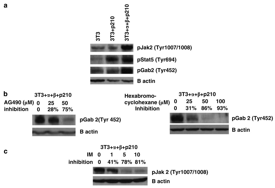
Figure 5.
Jak2, Gab2 and Stat5 are tyrosine phosphorylated in NIH 3T3 cells expressing α and β chains of the IL-3 receptor and P210 BCR-ABL. (a) The cell lysates from NIH 3T3, 3T3 + P210 and 3T3+ α + β + P210 cell lines were immunoblotted with phospho-Jak2 (Tyr 1007/1008) antibody, phospho-Gab2 (Tyr 452) antibody and phospho-Stat5 (Tyr 694) antibody. (b) 3T3+ α + β + P210 cells were treated with different concentrations of AG490 (left panel) or 1,2,3,4,5,6-hexabromocyclohexane (right panel) for 24 h. Cell lysates were immunoblotted with phospho-Gab2 (Tyr 452) antibody. (c) 3T3+ α + β + P210 cells were treated with different concentration of imatinib for 24h. Cell lysates were immunoblotted with phospho-Jak2 (Tyr 1007/1008) antibody.

