Abstract
Background and Purpose
Small, asymptomatic microbleeds commonly accompany larger symptomatic macrobleeds. It is unclear whether microbleeds and macrobleeds represent arbitrary categories within a single continuum versus truly distinct events with separate pathophysiologies.
Methods
We performed two complementary retrospective analyses. In a radiographic analysis, we measured and plotted the volumes of all hemorrhagic lesions detected by gradient-echo MRI among 46 consecutive patients with symptomatic primary lobar intracerebral hemorrhage diagnosed as probable or possible cerebral amyloid angiopathy (CAA). In a second neuropathologic analysis, we performed blinded qualitative and quantitative examinations of amyloid-positive vessel segments in 6 autopsied subjects whose MRI scans demonstrated particularly high microbleed counts (>50 microbleeds on MRI, n=3) or low microbleed counts (<3 microbleeds, n=3).
Results
Plotted on a logarithmic scale, the volumes of 163 hemorrhagic lesions identified on scans from the 46 subjects fell in a distinctly bimodal distribution with mean volumes for the two modes of 0.009 cm3 and 27.5 cm3. The optimal cut-point for separating the two peaks (determined by receiver operating characteristics) corresponded to a lesion diameter of 0.57 cm. On neuropathologic analysis, the high microbleed-count autopsied subjects showed significantly thicker amyloid-positive vessel walls than the low microbleed-count subjects (proportional wall thickness 0.53±0.01 versus 0.37±0.01, p<.0001, n=333 vessel segments analyzed).
Conclusions
These findings suggest that CAA-associated microbleeds and macrobleeds comprise distinct entities. Increased vessel wall thickness may predispose to formation of microbleeds relative to macrobleeds.
Keywords: Intracerebral hemorrhage, Microbleeds, Cerebral amyloid angiopathy
The widespread availability of MRI techniques with high sensitivity for hemosiderin has stimulated increasing interest in cerebral microbleeds.1-3 Microbleeds appear as small rounded hypointense lesions on gradient-echo T2*-weighted MRI sequences and as clusters of hemosiderin-containing macrophages, often perivascular, on histopathologic exam.4 Microbleeds can be seen in healthy aging,5-7 and at higher frequencies in ischemic stroke and particularly hemorrhagic stroke, where the prevalence is approximately 50 to 70%.1-3 Cerebral microbleeds have been implicated as a marker of, and possibly a contributor to,8-10 small vessel brain disease.
The cerebrovascular pathologies that give rise to microbleeds, such as hypertensive vasculopathy and cerebral amyloid angiopathy (CAA), are also responsible for larger symptomatic intracerebral hemorrhages (ICH). This raises the question of whether microbleeds and their larger macrobleed counterparts are simply two arbitrarily defined categories from what is actually a continuum of hemorrhage sizes resulting from vessel rupture. Alternatively, it is possible that there are key pathogenic steps that fundamentally differentiate a macrobleed from a microbleed. There is neuropathological evidence, for example, that the pathogenesis of large symptomatic ICH entails not only the initial rupture of a blood vessel, but also secondary mechanical shearing of surrounding blood vessels in order to reach full size.11
The current study sought to explore the relationship between microbleeds and macrobleeds in patients with CAA. We specifically addressed the following questions: 1) Among patients presenting with ICH, do hemorrhage volumes occur as a single unimodal distribution, or is there a natural division between the volumes of microbleeds and macrobleeds suggestive of distinct pathogeneses for these two types of hemorrhage? 2) Are there pathological characteristics of diseased blood vessels that predispose to microbleeds rather than macrobleeds (or vice versa)?
Materials and Methods
More detailed information on methods for measurement and quantitative analysis of hemorrhage volumes and arteriolar wall involvement can be found in the Supplementary Materials and Methods available on-line.
Measurement and Analysis of Hemorrhage Volumes
Analysis of hemorrhage volumes was performed on consecutive patients admitted to Massachusetts General Hospital (MGH) between January 1998 and December 2002 with symptomatic primary lobar intracerebral hemorrhage diagnosed as probable or possible CAA by the pathologically validated Boston criteria.12 Inclusion criteria were age >55 years and MRI with gradient-echo (GRE) sequences (performed as part of the admission evaluation as previously reported,13 using a 1.5T GE magnet with repetition time =750 msec, echo time =25 msec, flip angle =20°, NEX = 2, Matrix size 256×256, slice thickness 5 mm with 1 mm interslice gap) within 90 days of presentation. Subjects were excluded for unavailability of digital images or poor GRE image quality that precluded measurement of lesion volumes. Of 57 potentially eligible subjects with primary lobar ICH and gradient-echo MRI, 11 were excluded for poor image quality, yielding 46 subjects for analysis (Table 1). Clinical characteristics of subjects, and use of antiplatelet agents (aspirin in all cases) or anticoagulant agents (warfarin), were determined at the time of presentation as described.13
Table 1.
Subjects analyzed for hemorrhagic lesion volume
| Subjects analyzed (n=46) | |
|---|---|
| Age, mean ± standard deviation | 74.8 ± 7.7 |
| Female, n (%) | 23 (50) |
| Hypertension, n (%) | 26 (57) |
| Antiplatelet use, n (%) | 22 (48) |
| Anticoagulant use, n (%) | 4 (9) |
|
Number of hemorrhagic lesions per scan, median (25th, 75th percentiles) |
11 (3, 26) |
Age refers to time of MRI scan. Use of antiplatelet or anticoagulant agents refers to time of initial presentation with symptomatic intracerebral hemorrhage.
Hemorrhagic lesions were identified as hypointensities that were largest on the GRE images, excluding signal suggestive of vessel flow-voids, mineralization of the basal ganglia, cranial sinuses, or extension of a larger hemorrhage. Segmentation of hemorrhages, determination of hemorrhage volumes, statistical analysis of the distribution of hemorrhage volumes, and determination of the optimal cut-point between the two observed populations of hemorrhages are described in the Supplementary Materials and Methods.
Neuropathology of Cerebral Arterioles
Cerebral arteriolar pathology was analyzed in autopsied subjects for its association with the presence of high or low numbers of microbleeds. Neuropathological analysis was performed on consecutive patients admitted to MGH between January 1998 and December 2003 with symptomatic lobar ICH, GRE MRI performed prior to death demonstrating particularly high (defined as >50) or low (defined as <3) microbleed counts, and subsequent full brain autopsy confirming definite CAA-related ICH.12 Six such subjects were identified, three with high microbleed counts and three with low microbleed counts (Table 2). (Three of the six were among the 46 subjects analyzed for hemorrhage volume; the remaining three were not included in the hemorrhage volume analysis because of absence of adequate GRE MRI images as described above.)
Table 2.
Autopsied high- and low-microbleed count subjects
| Subject/Age/Sex | Antiplatelet Use | Hypertension | Microbleeds | Macrobleeds |
|---|---|---|---|---|
|
High microbleed count |
||||
| 1. 76F | − | + | 62 | 1 |
| 2. 75M | + | − | 58 | 1 |
| 3. 87M | + | + | 57 | 2 |
|
Low microbleed count |
||||
| 4. 67F | − | + | 0 | 6 |
| 5. 66F | + | + | 2 | 4 |
| 6. 76F | − | + | 2 | 1 |
Age refers to age at death. Antiplatelet use (aspirin in all cases) refers to time of initial presentation with symptomatic intracerebral hemorrhage. None of the autopsied subjects used anticoagulants. F=Female, M=Male.
Paraffin-embedded sections of occipital (Brodmann areas 17/18) and frontal cortex (areas 8/9) were stained for ß-amyloid (DAKO). Qualitative analysis of vessel characteristics was performed by review of all sections by a neuropathologist (M.P.F.) without knowledge of subject or neuroimaging information. In addition to this qualitative analysis, a single rater (P.D.) also blinded to subject information performed systematic quantitative analysis of CAA-positive vascular wall thickness and circumferential involvement. Methods for measurement of vascular wall thickness and circumferential involvement and statistical comparisons between the high and low microbleed-count subjects by linear mixed effects models are described in the Supplementary Materials and Methods.
Results
Volume of hemorrhagic lesions
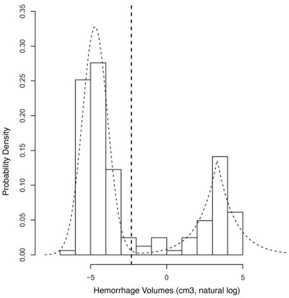
To assess whether CAA-related microbleeds and macrobleeds represent distinct size categories, we analyzed the distribution of hemorrhagic lesion volumes among 46 consecutive elderly subjects with probable or possible CAA and suitable GRE MRI images. Characteristics of the analyzed subjects are shown in Table 1 and the volumes of the 163 hemorrhages detected on these scans are plotted on a logarithmic scale as the histogram in Figure 1.
Figure 1.
Bimodal distribution of hemorrhage volumes. Volumes of 163 individual hemorrhages from 46 subjects with probable or possible CAA are plotted on a natural logarithmic scale and shown as a histogram of probability densities. The superimposed dashed curves represent a mixture model comprised of a normal distribution for the smaller volumes (mean and standard deviation of log volumes −4.7 and 0.83 respectively) and a double exponential curve (mean and standard deviation of log volumes 3.31 and 1.17) for the larger volumes, selected by goodness of fit testing (p=0.86). The dashed vertical line shows a threshold volume of 0.098 cm3 (log volume −2.319) that optimally classifies values in the two distributions. The null hypothesis that these populations of log hemorrhage volumes fit a unimodal normal distribution can be rejected (p<.0001).
Rather than forming a unimodal distribution (the null hypothesis of a single normal distribution could be rejected with p<.0001), the hemorrhagic lesion volumes distributed into two peaks. Mean volume of the higher-volume peak was 27.5 cm3 (corresponding to a diameter of 3.75 cm, assuming spherical shape). Mean volume of the lower volume peak was 0.009 cm3 (corresponding to a spherical diameter of 0.26 cm). The optimal threshold that correctly classified (with 99% probability) a hemorrhagic lesion from either subpopulation was 0.098 cm3, corresponding to a spherical diameter of 0.57 cm (vertical dashed line in Fig. 1). Exclusion of the 20 hemorrhagic lesions identified in 4 subjects taking warfarin at the time of presentation (Table 1) did not alter the bimodal appearance of the histogram (not shown).
Pathology of amyloid-positive vessels in high versus low microbleed-count CAA
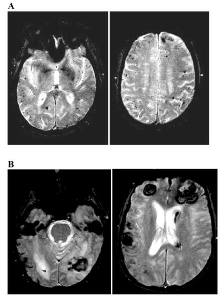
We next examined the structural basis by which CAA-affected vessels are predisposed to give rise to microbleeds as opposed to macrobleeds. We approached this question by identifying autopsied CAA subjects (Table 1) with large numbers of microbleeds (>50 on GRE MRI, see Fig. 2A) or few microbleeds (<3, see Fig. 2B). Visual inspection of sections of frontal and occipital cortex performed without knowledge of patient characteristics found noticeably increased wall thickness of amyloid-positive vessels among the high microbleed-count subjects relative to the low microbleed subjects (Fig. 3). No other qualitative pathological differences, such as the frequency or type of amyloid-positive vessels, or other secondary vascular changes such dyshoric amyloid deposits, concentric splitting of the vessel wall, or loss of smooth muscle cells,14 were observed.
Figure 2.
Representative MRI images from high and low microbleed-count subjects with subsequent neuropathological examination. Panel A shows axial GRE MRI images from subject 1 (62 microbleeds; see Table 2) and panel B from subject 4 (0 microbleeds). Each brain demonstrated definite CAA12 at autopsy.
Figure 3.

Representative vessel segments from high and low microbleed-count CAA subjects. Panel A shows cross-sectional vessel profiles from occipital cortex of high microbleed subjects (1, 2, and 3 from left to right; see Table 2), panel B from low microbleed subjects (4, 5, and 6 from left to right). Vessel walls from high microbleed subjects were significantly thicker than from low microbleed subjects. All specimens were stained by luxol fast blue-hematoxylin-eosin with original magnification 40x.
Increased wall thickness of amyloid-positive vessel segments in high microbleed versus low microbleed subjects was confirmed by blinded quantitative measurement. Proportional wall thickness (mean ± standard error) was 0.53 ± 0.01 for the high microbleed subjects versus 0.37 ± 0.01 for the low microbleed subjects (p<.0001). The association between high microbleed count and increased proportional wall thickness remained independent (p<.005) in an additional mixed effects model controlling for subject age and hypertension as fixed effects. A separate analysis of amyloid-negative white matter vessels found no difference in proportional wall thickness (0.40 ± 0.04 for high microbleed subjects, 0.36 ± 0.04 for low microbleed, p=0.45). Quantitative analysis of the circumferential extent of amyloid found circumferential involvement to be near complete in most amyloid-positive vessel (90% of 287 vessel analyzed segments scoring 12 on the 12-point scale) without significant difference between high and low microbleed subjects (p=0.19).
Discussion
We report two lines of evidence suggesting that CAA-related microbleeds and macrobleeds, though arising from a common vascular disease, represent distinct pathophysiologic events. Findings supporting this inference are that 1) hemorrhage volumes appear to fall into a bimodal distribution best modeled as a mixture of two separate populations, and 2) CAA subjects with many microbleeds demonstrate significantly thicker amyloid-positive vessels than those with few microbleeds. These essentially independent observations support a model by which symptomatic macrobleeding and asymptomatic microbleeding are distinct entities with characteristic pathophysiologies.
It is notable that the cut-point between the two hemorrhage volume populations determined by ROC analysis of the estimated two-component mixture model (Fig. 1) corresponded to a spherical diameter of 0.57 cm, a value very close to the upper size limits of 0.5 to 1 cm traditionally chosen to define microbleeds.1-3 The reasonably wide separation of the two peaks (plotted on a logarithmic scale in Fig. 1) suggests that the precise cut-point chosen probably has little effect on whether a given lesion will be classified as a microbleed or macrobleed. We note that the measurements on GRE MRI overestimate the true size of the microbleeds because of the susceptibility (“blooming”) artifact. A recent comparison of hemorrhage volumes on GRE MRI and CT suggested a correction factor of 0.8.15
The overall relationship between microbleeds and macrobleeds remains an active area of investigation. Some previous studies have suggested that the number of microbleeds predicts risk of future symptomatic ICH.13, 16 In the case of symptomatic hemorrhage following thrombolytic treatment for ischemic stroke, however, the presence of microbleeds appears not to be predictive.17, 18
The observation of increased vessel wall thickness among high relative to low microbleed-count subjects suggests that thicker vessels may render vessels more likely to produce microbleeding when they rupture. Thickening of the vessel wall and narrowing of the vascular lumen has long been noted as characteristic of CAA,14, 19 but the factors determining degree of wall thickness are largely unknown. Severe wall thickening occurs in Iowa-type hereditary CAA,20 a familial form of CAA also characterized by multiple microbleeds without symptomatic hemorrhage among 10 affected members of the originally identified pedigree. Whether similar considerations of microbleeding and vessel wall thickness apply to vasculopathies other than CAA, such as hypertensive hemorrhage,11 remains to be determined. In cerebral autosomal dominant arteriopathy with subcortical infarcts and leukoencephalopathy (CADASIL), another vascular pathology associated with severe thickening of the arterial wall and loss of normal wall elements, microbleeds are common but symptomatic macrobleeds are rare,9, 21-23 suggesting possible similarities to the CAA high microbleed group.
There are important limitations to the current analysis. Hemorrhages and hemorrhage volumes were measured by MRI (using relatively thick slices) rather than neuropathologically, yielding potential radiographic artifacts and mismeasurements, but also allowing systematic detection of hemorrhage throughout the full cerebral cortices. Also, since most of our CAA subjects are identified following presentation to a tertiary referral center with symptomatic ICH, our study cohort was likely biased to have more and larger macrobleeds than would be observed in a community-based analysis. Recent data from the population-based Brain Attack Surveillance in Corpus Christi (BASIC) study, for example, found a 25th percentile for ICH volumes of 3 cm3, indicating a substantial number of smaller ICHs.24 It is nonetheless unlikely that a bias towards more and larger macrobleeds would account for the overall bimodal distribution of hemorrhage volumes (Figure 1), which is caused by relatively underpopulated bins at even smaller volumes (approximately 0.14 to 2.7 cm3) than those detected in the BASIC study. We also note that the volumes of the presenting ICH were similar between subjects with and without accompanying microbleeds (p=0.62 by Wilcoxon rank sum test) and that there was no association between number of microbleeds and the volume of the presenting macrobleed (Kendall’s tau=−0.065, p=0.57), suggesting that the observed bimodal distribution was not the result of combining two separate subtypes of ICH patients. Finally, our neuropathologic analysis involved only a small number of subjects, though with a large enough number of vessel segments to allow robust statistical comparisons using a mixed-effects model to account for correlations within subjects and tissue sections. As this analysis was performed at a single time point for each subject, we cannot exclude the possibility that proportional wall thickness changes with increasing disease duration.
Summary
Volume measurements from subjects diagnosed with CAA suggest that microbleeds and macrobleeds represent two separate categories of hemorrhagic events. Subjects with the highest microbleed counts had significantly thicker amyloid-positive vessel wall segments than those with low microbleed counts, indicating that a vessel’s tendency to give rise to microbleeds may be driven by distinct pathologic features. These findings are particularly relevant to our understanding of the relationship between microbleeds and macrobleeds, a question of growing importance with increasing recognition of microbleeds in the healthy aging population.5-7
Supplementary Material
Acknowledgments and Funding
The authors thank David A. Schoenfeld, PhD for critical comments on the statistical methods and Alona Muzikansky, MS for additional statistical assistance. This work was supported by the National Institutes of Health (R01 AG026484, K24 NS056207) and the Harvard NeuroDiscovery Center.
Footnotes
The authors have no conflicts of interest.
References
- 1.Koennecke HC. Cerebral microbleeds on mri: Prevalence, associations, and potential clinical implications. Neurology. 2006;66:165–171. doi: 10.1212/01.wnl.0000194266.55694.1e. [DOI] [PubMed] [Google Scholar]
- 2.Viswanathan A, Chabriat H. Cerebral microhemorrhage. Stroke. 2006;37:550–555. doi: 10.1161/01.STR.0000199847.96188.12. [DOI] [PubMed] [Google Scholar]
- 3.Cordonnier C, Salman R Al-Shahi, Wardlaw J. Spontaneous brain microbleeds: Systematic review, subgroup analyses and standards for study design and reporting. Brain. 2007;130:1988–2003. doi: 10.1093/brain/awl387. [DOI] [PubMed] [Google Scholar]
- 4.Fazekas F, Kleinert R, Roob G, Kleinert G, Kapeller P, Schmidt R, Hartung HP. Histopathologic analysis of foci of signal loss on gradient-echo t2*-weighted mr images in patients with spontaneous intracerebral hemorrhage: Evidence of microangiopathy-related microbleeds. AJNR Am J Neuroradiol. 1999;20:637–642. [PMC free article] [PubMed] [Google Scholar]
- 5.Jeerakathil T, Wolf PA, Beiser A, Hald JK, Au R, Kase CS, Massaro JM, DeCarli C. Cerebral microbleeds: Prevalence and associations with cardiovascular risk factors in the framingham study. Stroke. 2004;35:1831–1835. doi: 10.1161/01.STR.0000131809.35202.1b. [DOI] [PubMed] [Google Scholar]
- 6.Sveinbjornsdottir S, Sigurdsson S, Aspelund T, Kjartansson O, Eiriksdottir G, Valtysdottir B, Lopez OL, van Buchem MA, Jonsson PV, Gudnason V, Launer LJ. Cerebral microbleeds in the population based ages reykjavik study: Prevalence and location. J Neurol Neurosurg Psychiatry. 2008;79:1002–1006. doi: 10.1136/jnnp.2007.121913. [DOI] [PMC free article] [PubMed] [Google Scholar]
- 7.Vernooij MW, van der Lugt A, Ikram MA, Wielopolski PA, Niessen WJ, Hofman A, Krestin GP, Breteler MM. Prevalence and risk factors of cerebral microbleeds: The rotterdam scan study. Neurology. 2008;70:1208–1214. doi: 10.1212/01.wnl.0000307750.41970.d9. [DOI] [PubMed] [Google Scholar]
- 8.Werring DJ, Frazer DW, Coward LJ, Losseff NA, Watt H, Cipolotti L, Brown MM, Jager HR. Cognitive dysfunction in patients with cerebral microbleeds on t2*-weighted gradient-echo mri. Brain. 2004;127:2265–2275. doi: 10.1093/brain/awh253. [DOI] [PubMed] [Google Scholar]
- 9.Viswanathan A, Guichard JP, Gschwendtner A, Buffon F, Cumurcuic R, Boutron C, Vicaut E, Holtmannspotter M, Pachai C, Bousser MG, Dichgans M, Chabriat H. Blood pressure and haemoglobin a1c are associated with microhaemorrhage in cadasil: A two-centre cohort study. Brain. 2006;129:2375–2383. doi: 10.1093/brain/awl177. [DOI] [PubMed] [Google Scholar]
- 10.Seo S Won, Lee B Hwa, Kim EJ, Chin J, Cho Y Sun, Yoon U, Na DL. Clinical significance of microbleeds in subcortical vascular dementia. Stroke. 2007;38:1949–1951. doi: 10.1161/STROKEAHA.106.477315. [DOI] [PubMed] [Google Scholar]
- 11.Fisher CM. Pathological observations in hypertensive cerebral hemorrhage. J Neuropathol Exp Neurol. 1971;30:536–550. doi: 10.1097/00005072-197107000-00015. [DOI] [PubMed] [Google Scholar]
- 12.Knudsen KA, Rosand J, Karluk D, Greenberg SM. Clinical diagnosis of cerebral amyloid angiopathy: Validation of the boston criteria. Neurology. 2001;56:537–539. doi: 10.1212/wnl.56.4.537. [DOI] [PubMed] [Google Scholar]
- 13.Greenberg SM, Eng JA, Ning M, Smith EE, Rosand J. Hemorrhage burden predicts recurrent intracerebral hemorrhage after lobar hemorrhage. Stroke. 2004;35:1415–1420. doi: 10.1161/01.STR.0000126807.69758.0e. [DOI] [PubMed] [Google Scholar]
- 14.Mandybur TI. Cerebral amyloid angiopathy: The vascular pathology and complications. J Neuropathol Exp Neurol. 1986;45:79–90. [PubMed] [Google Scholar]
- 15.Burgess RE, Warach S, Schaewe TJ, Copenhaver BR, Alger JR, Vespa P, Martin N, Saver JL, Kidwell CS. Development and validation of a simple conversion model for comparison of intracerebral hemorrhage volumes measured on ct and gradient recalled echo mri. Stroke. 2008;39:2017–2020. doi: 10.1161/STROKEAHA.107.505719. [DOI] [PMC free article] [PubMed] [Google Scholar]
- 16.Soo YO, Yang SR, Lam WW, Wong A, Fan YH, Leung HH, Chan AY, Leung C, Leung TW, Wong LK. Risk versus benefit of anti-thrombotic therapy in ischaemic stroke patients with microbleeds. J Neurology. 2009 doi: 10.1007/s00415-008-0967-7. in press. [DOI] [PubMed] [Google Scholar]
- 17.Kim HS, Lee DH, Ryu CW, Lee JH, Choi CG, Kim SJ, Suh DC. Multiple cerebral microbleeds in hyperacute ischemic stroke: Impact on prevalence and severity of early hemorrhagic transformation after thrombolytic treatment. AJR Am J Roentgenol. 2006;186:1443–1449. doi: 10.2214/AJR.04.1933. [DOI] [PubMed] [Google Scholar]
- 18.Fiehler J, Albers GW, Boulanger JM, Derex L, Gass A, Hjort N, Kim JS, Liebeskind DS, Neumann-Haefelin T, Pedraza S, Rother J, Rothwell P, Rovira A, Schellinger PD, Trenkler J. Bleeding risk analysis in stroke imaging before thrombolysis (brasil): Pooled analysis of t2*-weighted magnetic resonance imaging data from 570 patients. Stroke. 2007;38:2738–2744. doi: 10.1161/STROKEAHA.106.480848. [DOI] [PubMed] [Google Scholar]
- 19.Okoye MI, Watanabe I. Ultrastructural features of cerebral amyloid angiopathy. Hum Pathol. 1982;13:1127–1132. doi: 10.1016/s0046-8177(82)80251-7. [DOI] [PubMed] [Google Scholar]
- 20.Grabowski TJ, Cho HS, Vonsattel JPG, Rebeck GW, Greenberg SM. Novel amyloid precursor protein mutation in an iowa family with dementia and severe cerebral amyloid angiopathy. Ann Neurol. 2001;49:697–705. doi: 10.1002/ana.1009. [DOI] [PubMed] [Google Scholar]
- 21.Oberstein SA Lesnik, van den Boom R, van Buchem MA, van Houwelingen HC, Bakker E, Vollebregt E, Ferrari MD, Breuning MH, Haan J. Cerebral microbleeds in cadasil. Neurology. 2001;57:1066–1070. doi: 10.1212/wnl.57.6.1066. [DOI] [PubMed] [Google Scholar]
- 22.Dichgans M, Holtmannspotter M, Herzog J, Peters N, Bergmann M, Yousry TA. Cerebral microbleeds in cadasil: A gradient-echo magnetic resonance imaging and autopsy study. Stroke. 2002;33:67–71. doi: 10.1161/hs0102.100885. [DOI] [PubMed] [Google Scholar]
- 23.Ragoschke-Schumm A, Axer H, Fitzek C, Dichgans M, Peters N, Mueller-Hoecker J, Witte OW, Isenmann S. Intracerebral haemorrhage in cadasil. J Neurol Neurosurg Psychiatry. 2005;76:1606–1607. doi: 10.1136/jnnp.2004.059212. [DOI] [PMC free article] [PubMed] [Google Scholar]
- 24.Zahuranec DB, Gonzales NR, Brown DL, Lisabeth LD, Longwell PJ, Eden SV, Smith MA, Garcia NM, Hoff JT, Morgenstern LB. Presentation of intracerebral haemorrhage in a community. J Neurol Neurosurg Psychiatry. 2006;77:340–344. doi: 10.1136/jnnp.2005.077164. [DOI] [PMC free article] [PubMed] [Google Scholar]
Associated Data
This section collects any data citations, data availability statements, or supplementary materials included in this article.


