Abstract
Although functionally appealing in preserving the native knee, the condyle-sparing intercalary allograft of the distal femur may be associated with a higher risk of tumor recurrence and endoprosthetic replacement for malignant distal femoral bone tumors. We therefore compared the risk of local tumor recurrence between patients in these two types of reconstruction groups. We retrospectively reviewed 85 patients (mean age, 22 years; range, 4–82 years), 38 (45%) of whom had a condyle-sparing allograft and 47 (55%) of whom had endoprostheses. The minimum followup for both groups was 2 years (mean, 7 years; range, 2–19 years). Local recurrences occurred in 11% (five of 47) of the patients having implants versus 18% (seven of 38) of the patients having allografts. Using time to local recurrence as an end point, the Kaplan-Meier survivorship of the implant group was similar to that of the condyle-sparing allograft group at 2, 5, and 10 years (93% versus 87% at 2 years, 87% versus 81% at 5 years, and 87% versus 81% at 10 years, respectively). The condyle-sparing allograft procedure offers the potential advantage of retaining the native knee in a young patient population while incurring no greater risk of local recurrence as those offered the endoprosthetic procedure.
Level of Evidence: Level IV, therapeutic study. See Guidelines for Authors for a complete description of levels of evidence.
Introduction
In the past 20 years, neoadjuvant chemotherapy and limb salvage surgery have replaced amputation as the standard surgical treatment for bone sarcomas of the distal femur [5, 7, 9, 25–27, 31, 33]. The orthopaedic literature describes multiple surgical options for reconstruction after resection of a bone tumor of the distal femur, including custom total knee prosthesis [2, 3, 6–16, 19, 27, 33], osteoarticular allograft [1, 20, 25–27, 30], arthrodesis with intercalary bone grafting [9, 32], rotationplasty [10], and “condyle-sparing” intercalary allograft [23, 24]. Controversy remains concerning the optimal procedure with regard to functional outcome. Although endoprosthetic reconstruction is a well-accepted method for treatment of primary bone tumors of the distal femur, the long-term survival of these implants varies from 67% to 90% at 5 years [22]. The complications for this reconstruction option are aseptic loosening (3%–40%), infection (3%–28%), and mechanical wear/prosthetic or component fracture (1%–67%) [2, 6, 8, 12, 13, 15, 19, 27, 30, 33]. Intercalary allografts offer a joint-sparing reconstructive option, but nonunion, delayed union, infection, and graft fractures are well-described complications [20, 21, 23, 24].
Although a few studies compare amputation, rotationplasty, and arthrodesis with endoprosthetic reconstruction [9, 10, 25, 26, 29], even fewer compare endoprostheses with intercalary allografts, one in an animal model and the other for tumors in the proximal tibia [4, 17]. These few studies about the condyle-sparing or epiphyseal preservation technique have been case reports or series with less than 20 patients [4, 23] but raise concerns regarding the adequacy of surgical margins and the risk of local recurrence when preserving the distal femoral condyle [23]. The rate of local recurrence for published series on endoprosthestic reconstruction ranges from as low as “none reported” to as high as 6%, whereas the rate in the allograft literature ranges from 4% to 10% [1, 6, 15, 19, 21, 27, 33]. However, given the variations in the reports, it remains unclear whether the local recurrence rate or revision rate differs for endoprosthetic and condyle-sparing reconstruction.
We therefore compared (1) the rate of local recurrence after condyle-sparing intercalary resection with allograft reconstruction with endoprosthetic reconstruction for distal femoral malignant bone tumors; and (2) the revision rates for the allograft versus the implant group and the reasons for revisions in these two groups.
Patients and Methods
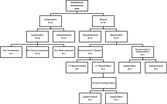
We retrospectively identified approximately 300 patients having distal femoral reconstruction with an endoprosthesis or a condyle-sparing (CS) intercalary allograft procedure for a bone sarcoma between January 1, 1988, and July 31, 2005. Patients were identified through numerous data collection sheets and an institutional database (begun in September 2001). We reviewed the surgical pathology reports for all patients to confirm diagnoses and, when available, recorded the data on distal and proximal osseous surgical margins for the condyle-sparing group only and pathologic response within the resected specimen, documenting the tumor proximity to the osteotomy. We were unable to locate tumor margin information for the endoprosthetic group. It can be presumed that the endoprosthestic group typically has closer tumor margins and thus requires wider surgical margins, which is why they are obvious candidates for the endoprosthesis and not the CS procedure. For those patients with histologic tumor data, response to chemotherapy was categorized using a modified Huvos score [18]. The Huvos score is a classification system that grades the histologic response of tumors to preoperative chemotherapy based on the percentage of tumor necrosis. We used a modified version of the original grading system by Lucas et al. [18] with “poor” = “≥ 50% viability,” “moderate” = “≥ 5% but < 50% viability,” and “excellent” = “0 ≤ 5% viability”. This was further modified to responders (< 50% tumor viability) and nonresponders (≥ 50% tumor viability). We included all patients who had their primary resection and reconstruction at one of two institutions and were followed for a minimum of 2 years (Fig. 1). Patients were included regardless of treatment status, including those who did and did not receive chemotherapy and/or radiation therapy. To be included as a candidate for the CS intercalary allograft procedure, or the “allograft” group, the distal extent of the tumor on preoperative MRI could not come within 2 cm of the joint line (nor the distal margin extend past 5 cm of the joint line to be considered “condyle-sparing”). Nine patients of the 38 patients in the CS allograft procedure were converted to an endoprosthesis after either one or two allograft reconstruction attempts; they were not included in the implant group (ie, they were censored after the first revision for their allograft and only the first revision was included in the survival curve). Two of 47 patients in the endoprosthesis group had tibial stems replaced, one after a total revision (4 years after the first revision) and the other after a femoral revision (8 years after the first revision). Their implants were not included a second time in the implant survival curve. Thus, there was no crossover between the two groups. We excluded patients from the CS allograft group if their distal osseous resection was greater than 5 cm from the medial joint line. Finally, we excluded patients who had their implant or allograft for a nonmalignant diagnosis (trauma, osteomyelitis, or benign bony disease). After exclusions, we were left with 85 patients: 47 patients with an endoprosthesis and 38 patients with a CS intercalary allograft. No patients in either group were lost to followup.
Fig. 1.
This flow chart represents our inclusion criteria for this study as well as describes the types of revision procedures patients underwent.
The CS allograft group consisted of 38 patients with a mean age of 16.3 years (range, 4.4–45.3 years) at the time of the index resection and reconstruction procedure. There were 20 (53%) males and 18 (47%) females. Patient diagnoses in this group included 28 high-grade osteosarcomas, three low-grade osteosarcomas (82% total osteosarcomas), five (13%) Ewing’s sarcomas, and two (5%) chondrosarcomas. Thirty-four (89%) of the patients had preoperative chemotherapy and two (5%) had postoperative radiation therapy. For the allograft group, the tumor response was as follows: 14 (50%) patients had excellent, five (18%) had moderate, and nine (32%) had poor response (Table 1). The mean resected length of femur was 15.3 cm (range, 6.5–31.7 cm). The mean proximal (tumor-free) osseous margin was 2.87 cm (range, 0.8–15.0 cm) and the mean distal (tumor-free) osseous margin was 1.7 cm (range, 0.3–5.0 cm). The endoprosthesis group was comprised of 47 patients, 24 (51%) males and 23 (49%) females. The mean age at the time of the index resection and reconstruction procedure was 28.1 years (range, 8.02–82.1 years). Patient diagnoses included 34 high-grade osteosarcomas, one low-grade osteosarcoma (74% total osteosarcomas), five (11%) chondrosarcomas, two (4%) Ewing’s sarcomas, four sarcomas not otherwise specified, and one leiomyosarcoma of bone (11% total “other”). Forty (85%) of the patients had neoadjuvant chemotherapy and three (8%) had adjuvant radiation therapy. The Huvos score in the endoprosthetic group was 12 (37%) had an excellent, nine (28%) had a moderate, and 11 (34%) had a poor response (Table 1). The mean resected length of femur was 16.7 cm (range, 9.0–39.3 cm). Osseous margins were not recorded for this group.
Table 1.
Demographics
| Demographics | N = 85 | Allograft (N = 38) | Implant (N = 47) |
|---|---|---|---|
| Age (mean, SD) | 16 years, 9.7 | 28 years, 19.9 | |
| < 18 years | 51 | 28 (74%) | 23 (49%) |
| ≥ 18 years | 34 | 10 (26%) | 24 (51%) |
| Gender | |||
| Female | 41 | 18 (47%) | 23 (49%) |
| Male | 44 | 20 (53%) | 24 (51%) |
| Chemotherapy | |||
| No | 11 | 4 (11%) | 7 (15%) |
| Yes | 74 | 34 (89%) | 40 (85%) |
| Radiation therapy* | |||
| No | 71 | 36 (95%) | 35 (92%) |
| Yes | 5 | 2 (5%) | 3 (8%) |
| Diagnosis | |||
| Osteosarcoma | 65 | 31(82%) | 35 (74%) |
| Chondrosarcoma | 7 | 2 (5%) | 5 (11%) |
| Ewing’s sarcoma | 7 | 5 (13%) | 2 (4%) |
| Other | 6 | — | 5 (11%) |
| Huvos score* | |||
| Poor (≥ 50% viability) | 20 | 9 (32%) | 11 (34%) |
| Moderate (≥ 5% but < 50 viability) | 14 | 5 (18%) | 9 (28%) |
| Excellent (≤ 5% viability) | 26 | 14 (50%) | 12 (37%) |
| Resection length* | |||
| < 15 cm | 42 | 17 (49%) | 25 (56%) |
| ≥ 15 cm | 38 | 18 (51%) | 20 (44%) |
| Allograft revision | |||
| No | — | 23 (61%) | — |
| Yes | — | 15 (39%) | — |
| Implant revision | |||
| No | — | — | 29 (62%) |
| Yes | — | — | 18 (38%) |
| Metastatic disease | |||
| No | 60 | 29 (76%) | 31 (66%) |
| Yes | 25 | 9 (24%) | 16 (34%) |
| Status | |||
| Alive with disease | 12 | 4 (11%) | 8 (17%) |
| Alive no disease | 51 | 26 (68%) | 25 (53%) |
| Died of disease | 17 | 8 (21%) | 9 (19%) |
| Died unknown cause | 5 | — | 5 (11%) |
* Missing data; SD = standard deviation.
We obtained positron emission tomography (PET) scans preoperatively on all patients with malignant, high-grade bone tumors beginning in 2000. Plain radiographs, MRI, and PET scans were ordered for all patients before each cycle of preoperative chemotherapy to assess tumor size and peritumoral inflammation. Patient with tumors that enlarged on coronal and axial, serial preoperative MRIs were excluded as CS candidates.
We performed endoprosthetic procedures after tumor resection through an anteromedial or anterolateral parapatellar surgical approach. Osseous margins were planned preoperatively by review of plain radiographs, MRI, and computed tomography scan. Plain radiographs were taken intraoperatively to confirm the placement of the osteotomy. Intraoperative frozen sections of marrow components at the margins of bone resection were evaluated routinely to assess negative marrow margins. All femoral implants had a rotating hinge design. Patients (83%, N = 39) had a Modular Replacement System implant (Howmedica, Mahwah, NJ) or an Orthopedic Salvage System implant (17%, N = 8) (Biomet, Warsaw, IN). We cemented femoral components in all but one case and cemented tibial components in all cases. Since 1996, our surgical implant technique has routinely included extracortical bone grafting performed with autologous bone graft placed at the junction of the host bone and the porous collar of the implant.
Most patients had supervised physical therapy with a hinged knee brace for 3 months and progressed to full weightbearing status 3 to 6 months postoperatively. We obtained radiographs every 3 months until we observed consolidation of the graft at the host-implant junction.
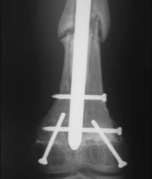
After “condyle-sparing” tumor resections, we used intercalary allografts to reconstruct the distal femur. In these CS procedures used a similar anteromedial or anterolateral parapatellar surgical approach. Using fluoroscopy with Steinman pins (Smith & Nephew, Memphis, TN) as markers for a cutting guide, we made the distal osteotomy less than or equal to 5 cm from the medial joint line. This allowed the surgeon bone stock distal to the true margin of the tumor to accomplish reconstruction after attempting adequate bony resection. Bone from the remaining distal femur as well as bone marrow from the proximal femur was routinely analyzed by frozen section intraoperatively to determine the adequacy of surgical margins. Frozen distal femoral allografts approximating the size of the patient’s femur were used to reconstruct the osseous defect with preservation of the articular cartilage of the remaining distal femoral condyles. We made 1-cm step cuts at the distal and proximal osseous graft junctions of the allograft to improve rotational stability and fixation of the graft. In some cases, the distal step cut was minimized to 5 mm when the remaining condyle fragment was small. Three of 38 (8%) allografts were fixed with plates, whereas the remaining 35 (92%) were secured with antegrade intramedullary nails with distal and proximal interlocking screws. We placed retrograde crossing screws in the medial and lateral condyle to provide distal condyle fixation in addition to fixation provided by the femoral rod (Figs. 2, 3).
Fig. 2.
An anteroposterior (AP) radiograph demonstrates a 1-cm step cut at the distal and proximal osseous graft junctions of the allograft and secured with an antegrade intramedullary nail. Note the retrograde crossing screws placed in the medial and lateral condyle to provide distal condyle fixation in addition to fixation provided by the femoral rod.
Fig. 3.
An anteroposterior (AP) radiograph shows the retrograde crossing screws placed in the medial and lateral condyle, the “condyle-sparing” approach in addition to the fixation provided by the intramedullary nail with a distal interlocking screw. There is nonunion of bone at the proximal junction.
With CS sparing surgery, patients had supervised physical therapy for 3 months and range of motion twice weekly for 6 to 12 weeks. Because allografts are slower to heal, we had patients toe-touch weightbearing for 6 weeks followed by partial weightbearing for another 6 weeks. We recommended return to full weightbearing no sooner than 6 months and in some patients 12 months when both the proximal and distal graft junction sites have healed radiographically.
Patients in both groups had routine tumor surveillance along with postoperative followup. Typically patients had clinical visits every 6 weeks for the first 6 months postoperatively and then every 3 to 6 months thereafter depending on their reconstruction procedure and tumor grade. We followed patients with a CS allograft more closely and with more imaging to check for bony healing and patients with high-grade tumors with imaging studies such as lung computed tomography and PET scans to monitor for tumor recurrence.
The primary outcome was the incidence of local recurrence. Time to local recurrence was defined as the date of surgery from the primary tumor resection and reconstruction to the time that a local recurrence was recognized clinically or radiographically, whichever came first. The secondary outcome was the incidence of allograft or implant revision. Time to allograft revision was defined as the time from date of index surgery to the time of allograft removal and replacement with either a second CS allograft or an endoprosthesis for any reason. Time to implant revision was defined as time from the date of index surgery to the time of revision of a femoral and/or tibial component for any reason. We generated Kaplan-Meier survivorship curves between the allograft and implant groups and compared them with the log rank test. All patients were entered at the date of the primary resection procedure. For the time to local recurrence survival curves, the date of local recurrence was used as the known end point and the remaining patients without a local recurrence were censored at the date of last known followup. For the revision survival curves, the date of allograft or implant revision or failure was used as the known end point, and the remaining patients without failure were censored at the date of last known followup, date of amputation from recurrent disease, or date of death. Patients who died or had an amputation for local recurrence less than 2 years after the index procedure were included in the revision analysis, because they were considered to have an intact prosthesis or allograft at the time of death or amputation.
Categorical data are presented as numbers (N) and percentages within groups. Odds ratios were calculated using the chi square test and, where cell counts (N) were low, Fisher’s exact tests are reported with 95% confidence intervals for the variables (age group [< 18 versus ≥ 18 years of age], gender, chemotherapy, radiation therapy, diagnosis, Huvos score, reconstruction group, and patient status) seen as risk factors for local recurrence (Table 2) and for revision (age group, gender, chemotherapy, radiation therapy, reconstruction group and length of resection) (Table 3). Multiple logistic regression analysis was not performed to investigate the independent factors associated with local recurrence because the post hoc power analysis suggested we had 16% power with a sample size of N1 = 38 and N2 = 47 to detect a difference of 7% in the local recurrence rate between the two groups for a two-sample t-test at alpha = 0.05. We used SAS software (Version 9.1.3; Cary, NC) for all analyses.
Table 2.
Odds ratio of risk factors and their association with local recurrence
| Risk factors | N = 85 | No local recurrence (N = 73) | Local recurrence (N = 12) | Odds ratio of local recurrence | 95% confidence interval |
|---|---|---|---|---|---|
| Age (mean, SD) | 16 years, 9.7 | 28 years, 19.9 | |||
| < 18 years | 51 | 46 (63%) | 5 (42%) | 1.00 | — |
| ≥ 18 years | 34 | 27 (37%) | 7 (58%) | 2.39 | 0.68–8.26 |
| Gender | |||||
| Female | 41 | 37 (51%) | 4 (33%) | 1.00 | — |
| Male | 44 | 36 (49%) | 8 (67%) | 2.06 | 0.57–7.43 |
| Chemotherapy | |||||
| No | 11 | 10 (14%) | 1 (8%) | 1.00 | — |
| Yes | 74 | 63 (86%) | 11 (92%) | 1.75 | 0.20–15.04 |
| Radiation therapy* | |||||
| No | 37 | 33 (89%) | 4 (80%) | 1.00 | — |
| Yes | 5 | 4 (11%) | 1 (20%) | 2.06 | 0.18–23.30 |
| Diagnosis | |||||
| Osteosarcoma | 65 | 56 (77%) | 9 (75%) | 1.00 | — |
| Chondrosarcoma | 7 | 17 (23%) | 3 (25%) | 0.91 | 0.22–3.75 |
| Ewing’s sarcoma | 7 | ||||
| Other | 6 | ||||
| Huvos score* | |||||
| Poor (≥ 50% viability) | 20 | 15 (29%) | 5 (34%) | 1.00 | — |
| Moderate (≥ 5% but < 50% viability) | 14 | 36 (71%) | 4 (44%) | 0.33 | 0.08–1.42 |
| Excellent (≤ 5% viability) | 26 | ||||
| Metastatic disease | |||||
| No | 60 | 56 (77%) | 4 (33%) | 1.00 | |
| Yes | 25 | 17 (23%) | 8 (67%) | 6.59 | 1.76–24.6 |
| Status | |||||
| Alive with disease | 12 | 60 (82%) | 3 (25%) | 1.00 | — |
| Alive no disease | 51 | ||||
| Died of disease | 17 | 13 (18%) | 9 (75%) | 13.84 | 3.29–58.3 |
| Died unknown cause | 5 | ||||
| Reconstruction group | |||||
| Allograft | 38 | 31 (42%) | 7 (58%) | 1.00 | |
| Implant | 47 | 42 (58%) | 5 (42%) | 0.53 | 0.15–1.82 |
* Missing data; SD = standard deviation.
Table 3.
Odds ratio of risk factors and their association with deep infection
| Risk Factors | N = 85 | No deep infection (N = 52) | Deep infection (N = 33) | Odds ratio of deep infection | 95% confidence interval |
|---|---|---|---|---|---|
| Reconstruction group | |||||
| Allograft | 38 | 31 (41%) | 7 (70%) | 1.00 | |
| Implant | 47 | 44 (59%) | 3 (30%) | 0.30 | 0.07–1.26 |
| Reconstruction failure | |||||
| Not Revised | 52 | 52 (50%) | 0 (57%) | 1.00 | — |
| Revised | 33 | 23 (50%) | 10 (43%) | 1.43 | 1.15–1.80 |
* Missing data.
Results
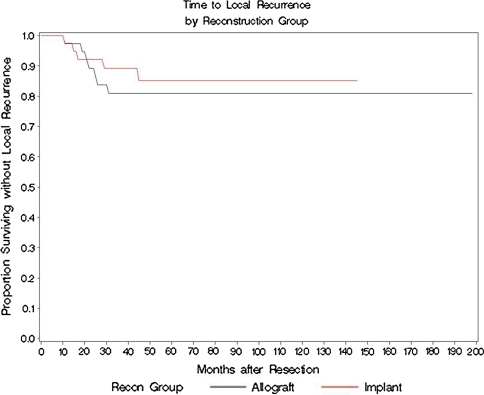
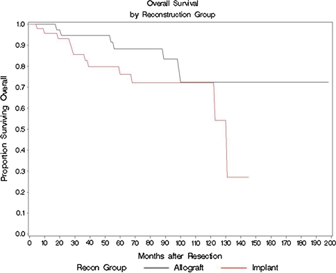
The percentage of local recurrences was similar (p = 0.36) in the patients having implants and those having allografts (five of 47 [11%] versus seven of 38 [18%], respectively). Patients who had a local recurrence were more than 6.5 times more likely to have subsequent metastatic disease (p = 0.002) and almost 14 times more likely to die of their disease (p < 0.0001) (Table 2). All other factors such as age group, gender, chemotherapy, radiation therapy, diagnosis, Huvos score, and revision group were similar for local recurrence. Of the seven of 38 patients with allografts who had local recurrences, six had amputations, and among the five patients out of the 47 with implants having a local recurrence, four had amputations. Using time to local recurrence as the end point, survivorship in the implant group versus the CS allograft group was similar (p = 0.18). The rates for survival at 2, 5, and 10 years for the implant group versus the CS allograft were as follows: 93% versus 89% at 2 years, 87% versus 81% at 5 years, and 87% versus 81% at 10 years, respectively (Fig. 4). The mean time to local recurrence for implants was 42.0 ± 1.5 months and for the CS allograft group, it was 28.5 months ± 0.7 months. Survivorship with the end point being date of death or date of last followup was also similar (p = 0.19) with survival of the implant group versus the CS allograft group being 94% versus 95% at 2 years, 75% versus 86% at 5 years, and 72% versus 72% at 10 years, respectively) (Fig. 5). The mean time to overall survival for the implant group was 103.9 ± 6.8 months and for the allograft group was 90.2 ± 4.0 months.
Fig. 4.
The Kaplan-Meier survival curve demonstrating similar time to local recurrence between the implant group and the condyle-sparing (CS) allograft group at 2, 5, and 10 years: 93% versus 89% at 2 years, 87% versus 81% at 5 years, and 87% versus 81% at 10 years, respectively.
Fig. 5.
The Kaplan-Meier overall survivorship (end point was date of death or date of last followup) showing similar survival of the implant group versus the condyle-sparing (CS) allograft group: 94% versus 95% at 2 years, 75% versus 86% at 5 years, and 72% versus 72% at 10 years, respectively.
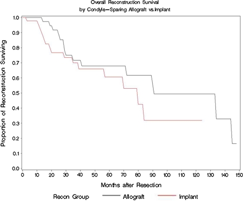
We observed similar (p = 0.99) percentages of revisions in patients with allografts and endoprostheses: 15 of 38 patients (39%) with allografts required revision of their first allograft and replacement with either a second allograft or reconstruction and 18 of 47 (38%) with endoprosthesis. Eight patients with CS allografts elected to have a second allograft procedure to replace their failed graft, whereas the remaining seven patients opted to replace their failed allograft with an endoprosthesis (Fig. 1). The most common reason for failure in the allograft group was the result of a deep infection (seven of 38 [47%]) and in the implant group it was aseptic loosening (18 of 47 [44%]) (Table 4). Patients with deep infection are 43% more likely to be revised than those without a deep infection (p < 0.0001) (Table 3). No other factors predicted revision (Table 5). Kaplan-Meier survivorship was similar (p = 0.18) when comparing the survival of the surgical reconstruction using revision as an end point for the allograft versus implant groups, which was 92% versus 76% at 2 years, 70% versus 64% at 5 years, and 53% versus 36% at 10 years, respectively (Fig. 6). The mean allograft revision survival time was 97.0 ± 9.9 months compared with the mean implant revision survival time of 61.8 ± 5.2 months.
Table 4.
Reasons for failure
| Reasons for failure | Allograft group (N = 15) | Implant group (N = 18) |
|---|---|---|
| Aseptic loosening | — | 8 (44%) |
| Deep infection | 7 (47%) | 3 (17%) |
| Graft/tibial/stem fracture | 1 (7%) | 3 (17%) |
| Local recurrence | 1 (7%) | 1 (6%) |
| Malalignment | 3 (17%) | |
| Resulting from tibial polyethylene wear | 2 | |
| True malalignment | 1 | |
| Persistent distal nonunion | 6 (40%) | — |
Table 5.
Odds ratio of risk factors and their association with revision status
| Risk factors | N = 85 | Not revised (N = 52) | Revised (N = 33) | Odds ratio of revision | 95% confidence interval |
|---|---|---|---|---|---|
| Age | |||||
| < 18 years | 51 | 30 (58%) | 21 (64%) | 1.00 | — |
| ≥ 18 years | 34 | 22 (42%) | 12 (36%) | 0.78 | 0.32–1.91 |
| Gender | |||||
| Female | 41 | 25 (48%) | 16 (48%) | 1.00 | — |
| Male | 44 | 27 (52%) | 17 (52%) | 0.98 | 0.41–2.36 |
| Chemotherapy | |||||
| No | 11 | 7 (13%) | 4 (12%) | 1.00 | — |
| Yes | 74 | 45 (87%) | 29 (88%) | 1.13 | 0.30–4.20 |
| Radiation therapy* | |||||
| No | 37 | 23 (82%) | 14 (100%) | 1.00 | — |
| Yes | 5 | 5 (18%) | 0 (0%) | 0.15 | 0.008–2.87 |
| Reconstruction group | |||||
| Allograft | 38 | 23 (44%) | 15 (45%) | 1.00 | |
| Implant | 47 | 29 (56%) | 18 (56%) | 0.95 | 0.40–2.29 |
| Resection length* | |||||
| < 15 cm | 42 | 25 (50%) | 17 (57%) | 1.00 | — |
| ≥ 15 cm | 38 | 25 (50%) | 13 (43%) | 0.76 | 0.31–1.90 |
* Missing data.
Fig. 6.
The Kaplan-Meier survivorship showing similar survival of the surgical reconstruction using revision as an end point for the allograft versus implant groups: 92% versus 76% at 2 years, 70% versus 64% at 5 years, and 53% versus 36% at 10 years, respectively.
Discussion
The treatment of malignant tumors of the distal femur has evolved considerably over the past 20 years. As a result of the implementation of neoadjuvant chemotherapy, improvement in orthopaedic implant technology, and the refinement of surgical indications and techniques, limb salvage procedures are now commonplace. Endoprosthetic reconstructions replace the native articular surface of the knee with a prosthetic implant that has a higher failure rate than conventional arthroplasty of the knee [22, 28]. CS procedures preserve the native articular surface and avoid the need for arthroplasty of the knee with a possible increase in local recurrence and graft healing complications [21, 24]. Comparison of these two techniques reflects the decisions tumor surgeons face: the decision to provide adequate surgical margins to prevent local recurrence while salvaging enough bone to preserve longevity and function of the affected limb. We therefore compared the recurrence and revision rates of endoprosthetic reconstruction and “condyle-sparing” intercalary allograft in patients treated with malignant tumors of the distal femur.
This study had several limitations in design. First, a matched case-control based on age, tumor diagnosis, grade, and treatment would have reduced confounding but based on our post hoc power analysis, we only had a 16% chance of obtaining a significant result at the 5% level so all significant results should be interpreted with caution. However, when the incidence of a sarcoma is approximately 10,000 new cases each year (less than 1% of all adult cancers diagnosed), it is difficult to obtain a large sample size unless one attempts a multi-institutional trial. Second, the retrospective nature of the study underscores the evolution of the CS technique as it was implemented and refined by the senior surgeon. Although one surgeon performed the surgery, surgical technique was not strictly controlled and there was selection bias in regard to diagnostic categories and the extent of osseous resection. We did not control for the use of different implant manufacturers, implant design, and stem geometry in this study. Body mass index and other comorbidities (such as the use of certain types of chemotherapeutic drugs [MAID {combined mesna, doxorubicin, ifosfamide, and dacarbazine} or AIM {doxorubicin, ifosfamide, and mesna}, radiation treatments {external beam verses brachytherapy}]) also may affect the validity of these results because they were not collected and thus not eliminated as confounding variables. Third, we did not randomly select patients to enter either treatment arm, and the decision to perform a CS procedure was only offered to those patients whose distal extent of the tumor on MRI was more than 1 cm from the distal physis. Patients and their families were informed preoperatively of the potentially higher risk of the CS procedure and were given the option of the more “traditional” endoprosthetic procedure or amputation after tumor resection. Hence, the patient populations constituting these two cohorts are not truly equal.
We observed no difference in the local recurrence rate between patients who had endoprosthetic reconstructions (11%) and those who had the CS procedure (18%). Local recurrence with limb salvage is reportedly between 5% and 10% [28]. The literature from the last 10 years regarding the risk of local recurrences with limb salvage demonstrates a mean local recurrence rate of 6.9% (range, 0%–18%) (Table 6). In the same studies, the average local recurrence rates for implants (6.2%) versus allografts (7.6%) demonstrated a slight difference between the two groups. Overall patient survival was equal (72%) at 10 years between the CS allograft group and the endoprosthetic group. Patients with metastatic disease were more likely to experience a local recurrence regardless of their reconstruction treatment group.
Table 6.
Comparison of implant and allograft local recurrence and complication rates
| Year | Author, citation | Type of limb salvage | Cohort | Age | Length of followup | Local recurrence rate | Complication rate |
|---|---|---|---|---|---|---|---|
| 1991 | Eckardt et al. [6] | Implant | 52 DF, 78 Total | Mean: 17 years (6–75 years) | Estimated 2–198 months | 5% | Aseptic loosening—3% |
| Infection—3% | |||||||
| Mechanical/fracture—8% | |||||||
| 1994 | Safran et al. [27] | Implant | 81 DF, 151 Total | 4–83 years | Mean: 52 months (24–114 months) | 6% | Aseptic loosening—4% |
| Infection—5% | |||||||
| Mechanical/fracture—12% | |||||||
| 1995 | Malawer and Chou [19] | Implant | 31 DF, MF, 82 Total | Median: 19 years (5–78 years) | 36–l20 months | 6% | Aseptic loosening—5% |
| Infection—13% | |||||||
| Mechanical/fracture—4% | |||||||
| 1998 | Kawai et al. [12, 15] | Implant | 40 DF total | Mean: 26 years (12–68 years) | Mean: 100 months (60–120 + months) | None observed | Aseptic loosening—40% |
| Infection—10% | |||||||
| Mechanical/fracture—25% | |||||||
| 1999 | Kawai et al. [13, 14] | Implant | 82 DF, 111 total | Mean: 27 years (12–71 years) | Mean: 67 months (24–204 months) | 2% (2/111) | Aseptic loosening—22% |
| Infection—6% | |||||||
| Mechanical/fracture—18% | |||||||
| 2002 | Bickels et al. [2] | Implant | 110 DF total | Median: 21 years (10–80 years) | Median: 94 months (24–198 months) | 5% | Aseptic loosening—5% |
| Infection—5% | |||||||
| Mechanical/fracture—5% | |||||||
| 2004 | Zeegen et al. [33] | Implant | 55 DF, 141 total | Mean: 49 years (8–87 years) | Mean: 18 months (6–96 months) | 4% | Aseptic loosening—8% |
| Infection—10% | |||||||
| Mechanical/fracture—2% | |||||||
| 2006 | Morgan et al. [22] | Implant | 76 DF, 105 total | Median: 22 years (9–86 years) | Median: 57 months (1–235 months) | 2% | Aseptic loosening—17% |
| Infection—7% | |||||||
| Mechanical/fracture—7% | |||||||
| 2009 | Zimel et al. [current study] | Implant | 47 DF | Mean: 28 years (9–82 years) | Mean: 68 months (1–175 months) | 11% | Aseptic loosening—17% |
| Infection—7% | |||||||
| Mechanical/fracture—11% | |||||||
| Summary | Implant | 64 DF, 103 total | Mean: 26 years (17–49 years) | Mean: 65 months (18–100 months) | 5% | Aseptic loosening—13% | |
| Infection—7% | |||||||
| Mechanical/fracture—10% | |||||||
| 1995 | Alman et al. [1] | Allograft | 8 F, 26 total | Mean: 12 years (5–17 years) | Mean: 63 months (3–134 months) | 4% | Nonunion—15% |
| Infection—12% | |||||||
| Fracture—55% | |||||||
| 1996 | Mankin et al. [21] | Allograft | 271 DF, 85 F, 818 total | Mean: 32 years (4–80 years) | Mean: 72 months (24–245 months) | 10% | Nonunion—17% |
| Infection—11% | |||||||
| Fracture—19% | |||||||
| 1997 | Ortiz-Cruz et al. [24] | Allograft | 39 F, 104 total | Mean: 28 years (4–69 years) | Mean: 73 months (24–220 months) | 9% | Nonunion—30% |
| Infection—12% | |||||||
| Fracture—17% | |||||||
| 2004 | Muscolo et al. [23] | Allograft | 13 Condyle-sparing | Mean: 18 years (9–40 years) | Mean: 63 months (24–144 months) | 8% | Nonunion—15% |
| Infection—8% | |||||||
| Fracture—23% | |||||||
| 2009 | Zimel et al. [current study] | Allograft | 38 Condyle-sparing | Mean: 16 years (4–42 years) | Mean: 105 months (14–231 months) | 18% | Nonunion—16% |
| Infection—18% | |||||||
| Fracture—3% | |||||||
| Summary | Allograft | 91 F, 200 total | Mean: 21 years (12–32 years) | Mean: 75 months (63–105 months) | 10% | Nonunion—19% | |
| Infection—12% | |||||||
| Fracture—23% |
F = femur; DF = distal femur; MF = midfemur; Total = total patients in cohort.
The CS procedure requires careful attention to the preoperative evaluation of the distal osseous surgical margins with preoperative MRI. As reported by Muscolo et al. [23], with the advances in imaging, this careful attention to the bony surgical margins during preoperative planning gives us the ability to perform this technique. Inherent with the CS technique is a smaller osseous surgical margin that assumes a theoretical increased risk of local recurrence [23]. Although we found no increased risk of local recurrence, we believe the point remains important point of consideration for both the surgeon and the patient. All patients should be carefully informed preoperatively of these techniques and their risks.
We found the rates of revision similar in the two groups, 39% in the allograft group and 38% in the implant group. Revision rates in the literature range from 4% to 41% for implants and from 15% to 27% for the allograft group (Table 5). The reconstruction survival at 10 years was 53% for the allograft group and 36% for the implant group. Given the similar rates of revision in this series, retaining the native knee is preferable, especially in a younger population whose growth plates are still open. The most likely explanation for the higher allograft revision rate in this group compared with allograft complication rates reported in the literature is that the graft junction site approximating the condyle is riskier than a middiaphyseal segment or condyle-replacing procedure.
We report a retrospective review comparing the local recurrence and revision rates for the orthopaedic management of malignant tumors of the distal femur. Patients undergoing the CS procedure had similar rates of local recurrence and revision compared with patients having the endoprosthetic procedure. The CS allograft procedure offers the potential advantage of retaining the native knee in a young patient population while incurring similar risk of local recurrence as those offered the endoprosthetic procedure.
Acknowledgments
We thank Thomas Scharschmidt, MD, and Jedediah White for their assistance in preparing the manuscript for publication.
Footnotes
One of the authors (EUC) has received funding from Stryker Orthopaedics (Mahwah, NJ).
Each author certifies that his or her institution has approved the human protocol for this investigation, that all investigations were conducted in conformity with ethical principles of research, and that informed consent for participation in the study was obtained.
This work was performed at the University of Washington Medical Center, Seattle, WA.
References
- 1.Alman BA, De Bari A, Krajbich JI. Massive allografts in the treatment of osteosarcoma and Ewing sarcoma in children and adolescents. J Bone Joint Surg Am. 1995;77:54–64. [DOI] [PubMed]
- 2.Bickels J, Wittig JC, Kollender Y, Henshaw RM, Kellar-Graney KL, Meller I, Malawer MM. Distal femur resection with endoprosthetic reconstruction: a long-term followup study. Clin Orthop Relat Res. 2002;400:225–235. [DOI] [PubMed]
- 3.Bradish CF, Kemp HB, Scales JT, Wilson JN. Distal femoral replacement by custom-made prostheses. Clinical follow-up and survivorship analysis. J Bone Joint Surg Br. 1987;69:276–284. [DOI] [PubMed]
- 4.Brien E, Terek R, Healey J, Lane J. Allograft reconstruction after proximal tibial resection for bone tumors. An analysis of function and outcome comparing allograft and prosthetic reconstructions. Clin Orthop Relat Res. 1994;303:116–127. [PubMed]
- 5.Dubousset J, Missenard G, Kalifa C. Management of osteogenic sarcoma in children and adolescents. Clin Orthop Relat Res. 1991;270:52–59. [PubMed]
- 6.Eckardt JJ, Eilber FR, Rosen G, Mirra JM, Dorey FJ, Ward WG, Kabo JM. Endoprosthetic replacement for stage IIB osteosarcoma. Clin Orthop Relat Res. 1991;270:202–213. [PubMed]
- 7.Finn HA, Simon MA. Limb-salvage surgery in the treatment of osteosarcoma in skeletally immature individuals. Clin Orthop Relat Res. 1991;262:108–118. [PubMed]
- 8.Ham SJ, Schraffordt Koops H, Veth RP, van Horn JR, Molenaar WM, Hoekstra HJ. Limb salvage surgery for primary bone sarcoma of the lower extremities: long-term consequences of endoprosthetic reconstructions. Ann Surg Oncol. 1998;5:423–436. [DOI] [PubMed]
- 9.Harris IE, Leff AR, Gitelis S, Simon MA. Function after amputation, arthrodesis, or arthroplasty for tumors about the knee. J Bone Joint Surg Am. 1990;72:1477–1485. [PubMed]
- 10.Hillmann A, Hoffmann C, Gosheger G, Krakau H, Winkelmann W. Malignant tumor of the distal part of the femur or the proximal part of the tibia: endoprosthetic replacement or rotationplasty. Functional outcome and quality-of-life measurements. J Bone Joint Surg Am. 1999;81:462–468. [DOI] [PubMed]
- 11.Horowitz SM, Glasser DB, Lane JM, Healey JH. Prosthetic and extremity survivorship after limb salvage for sarcoma. How long do the reconstructions last? Clin Orthop Relat Res. 1993;293:280–286. [PubMed]
- 12.Kawai A, Backus SI, Otis JC, Healey JH. Interrelationships of clinical outcome, length of resection, and energy cost of walking after prosthetic knee replacement following resection of a malignant tumor of the distal aspect of the femur. J Bone Joint Surg Am. 1998;80:822–831. [DOI] [PubMed]
- 13.Kawai A, Healey JH, Boland PJ, Athanasian EA, Jeon DG. A rotating-hinge knee replacement for malignant tumors of the femur and tibia. J Arthroplasty. 1999;14:187–196. [DOI] [PubMed]
- 14.Kawai A, Lin PP, Boland PJ, Athanasian EA, Healey JH. Relationship between magnitude of resection, complication, and prosthetic survival after prosthetic knee reconstructions for distal femoral tumors. J Surg Oncol. 1999;70:109–115. [DOI] [PubMed]
- 15.Kawai A, Muschler GF, Lane JM, Otis JC, Healey JH. Prosthetic knee replacement after resection of a malignant tumor of the distal part of the femur. Medium to long-term results. J Bone Joint Surg Am. 1998;80:636–647. [DOI] [PubMed]
- 16.Krepler P, Dominkus M, Toma CD, Kotz R. Endoprosthesis management of the extremities of children after resection of primary malignant bone tumors [in German]. Orthopade. 2003;32:1013–1019. [DOI] [PubMed]
- 17.Liptak J, Dernell W, Ehrhart N, Lafferty M, Monteith G, Withrow S. Cortical allograft and endoprosthesis for limb-sparing surgery in dogs with distal radial osteosarcoma: a prospective clinical comparison of two different limb-sparing techniques. Vet Surg. 2006;35:518–533. [DOI] [PubMed]
- 18.Lucas D, Kshirsagar M, Biermann J, Hamre M, Thomas D, Schuetze S, Baker L. Histologic alterations from neoadjuvant chemotherapy in high-grade extremity soft tissue sarcoma: clinicopathological correlation. Oncologist. 2008;13:451–458. [DOI] [PubMed]
- 19.Malawer MM, Chou LB. Prosthetic survival and clinical results with use of large-segment replacements in the treatment of high-grade bone sarcomas. J Bone Joint Surg Am. 1995;77:1154–1165. [DOI] [PubMed]
- 20.Mankin HJ. The changes in major limb reconstruction as a result of the development of allografts. Chir Organi Mov. 2003;88:101–113. [PubMed]
- 21.Mankin HJ, Gebhardt MC, Jennings LC, Springfield DS, Tomford WW. Long-term results of allograft replacement in the management of bone tumors. Clin Orthop Relat Res. 1996;324:86–97. [DOI] [PubMed]
- 22.Morgan HD, Cizik AM, Leopold SS, Hawkins DS, Conrad EU 3rd. Survival of tumor megaprostheses replacements about the knee. Clin Orthop Relat Res. 2006;450:39–45. [DOI] [PubMed]
- 23.Muscolo DL, Ayerza MA, Aponte-Tinao LA, Ranalletta M. Partial epiphyseal preservation and intercalary allograft reconstruction in high-grade metaphyseal osteosarcoma of the knee. J Bone Joint Surg Am. 2004;86:2686–2693. [DOI] [PubMed]
- 24.Ortiz-Cruz E, Gebhardt MC, Jennings LC, Springfield DS, Mankin HJ. The results of transplantation of intercalary allografts after resection of tumors. A long-term follow-up study. J Bone Joint Surg Am. 1997;79:97–106. [DOI] [PubMed]
- 25.Renard AJ, Veth RP, Schreuder HW, van Loon CJ, Koops HS, van Horn JR. Function and complications after ablative and limb-salvage therapy in lower extremity sarcoma of bone. J Surg Oncol. 2000;73:198–205. [DOI] [PubMed]
- 26.Rougraff BT, Simon MA, Kneisl JS, Greenberg DB, Mankin HJ. Limb salvage compared with amputation for osteosarcoma of the distal end of the femur. A long-term oncological, functional, and quality-of-life study. J Bone Joint Surg Am. 1994;76:649–656. [DOI] [PubMed]
- 27.Safran MR, Kody MH, Namba RS, Larson KR, Kabo JM, Dorey FJ, Eilber FR, Eckardt JJ. 151 endoprosthetic reconstructions for patients with primary tumors involving bone. Contemp Orthop. 1994;29:15–25. [PubMed]
- 28.Simon MA, Aschliman MA, Thomas N, Mankin HJ. Limb-salvage treatment versus amputation for osteosarcoma of the distal end of the femur. J Bone Joint Surg Am. 1986;68:1331–1337. [PubMed]
- 29.Simon MA, Aschliman MA, Thomas N, Mankin HJ. Limb-salvage treatment versus amputation for osteosarcoma of the distal end of the femur. 1986. J Bone Joint Surg Am. 2005;87:2822. [DOI] [PubMed]
- 30.Tunn PU, Schmidt-Peter P, Pomraenke D, Hohenberger P. Osteosarcoma in children: long-term functional analysis. Clin Orthop Relat Res. 2004;421:212–217. [DOI] [PubMed]
- 31.Weiner SD, Scarborough M, Vander Griend RA. Resection arthrodesis of the knee with an intercalary allograft. J Bone Joint Surg Am. 1996;78:185–192. [DOI] [PubMed]
- 32.Wittig JC, Bickels J, Priebat D, Jelinek J, Kellar-Graney K, Shmookler B, Malawer MM. Osteosarcoma: a multidisciplinary approach to diagnosis and treatment. Am Fam Physician. 2002;65:1123–1132. [PubMed]
- 33.Zeegen EN, Aponte-Tinao LA, Hornicek FJ, Gebhardt MC, Mankin HJ. Survivorship analysis of 141 modular metallic endoprostheses at early followup. Clin Orthop Relat Res. 2004;420:239–250. [DOI] [PubMed]


